. 2014 Jul;4(7):140063.
doi: 10.1098/rsob.140063.

Optimization of the analogue-sensitive Cdc2/Cdk1 mutant by in vivo selection eliminates physiological limitations to its use in cell cycle analysis

Affiliations

Optimization of the analogue-sensitive Cdc2/Cdk1 mutant by in vivo selection eliminates physiological limitations to its use in cell cycle analysis

Yuki Aoi et al. Open Biol. 2014 Jul.

Abstract

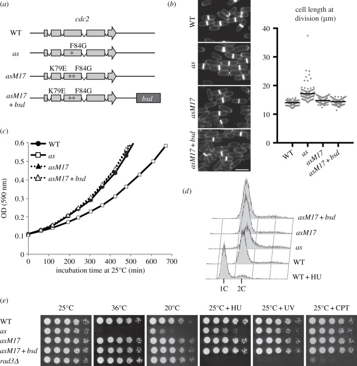
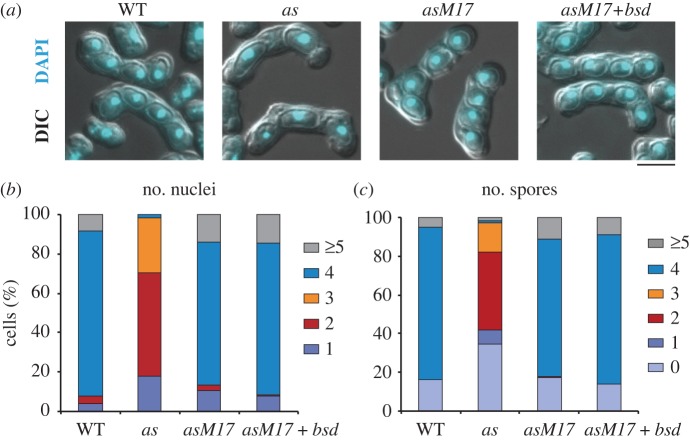
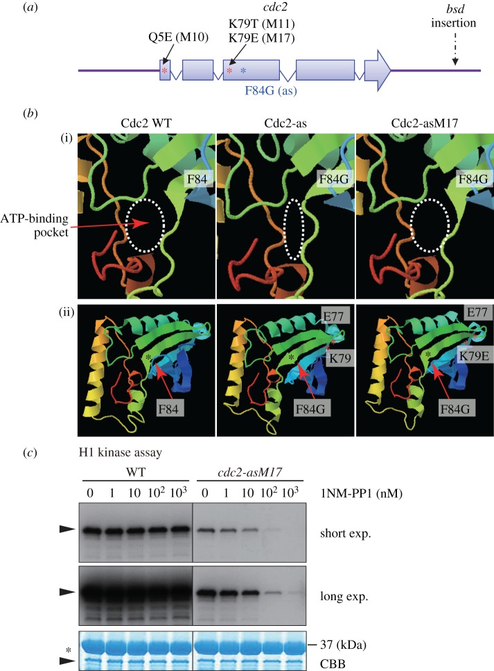
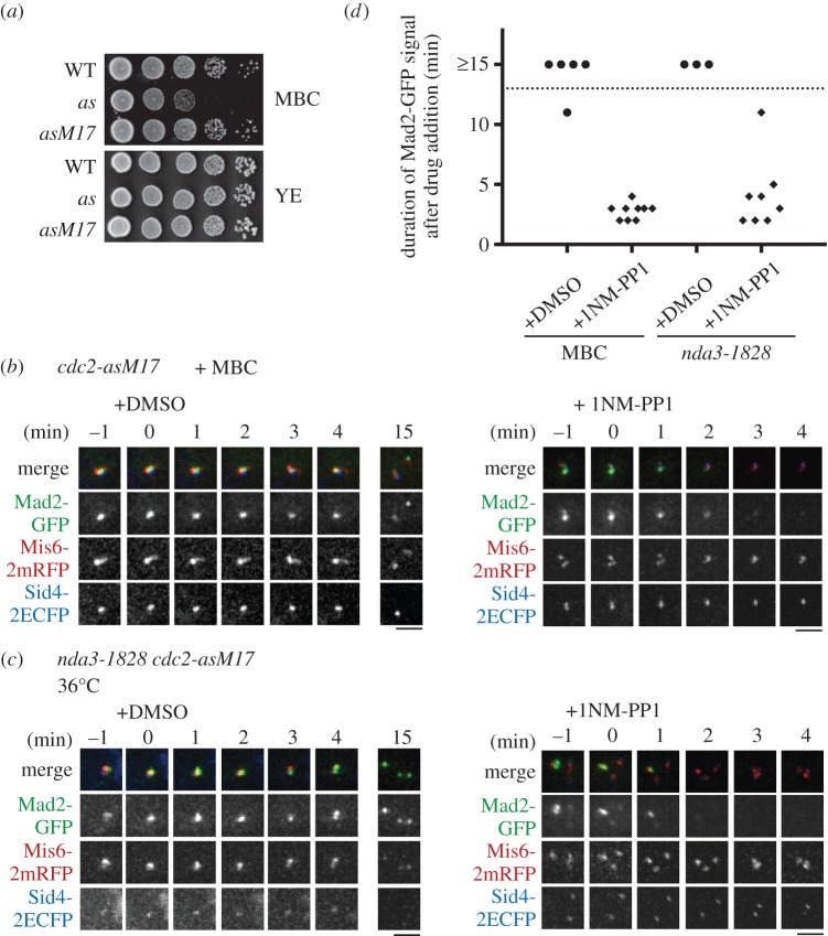
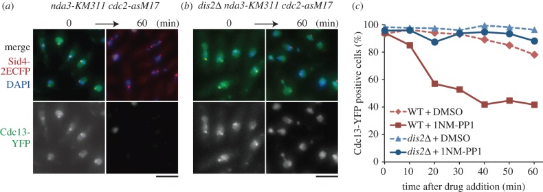
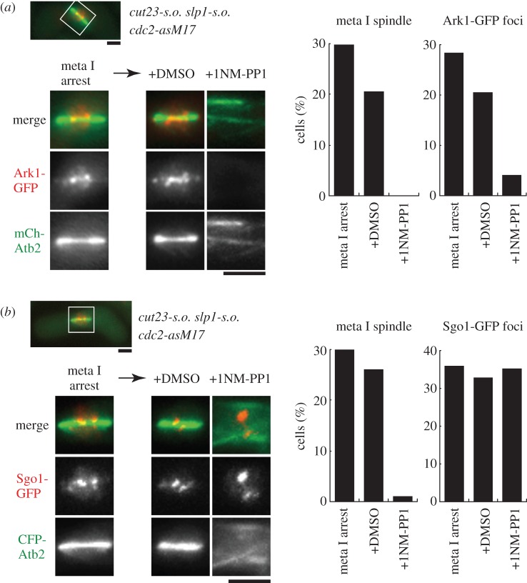
Analogue-sensitive (as) mutants of kinases are widely used to selectively inhibit a single kinase with few off-target effects. The analogue-sensitive mutant cdc2-as of fission yeast (Schizosaccharomyces pombe) is a powerful tool to study the cell cycle, but the strain displays meiotic defects, and is sensitive to high and low temperature even in the absence of ATP-analogue inhibitors. This has limited the use of the strain for use in these settings. Here, we used in vivo selection for intragenic suppressor mutations of cdc2-as that restore full function in the absence of ATP-analogues. The cdc2-asM17 underwent meiosis and produced viable spores to a similar degree to the wild-type strain. The suppressor mutation also rescued the sensitivity of the cdc2-as strain to high and low temperature, genotoxins and an anti-microtubule drug. We have used cdc2-asM17 to show that Cdc2 activity is required to maintain the activity of the spindle assembly checkpoint. Furthermore, we also demonstrate that maintenance of the Shugoshin Sgo1 at meiotic centromeres does not require Cdc2 activity, whereas localization of the kinase aurora does. The modified cdc2-asM17 allele can be thus used to analyse many aspects of cell-cycle-related events in fission yeast.

Keywords: analogue-sensitive mutant; cell cycle; chemical genetics; cyclin-dependent kinase; fission yeast.


Figures

Figure 1.
Figure 1.
Characterization of the cdc2-asM17 mutant in mitotic cell cycle. (a) Schematic of wild-type cdc2 (WT), cdc2-as (as), cdc2-asM17 (asM17) and cdc2-asM17 + bsd (asM17 + bsd) mutant genes. The bsd marker was inserted in the downstream of the cdc2 coding sequence. (b) Calcofluor staining of vegetative cells at 25°C. Scale bar, 10 µm. The scatter-dot plot indicates distribution of cell length at cell division (µm; n ≥ 100). Black bars indicate mean values (mean ± s.e.: WT = 14.0 ± 0.1, as = 17.2 ± 0.3, asM17 = 14.8 ± 0.1, asM17 + bsd = 14.3 ± 0.1). (c) OD (590 nm) measurement of log-phase cultures at 25°C. (d) FACS results showing the DNA content of vegetative cells at 25°C. For control of 1C DNA content, 12 mM HU was added to the WT culture (WT + HU). (e) Fivefold dilutions of the indicated strains were spotted onto the following media: YE containing 5 mM HU or 5 µM CPT, YE irradiated with 100 J m−2 UV. Plates were incubated at the indicated temperature for 3–6 days.
Figure 2.
Figure 2.
Characterization of the cdc2-asM17 mutant in meiosis. (a) DAPI staining of cells that underwent mating, meiosis and sporulation. DAPI and differential interference contrast (DIC) images are shown merged. Scale bar, 5 µm. (b,c) The numbers of (b) nuclei and (c) spores in each ascus shown in (a) were counted for the indicated strains and the percentages are shown. (n > 200).
Figure 3.
Figure 3.
Intragenic suppressor mutations of the isolated cdc2 mutants. (a) The schematic of the cdc2 gene. The analogue-sensitive mutation is F84G [17]. The suppressor mutant M10 contained the Q5E substitution. The M11 and M17 mutants shared the mutation site K79 to T (M11) and to E (M17) in the open reading frame of the cdc2 gene, in addition to the F84G mutation. (b) The prediction of secondary and tertiary structure of Cdc2 WT (wild-type), Cdc2-as and Cdc2-asM17 proteins made by the Phyre program. (i) Magnified view around the ATP-binding pocket and the gatekeeper residue F84 (or F84G). (ii) The overview for (i). The mutation site K79E and a reference site E77 are shown with red asterisks, and F84G is shown with black asterisks. (c) In vitro kinase assay using Cdc2 WT and Cdc2-asM17 proteins purified from WT and the mutant strains, respectively. The Cdk1 substrate histone H1 was incubated with purified Cdc2 proteins and [32P]-α-ATP in the indicated concentration of 1NM-PP1. Autoradiograph images with a short and long exposure are shown. CBB; Coomassie brilliant blue straining for the loading control. Arrowheads indicate the position of histone H1, and the asterisk band corresponds to GST-tagged p13Suc1 derived from Suc1-beads used in affinity purification.
Figure 4.
Figure 4.
The requirement of Cdc2 in SAC maintenance was revealed by use of the cdc2-asM17 mutant. (a) The original cdc2-as mutant (as) was sensitive to a low dose of the microtubule drug MBC (10 µg ml−1), whereas the revived mutant cdc2-asM17 (asM17) was not. This enabled use of the analogue-sensitive cdc2 mutant in the presence of MBC. (b) The cdc2-asM17 mutant was used in combination with MBC treatment. cdc2-asM17 cells were treated with MBC to arrest cells at metaphase without spindles. Cells with Mad2-GFP dots at kinetochores were chosen for filming. After 1NM-PP1 addition (t = 0 min), Mad2-GFP dots disappeared within 4 min. DMSO was added as negative control. Mis6-2mRFP, a kinetochore marker; Sid4-2ECFP, an SPB marker. (c) The cdc2-asM17 mutation was combined with the β-tubulin ts mutation nda3/alp12–1828. Cells were arrested at metaphase at 36°C. Filming was done similarly to (b). After 1NM-PP1 addition (t = 0 min), Mad2-GFP dots disappeared within 3 min. DMSO was added as negative control. Scale bars, 2 µm. (d) Duration of Mad2-GFP dots residence at kinetochores after addition of DMSO or 1NM-PP1 in (b,c) was measured.
Figure 5.
Figure 5.
Dis2/PP1 is required for checkpoint inactivation triggered by Cdc2 inhibition. (a) The cdc2-asM17 mutation was combined with the cs β-tubulin mutation nda3-KM311. The mutant was arrested at metaphase at 18°C with high concentration of cyclin B1/Cdc13-YFP in the nucleus and at SPBs (0 min). Then, 1NM-PP1 was added to the culture. After 60 min, the percentage of cells with Cdc13-YFP decreased. Sid4–2ECFP (SPB) and DAPI are also shown. (b) Similar experiments were done with the triple mutant dis2Δ nda3-KM311 cdc2-asM17. Cells were cultured at 18°C (0 min) and then 1NM-PP1 was added. In contrast to dis2+ cells (a), Cdc13-YFP remained in the nucleus. Scale bars, 5 µm. (c) Frequencies of cells with nuclear Cdc13-YFP signal in dis2+ (WT) and dis2Δ strains in the nda3-KM311 cdc2-asM17 background were plotted in response to 1NM-PP1 (or DMSO for negative control) addition (n ≥ 100).
Figure 6.
Figure 6.
Cdc2 activity is required for aurora-B localization but not for the Shugoshin Sgo1 during meiosis I. (a) The cdc2-asM17 mutation was combined with slp1-s.o. and cut23-s.o. mutations, in which meiotic transcription of these APC/C factors is shut off and cells are arrested in metaphase I. The rectangular region was magnified and is shown below. The aurora B/Ark1-GFP localized to centromeres on the metaphase spindle, which was visualized by CFP-Atb2 (α2-tubulin). Ark1-GFP foci dispersed after 1NM-PP1 treatment, but not after DMSO treatment (negative control). Images corresponding to the nuclear region were taken 1 h after addition of 1NM-PP1 or DMSO. The frequencies of cells with the metaphase I spindle (the left graph) and with Ark1-GFP foci (the right graph) are also shown (n > 100). (b) Similar experiments to (a) were done for Sgo1-GFP. Sgo1-GFP foci at centromeres did not disperse even 1 h after 1NM-PP1 addition (n > 100). Scale bars, 2 µm.

References

    1. Wood V, et al. 2012. PomBase: a comprehensive online resource for fission yeast. Nucleic Acids Res. 40, D695–D699. (doi:10.1093/nar/gkr853) - DOI - PMC - PubMed
    1. Kawashima SA, Takemoto A, Nurse P, Kapoor TM. 2013. A chemical biology strategy to analyze rheostat-like protein kinase-dependent regulation. Chem. Biol. 20, 262–271. (doi:10.1016/j.chembiol.2013.01.003) - DOI - PMC - PubMed
    1. Bishop AC, et al. 2000. A chemical switch for inhibitor-sensitive alleles of any protein kinase. Nature 407, 395–401. (doi:10.1038/35030148) - DOI - PubMed
    1. Ubersax JA, Woodbury EL, Quang PN, Paraz M, Blethrow JD, Shah K, Shokat KM, Morgan DO. 2003. Targets of the cyclin-dependent kinase Cdk1. Nature 425, 859–864. (doi:10.1038/nature02062) - DOI - PubMed
    1. Kung C, Kenski DM, Dickerson SH, Howson RW, Kuyper LF, Madhani HD, Shokat KM. 2005. Chemical genomic profiling to identify intracellular targets of a multiplex kinase inhibitor. Proc. Natl Acad. Sci. USA 102, 3587–3592. (doi:10.1073/pnas.0407170102) - DOI - PMC - PubMed

Publication types

MeSH terms

Substances