. 2014 Sep;15(9):982-90.
doi: 10.15252/embr.201438463. Epub 2014 Jul 2.

Mitochondrial apoptosis is dispensable for NLRP3 inflammasome activation but non-apoptotic caspase-8 is required for inflammasome priming

Affiliations

Mitochondrial apoptosis is dispensable for NLRP3 inflammasome activation but non-apoptotic caspase-8 is required for inflammasome priming

Ramanjaneyulu Allam et al. EMBO Rep. 2014 Sep.

Abstract

A current paradigm proposes that mitochondrial damage is a critical determinant of NLRP3 inflammasome activation. Here, we genetically assess whether mitochondrial signalling represents a unified mechanism to explain how NLRP3 is activated by divergent stimuli. Neither co-deletion of the essential executioners of mitochondrial apoptosis BAK and BAX, nor removal of the mitochondrial permeability transition pore component cyclophilin D, nor loss of the mitophagy regulator Parkin, nor deficiency in MAVS affects NLRP3 inflammasome function. In contrast, caspase-8, a caspase essential for death-receptor-mediated apoptosis, is required for efficient Toll-like-receptor-induced inflammasome priming and cytokine production. Collectively, these results demonstrate that mitochondrial apoptosis is not required for NLRP3 activation, and highlight an important non-apoptotic role for caspase-8 in regulating inflammasome activation and pro-inflammatory cytokine levels.

Keywords: NLRP3; apoptosis; caspase‐8; inflammasome; mitochondria.


Figures

Figure 1
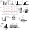
Figure 1. The mitochondrial apoptotic pathway is dispensable for NLRP3 activation
A, B Bone marrow-derived macrophages (BMDMs) from control (WT) and H2K-Bcl-2 transgenic mice were primed with LPS (20 ng/ml) for 3 h and then stimulated with ATP (A) or nigericin (B) for the indicated times. Cell supernatants were collected and analysed for IL-1β secretion by ELISA. The mean (bar) and measurements of triplicate experiments are shown for two (A) or one (B) experiment. C, D LPS-primed BMDMs from control (WT) and Vav-Bcl-2 transgenic mice were stimulated with ATP (5 mM, 1.5 h), nigericin (NIG, 5 μM, 2 h) and alum (300 μg/ml, 5 h) (C). Alternatively, BMDMs were primed with LPS for 2.5 h then treated with ABT-737 (10 μM) or DMSO (control) for 0.5 h prior to ATP stimulation for 1.5 h (D). Cell supernatants were collected and analysed for IL-1β secretion by ELISA. Error bars represent the SD of assays on cells derived from 4 mice of each genotype. E, F BMDMs from control (WT) and Bak−/−Bax−/− mice were primed with LPS as indicated and then stimulated with alum (250 μg/ml), R837 (15 μg/ml) or staurosporine (STS) for 6 h, or ATP (5 mM) and nigericin (Nig., 5 μM) for 1 h. Cell supernatants (E) and lysates (F) were analysed by Western blot. G LPS-primed BMDMs from control (WT) and Bak−/−Bax−/− mice were stimulated with ATP (5 mM), nigericin (5 μM), staurosporine (STS, 1 μM), cycloheximide (CHX, 20 μg/ml) or UVB irradiation (500 mJ/cm2) for 1 h (ATP and nigericin) or 6 h (other stimuli). Cell supernatants were collected and analysed for IL-1β content by ELISA. Error bars represent the SD of measurements from cells derived from 3 mice of each genotype. H BMDMs from Bok−/− mice and littermate (WT) controls were stimulated with ATP (5 mM), nigericin (5 μM) for 40 min, or alum for 5 h, and IL-1β secretion into the cell supernatant measured by ELISA. Error bars represent the SD of measurements from cells derived from 3 mice of each genotype. Source data are available online for this figure.
Figure 2
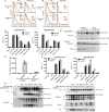
Figure 2. NLRP3 activation can occur in the absence of MPT and cyclophilin D
A LPS-primed BMDMs from control (WT) and Nlrp3-deficient mice were stimulated for 4 h with staurosporine (STS) at the indicated concentrations. Cell supernatants were collected and analysed for IL-1β content by ELISA. Error bars represent the SEM from 3 independent experiments, each performed in triplicate. B BMDMs from WT and Caspase-9−/− mice were primed with LPS and stimulated with alum, staurosporine (STS) and cycloheximide (CHX) for 6 h or ATP for 1 h and cell supernatants analysed for IL-1β by ELISA. Error bars represent the SD of measurements from cells derived from 3 mice of each genotype. C, D LPS-primed BMDMs from control (WT) mice or mice lacking Nlrp3, Pycard (C) or Caspase-1 (D) were stimulated for 2 h with ATP (5 mM) or nigericin (5 μM). Cell supernatants were collected and analysed for LDH release (a measure of plasma membrane rupture). Error bars represent the SEM from 3 independent experiments, each performed in triplicate. E BMDMs were pre-treated with the indicated stimuli for 10-15 min and then stained with TMRE (tetramethylrhodamine, ethyl ester) for a further 20-25 min and cells analysed by flow cytometry. Dashed line represents non-stimulated background controls. Data derived from 3 different mice (numbered) are shown. F, G BMDMs were primed with LPS and then stimulated with ATP (5 mM) or nigericin (10 μM) for 40 min (F) or alum (300 μg/ml) and staurosporine (500 nM) (G) for 5 h. Mitochondria containing membranes and cytosolic content were separated and analysed by Western blot. H BMDMs were primed with LPS and then treated as indicated in the presence or absence of cyclosporin A. Cell supernatants were analysed for IL-1β content by ELISA. Error bars represent the SD of measurements from cells derived from 3 mice of each genotype. I Macrophages from foetal liver of independent (numbered) WT and cyclophilin D-deficient mice were analysed by Western blotting for cyclophilin D expression as indicated. J, K Foetal liver (J) or bone marrow (K)-derived macrophages of the indicated genotypes were primed with LPS and treated as indicated for 1 h (ATP, Nigericin) or 6 h (alum). Error bars represent the SD of measurements from cells derived from 3–5 mice or embryos of each genotype. L Cell supernatants from (J) were analysed by Western blot for caspase-1 processing and secretion. Source data are available online for this figure.
Figure 3
Figure 3. MAVS and Parkin are dispensable for canonical NLRP3 activation
A, B BMDMs from WT and Parkin (Park2)-deficient mice were primed with LPS (3 h), then stimulated with or without CCCP (10 μM) for 1 h. Cells were subsequently stained with 200 nM MitoTracker Green (to measure mitochondrial mass) (A) or 200 nM MitoTracker Red (to measure respiring mitochondria) (B) for 20 min and analysed by flow cytometry. C WT and Parkin (Park2)-deficient BMDMs were stimulated as indicated after LPS priming. Cell supernatants were analysed for IL-1β content by ELISA. Bars indicate the mean of duplicate experiments (symbols). D BMDMs from WT and Parkin (Park2)-deficient mice were stimulated as indicated after LPS priming and cell supernatants and lysates examined by Western blot. E BMDMs from WT and MAVS-deficient mice were transfected with poly(I:C) for 6 h. IFN-β mRNA was measured by RT-PCR and quantified relative to 18S rRNA expression. The mean and SD of two independent experiments are shown (each performed in duplicate; symbols). F BMDMs from WT and MAVS-deficient mice were stimulated as indicated with ATP or nigericin for 1 h or with MSU or silica for 6 h. Cell supernatants were analysed for IL-1β content by ELISA. Bars indicate the mean of experiments performed in triplicate (symbols). G BMDMs from WT and MAVS-deficient mice were stimulated as described in (F) and cell supernatants and lysates examined by Western blotting as indicated. Source data are available online for this figure.
Figure 4
Figure 4. Combined loss of RIPK3 and caspase-8 limits TLR-induced cytokine production
A BMDMs of the indicated genotypes were analysed by Western blot. B BMDMs of the indicated genotypes (3 mice each; numbered) were stimulated with LPS and pro-IL-1β measured by Western blot. C BMDMs of the indicated genotypes were treated with the TLR-ligands LPS (20 ng/ml), Pam3Cys4 (2.5 μg/ml) or poly(I:C) (5 μg/ml) and TNF secretion into the supernatant determined by ELISA after 3 h. Error bars are the SEM from 3 mice of each genotype. D BMDMs of the indicated genotypes were primed with LPS (20 ng/ml) for 3 h and stimulated as indicated. IL-1β secretion into the supernatant was measured by ELISA. Error bars are the SEM from 3 mice of each genotype. E BMDMs of the indicated genotypes were stimulated with LPS (50 ng/ml) as indicated and NF-κB activation assessed by Western blot. F-H Mice of the indicated genotypes were injected i.p. with LPS (100 μg). Serum cytokine levels (F, IL-1; G, TNF; H, IL-6) were measured in mice before challenge (0) and 6 h post-LPS injection. Each symbol represents one mouse (5 – 6 per genotype). The mean and SEM are shown. Source data are available online for this figure.

References

    1. Croker BA, O’Donnell JA, Gerlic M. Pyroptotic death storms and cytopenia. Curr Opin Immunol. 2014;26:128–137. - PubMed
    1. Khan N, Lawlor KE, Murphy JM, Vince JE. More to life than death: molecular determinants of necroptotic and non-necroptotic RIP3 kinase signaling. Curr Opin Immunol. 2014;26:76–89. - PubMed
    1. Lawlor KE, Vince JE. Ambiguities in NLRP3 inflammasome regulation: is there a role for mitochondria? Biochim Biophys Acta. 2013;1840:1433–1440. - PubMed
    1. Shimada K, Crother TR, Karlin J, Dagvadorj J, Chiba N, Chen S, Ramanujan VK, Wolf AJ, Vergnes L, Ojcius DM, et al. Oxidized mitochondrial DNA activates the NLRP3 inflammasome during apoptosis. Immunity. 2012;36:401–414. - PMC - PubMed
    1. Nakahira K, Haspel JA, Rathinam VA, Lee SJ, Dolinay T, Lam HC, Englert JA, Rabinovitch M, Cernadas M, Kim HP, et al. Autophagy proteins regulate innate immune responses by inhibiting the release of mitochondrial DNA mediated by the NALP3 inflammasome. Nat Immunol. 2011;12:222–230. - PMC - PubMed

Publication types

MeSH terms