. 2014 Jul 3;511(7507):41-5.
doi: 10.1038/nature13496. Epub 2014 Jun 18.

Patterning and post-patterning modes of evolutionary digit loss in mammals

Affiliations

Patterning and post-patterning modes of evolutionary digit loss in mammals

Kimberly L Cooper et al. Nature. .

Abstract

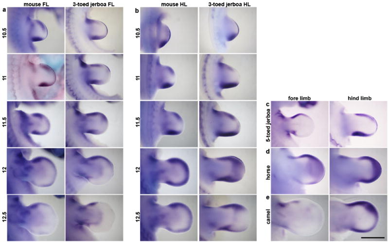
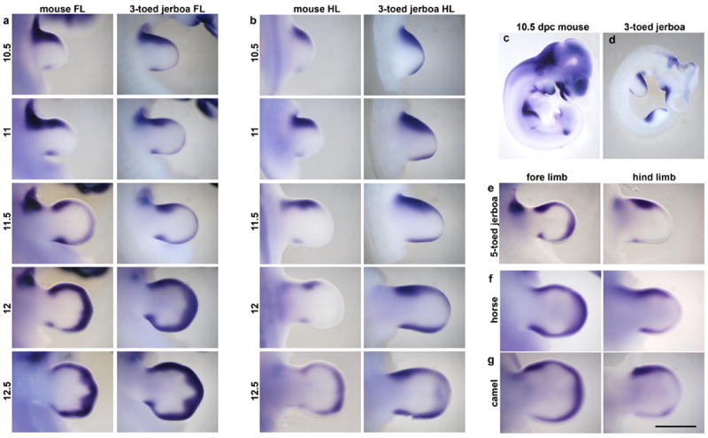
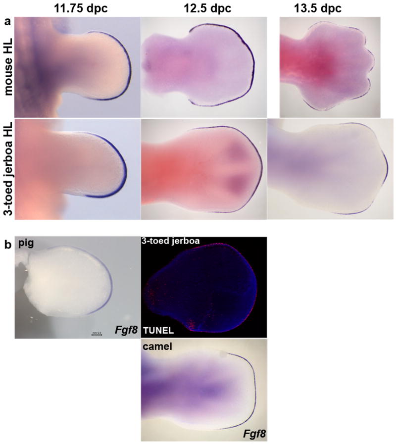
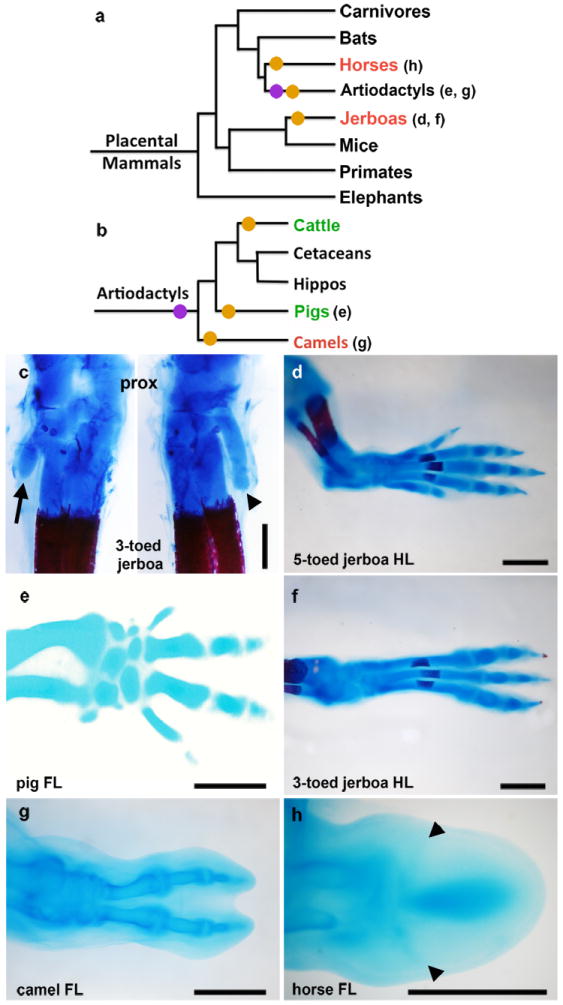
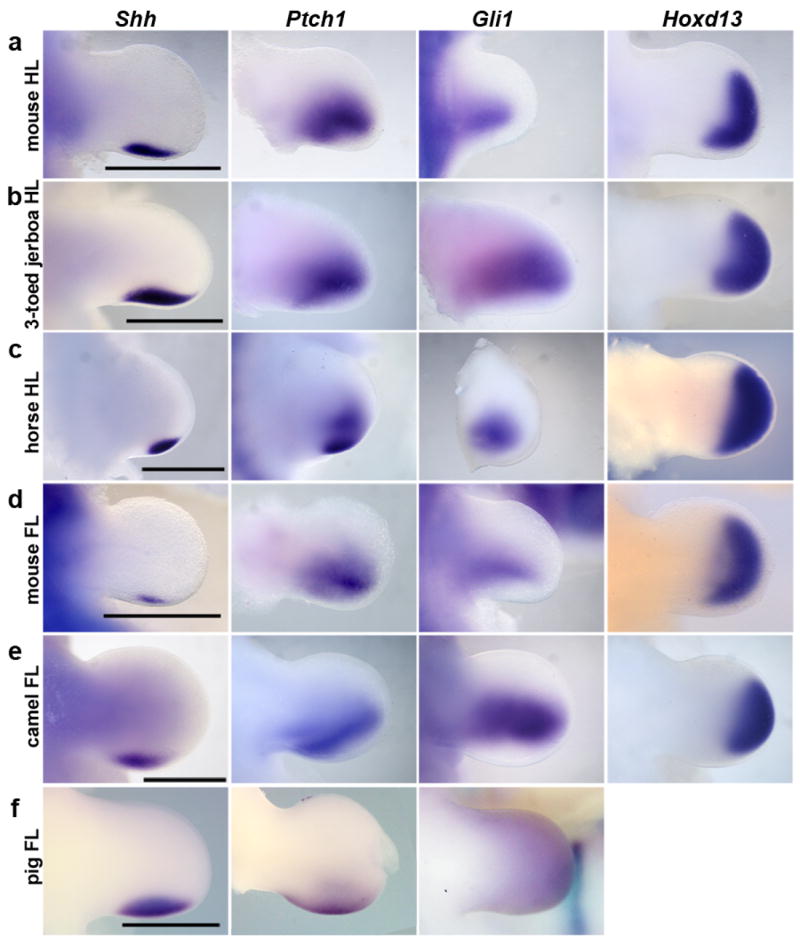
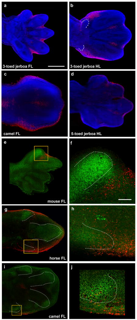
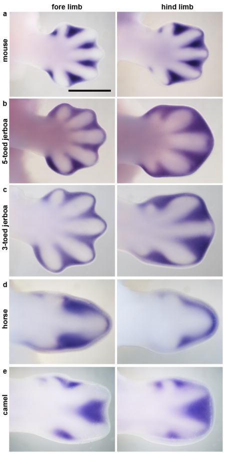
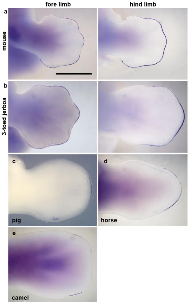
A reduction in the number of digits has evolved many times in tetrapods, particularly in cursorial mammals that travel over deserts and plains, yet the underlying developmental mechanisms have remained elusive. Here we show that digit loss can occur both during early limb patterning and at later post-patterning stages of chondrogenesis. In the 'odd-toed' jerboa (Dipus sagitta) and horse and the 'even-toed' camel, extensive cell death sculpts the tissue around the remaining toes. In contrast, digit loss in the pig is orchestrated by earlier limb patterning mechanisms including downregulation of Ptch1 expression but no increase in cell death. Together these data demonstrate remarkable plasticity in the mechanisms of vertebrate limb evolution and shed light on the complexity of morphological convergence, particularly within the artiodactyl lineage.


Figures

Extended Data Figure 1
Extended Data Figure 1. The proximal remnants of truncated skeletal elements in D sagitta are correctly patterned
Alcian blue and alizarin red stained skeletons of postnatal day 0 mouse (left) and three-toed jerboa, D sagitta (right) with proximal (ankle) at the top. a, Anterior view highlights the first metatarsal (arrow head). b, Posterior view highlights the fifth metatarsal (arrow). c, Dissected first tarsal-metatarsal elements demonstrate the morphology of the truncated first metatarsal of D sagitta (right, arrow head) compared to mouse (left). Joint interzone indicated by white dashed line. Scalebars = 0.5 mm.
Extended Data Figure 2
Extended Data Figure 2. The shape of the three-toed jerboa hind limb differs from the mouse as early as 11.5 dpc
Trace outlines of limb buds of the mouse (black) and three-toed jerboa, D sagitta (green) over a developmental time series.
Extended Data Figure 3
Extended Data Figure 3. Proliferation is unchanged in the D sagitta hind limb bud
Phospho-histone H3 detection in sections of mouse and three-toed jerboa, D sagitta, limb buds. a, fore limbs; b, hind limbs.
Extended Data Figure 4
Extended Data Figure 4. Developmental time course and species comparisons of Bmp4 expression
a, Fore limb buds (FL) and b, hind limb buds (HL) of mouse and the three-toed jerboa, Dipus sagitta, at 10.5, 11, 11.5, 12, and 12.5 dpc. c, Fore limb and hind limb of the five-toed jerboa, A elater, at approximately 12.25 dpc. d, Fore limb and hind limb of the horse at 30 dpc (approximately equivalent to mouse 12 dpc). e, Fore limb and hind limb of the camel at 38 dpc (approximately equivalent to mouse 12.5 dpc). Scalebar = 1 mm for D sagitta, A elater, horse, and camel and 0.8 mm for mouse limbs.
Extended Data Figure 5
Extended Data Figure 5. a, Developmental time course and species comparisons of Msx2 expression
a, Fore limb buds (FL) and b, hind limb buds (HL) of mouse and the three-toed jerboa, Dipus sagitta, at 10.5, 11, 11.5, 12, and 12.5 dpc. c, d, Msx2 expression in the (c) mouse and (d) D sagitta embryo at 10.5 dpc. e, Fore limb and hind limb of the five-toed jerboa, A elater, at approximately 12.25 dpc. f, Fore limb and hind limb of the horse at 30 dpc (approximately equivalent to mouse 12 dpc). g, Fore limb and hind limb of the camel at 38 dpc (approximately equivalent to mouse 12.5 dpc). Scalebar = 1 mm for D sagitta, A elater, horse, and camel and 0.8 mm for mouse limbs.
Extended Data Figure 6
Extended Data Figure 6. Developmental time course of Fgf8 expression and early TUNEL in the jerboa hind limb
a, Time series of Fgf8 expression in the mouse and three-toed jerboa, D sagitta, hind limb. b, Fgf8 expression in the pig (25 dpc) and camel (42 dpc) hind limbs of embryos in Figure 7. TUNEL labeling of cell death in the 12.5 dpc D sagitta hind limb. Limbs in (b) are aligned with the closest stage matched embryos in (a).
Figure 1
Figure 1. Convergent evolution of the embryonic limb skeleton in multiple mammal species
a-b, Phylogeny of (a) mammals and of (b) artiodactyls representing the major groups that have independently lost digits, based on Meredith et al. Parenthetical lettering references skeletons in accompanying panels. Orange circles indicate an evolutionary incidence of digit loss. Purple circles represent the shift from mesaxonic to paraxonic limbs in basal artiodactyls. Species that sculpt the limb by cell death are highlighted in red, and those that show a restriction of Ptch1 expression are highlighted in green. C Alcian blue and alizarin red stained skeleton of postnatal day 0 three-toed jerboa, D sagitta with the ankle (proximal) at the top. Posterior view (left) highlights the fifth metatarsal (arrow). Anterior view (right) highlights the first metatarsal (arrow head). d, Alcian blue stained skeletons of the approximately 16 dpc five-toed jerboa, A elater, hind foot; e, 30 dpc pig fore foot; f, approximately 16 dpc D sagitta hind foot; g, 50 dpc camel hind foot; h, 34 dpc horse fore foot; c, d, f, scalebar = 0.5 mm. e, g, h, scalebar = 1 mm.
Figure 2
Figure 2. Expression of early patterning genes: Shh, Ptch1, Gli1, and HoxD13
a, mouse hind limb (HL). b, three-toed jerboa, D sagitta, hind limb. c, horse hind limb. d, mouse fore limb (FL). e, camel fore limb. f, pig fore limb. Scalebars = 1 mm.
Figure 3
Figure 3. Patterns of cell death
DAPI (blue), Sox9 IHC (green), TUNEL (red). a, approximately 13.5 dpc three-toed jerboa, D sagitta, fore limb; b, approximately 13.5 dpc D sagitta hind limb (white dashed line indicates truncated metatarsals I and V); c, 45 dpc camel fore limb; d, approximately 13.5 dpc five-toed jerboa, A elater, hind limb e, mouse E13.5 with Sox9 and TUNEL; f, magnification of boxed region in (e); g, 34 dpc horse fore limb; h, magnification of boxed region in (g); i, 42 dpc camel fore limb; j, magnification of boxed region in (i). Scalebar in (a) = 0.5 mm for a-d, e, g, and i. Scalebar in (f) = 0.1 mm for f, h, and j.
Figure 4
Figure 4. Expression of Msx2 at the start of digit chondrogenesis
Fore limb and hind limb of a, 13 dpcmouse; b, approximately 13 dpc five-toed jerboa, A elater; c, approximately 13 dpc three-toed jerboa, D sagitta; d, 34 dpc horse; e, 42 dpc camel. Scalebar = 1 mm.
Figure 5
Figure 5. Fgf8 expression is restricted to the AER overlying nascent digits
Fore limb and hind limb of a, 13 dpc mouse; b, approximately 13 dpc three-toed jerboa, D sagitta; c, 25 dpc pig; d, 34 dpc horse hind limb; e, 42 dpc camel fore limb. Scalebar = 1 mm.

Comment in

References

    1. Clack JA. The Fish–Tetrapod Transition: New Fossils and Interpretations. Evol Educ Outreach. 2009;2:213–223.
    1. Skinner A, Lee MS, Hutchinson MN. Rapid and repeated limb loss in a clade of scincid lizards. BMC Evol Biol. 2008;8:310. - PMC - PubMed
    1. Shapiro M. Developmental morphology of limb reduction inhemiergis (squamata: scincidae): chondrogenesis, osteogenesis, and heterochrony. J Morphol. 2002;254:211–231. - PubMed
    1. Shapiro MD, Hanken J, Rosenthal N. Developmental basis of evolutionary digit loss in the Australian lizard Hemiergis. J Exp Zool B Mol Dev Evol. 2003;297:48–56. - PubMed
    1. Harfe BD, et al. Evidence for an expansion-based temporal Shh gradient in specifying vertebrate digit identities. Cell. 2004;118:517–28. - PubMed

Publication types

MeSH terms