Review
. 2014 Aug 4;20(32):9840-50.
doi: 10.1002/chem.201402474. Epub 2014 Jul 2.

Detecting enzyme activities with exogenous MRI contrast agents

Affiliations
Review

Detecting enzyme activities with exogenous MRI contrast agents

Dina V Hingorani et al. Chemistry. .

Abstract

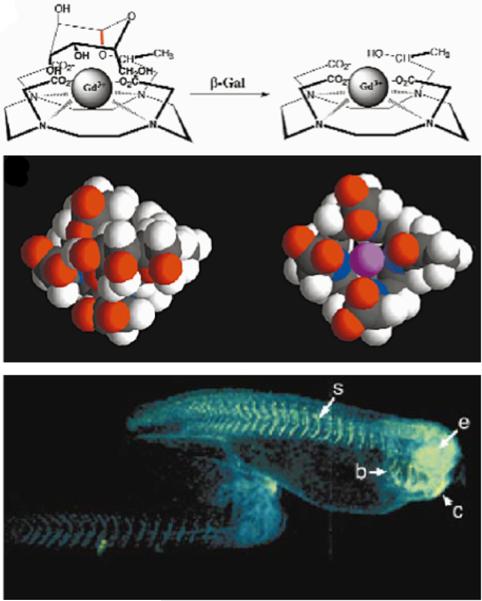
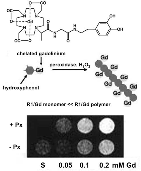
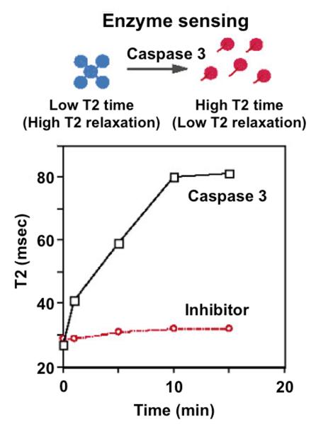
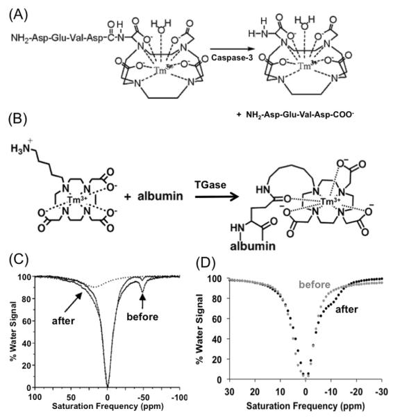
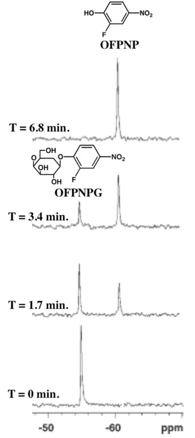
This review focuses on exogenous magnetic resonance imaging (MRI) contrast agents that are responsive to enzyme activity. Enzymes can catalyze a change in water access, rotational tumbling time, the proximity of a (19)F-labeled ligand, the aggregation state, the proton chemical-exchange rate between the agent and water, or the chemical shift of (19)F, (31)P, (13)C or a labile (1)H of an agent, all of which can be used to detect enzyme activity. The variety of agents attests to the creativity in developing enzyme-responsive MRI contrast agents.

Keywords: CEST MRI; MR spectroscopy; enzymes; imaging agents; relaxation-based MRI.


Figures

Figure 1
Figure 1
Detecting enzyme activities through changes in T1 relaxation caused by alterations in water accessibility. Top panel: The cleavage of a galactopyranosyl ring of the T1 contrast agent, EgadMe, causes the inner sphere coordination site of the Gd3+ ion to become more accessible to water. Middle panel: A space-filling molecular model illustrates the increased accessibility of the Gd3+ ion (magenta) upon cleavage. Bottom Panel: Two living X. laevis embryos were injected with EgadMe, and the embryo shown on the right was also injected with β-galactosidase mRNA. The pseudocolor rendering of MR images shows that the signal strength is 45–65% greater in the embryo containing β-galactosidase mRNA, demonstrating the detection of β-galactosidase activity. Labeled anatomy: e, eye; c, cement gland; s, somite; b, brachial arches. Reproduced with permission from (22).
Figure 2
Figure 2
Detecting enzyme activities through changes in T1 relaxation time caused by alterations in rotational tumbling times. Top Panel: The contrast agent, hydroxytyraminyl-glycylmethylDOTA [D-DOTA(Gd)]; Bottom Panel: Polymerization of the contrast agent by perixodiase slows the rotational tumbling time of the agent, which causes a decrease in the T1 relaxation time of the agent on a per-Gd basis. (C) MR images of the contrast agent at various gadolinium concentrations in the presence of peroxidase (+ Px) or in the absence of peroxidase (− Px) and hydrogen peroxide, demonstrating the detection of peroxidase enzyme activity. Reproduced with permission from (31a).
Figure 3
Figure 3
Detecting enzyme activities via changes in T2 relaxation. Top Panel: A nanoassembly of Gd-based contrast agents that are linked with DEVD peptides can be disaggregated following DEVD peptide cleavage by caspase-3. Bottom Panel: The incubation of the DEVD-linked nanoassembly with caspase-3 showed an increase in T2 relaxation time, caused by disassembly. Incubation of the nanoassembly with the enzyme and a caspase-3 inhibitor further confirmed that this approach detected caspase-3 enzyme activity. Reproduced with permission from (43a).
Figure 4
Figure 4
Detecting enzyme activities with CEST MR methods. (A): The cleavage of a peptidyl ligand of a CEST agent, DEVD-amido-(Tm-DOTA), by caspase-3 converts an amide to an amine. (B): The conjugation a CEST agent, Tm-DO3A-cadaverine, to a protein’s glutamine side chain by TGase converts an amine to an amide. (C): The CEST spectra of DEVD-amido-(Tm-DOTA) before (black) and after (gray) incubation with caspase-3 showed a decrease in CEST at μ̵11 ppm and the appearance of CEST at +8 ppm. This appearance of CEST was further confirmed by deconvoluting the CEST spectrum of the product (dashed line), which confirmed the detection of caspase-3 enzyme activity. (D): The CEST spectra of a mixture of albumin, Tm-DO3A-cadaverine and L-glutathione before (black) and after (gray) incubation with TGase showed a decrease in CEST at +4.6 ppm and the appearance of CEST at −9.2 ppm, which demonstrated detection of TGase activity. Reproduced with permission from (55) and (53).
Figure 5
Figure 5
Detecting enzyme activities with MR spectroscopy. A series of 19F-NMR spectra were acquired of OFPNPG in PBS (0.1M, pH 7.4, 600mL) before and after addition of β-galactosidase. Rapid hydrolysis of OFPNPG to form OFPNP was monitored by observing a new 19F-NMR signal for the aglycone, which was used to monitor the enzyme activity. The chemical structures of OFPNPG and OFPNP are shown. Reproduced with permission from (68).

Similar articles

Cited by

References

    1. Sittampalam GS. Assay Guidance Manual. Eli Lilly & Company and the National Center for Advancing Translational Sciences; Bethesda MD: 2004. - PubMed
    1. Messerschmidt A, editor. Handbook of Metalloproteases. 2011.
    2. Adam JA. Chem. Rev. 2001,;101:2271–2290. - PubMed
    3. Barrett AJ, Rawlings ND, Woessner JF, editors. Handbook of Proteolytic Enzymes. Academic Press; London: 1998.
    1. Gatti-Lafranconi P, Hollfelder F. ChemBioChem. 2013;14:285–292. - PubMed
    1. Moffitt JR, Chemla YR, Bustamante C. Methods in Enzymology (Single Molecule Tools, Part B) 2010:221–257. - PubMed
    1. Maeng HJ, Chow ECY, Fan J, Pang KS. In: Encyclopedia of Drug Metabolism and Interactions. Lyubimov AV, Rodrigues AD, Sinz MA, editors. 2012. pp. 637–684.

Publication types

LinkOut - more resources