Case Reports
. 2014 Jul 2:2014:bcr2014011273.
doi: 10.1136/bcr-2014-011273.

Acute subarachnoid hemorrhage in posterior condylar canal dural arteriovenous fistula: imaging features with endovascular management

Affiliations
Case Reports

Acute subarachnoid hemorrhage in posterior condylar canal dural arteriovenous fistula: imaging features with endovascular management

Prabath Kumar Mondel et al. BMJ Case Rep. .

Abstract

A 43-year-old man presented with acute subarachnoid hemorrhage. He was investigated and found to have a rare posterior condylar canal dural arteriovenous fistula (DAVF). DAVFs of the posterior condylar canal are rare. Venous drainage of the DAVF was through a long, tortuous, and aneurysmal bridging vein. We describe the clinical presentation, cross sectional imaging, angiographic features, and endovascular management of this patient. The patient was treated by transarterial embolization of the fistula through the ascending pharyngeal artery. This is the first report of an acutely bled posterior condylar canal DAVF treated by transarterial Onyx embolization with balloon protection in the vertebral artery. The patient recovered without any neurological deficit and had an excellent outcome. On 6 month follow-up angiogram, there was stable occlusion of the dural fistula.

Keywords: Balloon; CT Angiography; Fistula; Hemorrhage; Liquid Embolic Material.


Figures

Figure 1
Figure 1
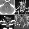
(A) Non-contrast CT scan at the level of the mid-pons shows the fourth ventricular hemorrhage (asterisk). (B) Coronal CT angiogram through the posterior condylar foramina (black arrows) shows a right posterior condylar canal dural arteriovenous fistula (arrow). A complex tangle of dilated and tortuous veins with a large venous aneurysm in the fourth ventricle and a daughter sac is seen (arrowhead). (C) Coronal (volume rendered) CT angiogram image after bone removal shows a right posterior condylar dural fistula (arrow). The daughter sac points posterosuperiorly (arrowhead). (D) A high resolution CT scan of the skull base through both the posterior condylar foramina (white arrows) after transarterial embolization shows the Onyx cast in the right posterior condylar canal.
Figure 2
Figure 2
(A, B) Axial T2 weighted and time of flight MR angiogram shows multiple convoluted and enlarged vascular channels extending from the right posterior condylar foramen into the cerebello-medullary cistern (arrow). (C) Sagittal T2 weighted image shows a large venous pouch (arrowhead) located in the fourth ventricle with multiple flow voids in the cerebello-medullary cistern. (D) Post embolization, axial T2 weighted image through the skull base shows Onyx cast in the right posterior condylar canal dural arteriovenous fistula extending into the cerebello-medullary cistern (long arrow). The asterisks denote occipital condyles and the short white arrows the posterior condylar canals.
Figure 3
Figure 3
(A–C) Right vertebral, left vertebral, and ascending pharyngeal angiograms reveal a right posterior condylar canal dural arteriovenous fistula (DAVF) (arrows) supplied by an enlarged neuromeningeal trunk, C1 cervical dural branches from both vertebral arteries, and left posterior meningeal artery. The fistula is drained by a single long and tortuous medullary bridging vein that drains to the vein of lateral recess of the fourth ventricle with a large venous pouch (arrowhead). (D, E) Microcatheter angiograms in Towne's and lateral view show the location of the right posterior condylar canal DAVF (arrow). A complex tangle of dilated and tortuous veins with a large venous pouch in the fourth ventricle is seen (arrowhead). (F) Fluoroscopic lateral view of the skull base shows Onyx cast in the right posterior condylar canal (black arrow). A balloon (asterisk) is seen inflated across the origin of the dural branches of the right vertebral artery with the microcatheter in the right ascending pharyngeal artery.
Figure 4
Figure 4
Diagrammatic representation of the posterior fossa illustrating the arterial blood supply and venous outflow of the posterior condylar canal dural arteriovenous fistula. (1) V3 segment of the vertebral artery. (2) C1 cervical dural branches (asterisks denote posterior condylar canals). (3) Posterior meningeal artery. (4) Bridging vein (blood flow depicted by white arrows). (5) Vein of lateral recess of the fourth ventricle (blood flow depicted by green arrows). (6) Transverse nodulo-uvular vein. (7) Vein of inferior cerebellar peduncle (blood flow depicted by orange arrows). (8) Posterior median medullary vein. (9) Perimedullary venous plexus. (10) Lateral medullary vein→lateral pontine vein→anterior cerebellar vein (blood flow depicted by yellow arrows). (11) Petrosal vein. (12) Superior petrosal sinus. (13) Lateral mesencephalic vein. (14) Posterior mesencephalic vein. (15) Straight sinus and vein of Galen. ACC, anterior condylar confluent; BA, basilar artery; IJV, internal jugular vein; JB, jugular bulb; PCA, posterior cerebral artery; PCV, posterior condylar vein; SS, sigmoid sinus; TS, transverse sinus; VA, vertebral artery.

Similar articles

Cited by

References

    1. Kiyosue H, Okahara M, Sagara Y, et al. Dural arteriovenous fistula involving the posterior condylar canal. AJNR Am J Neuroradiol 2007;28:1599–601 - PMC - PubMed
    1. Boyd GI. The emissary foramina of the cranium in man and the anthropoids. J Anat 1930;65(Pt 1):108–21 - PMC - PubMed
    1. San Millan Ruiz D, Gailloud P, Rufenacht DA, et al. The craniocervical venous system in relation to cerebral venous drainage. AJNR Am J Neuroradiol 2002;23:1500–8 - PMC - PubMed
    1. Martins C, Yasuda A, Campero A, et al. Microsurgical anatomy of the dural arteries. Neurosurgery 2005;56(Suppl 2):211–51 - PubMed
    1. Mitsuhashi Y, Aurboonyawat T, Pereira VM, et al. Dural arteriovenous fistulas draining into the petrosal vein or bridging vein of the medulla: possible homologs of spinal dural arteriovenous fistulas. Clinical article. J Neurosurg 2009;111:889–99 - PubMed

Publication types

MeSH terms