Review
. 2014 Sep 15;307(6):R571-84.
doi: 10.1152/ajpregu.00142.2014. Epub 2014 Jul 2.

Central role of the BK channel in urinary bladder smooth muscle physiology and pathophysiology

Affiliations
Review

Central role of the BK channel in urinary bladder smooth muscle physiology and pathophysiology

Georgi V Petkov. Am J Physiol Regul Integr Comp Physiol. .

Abstract

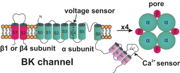
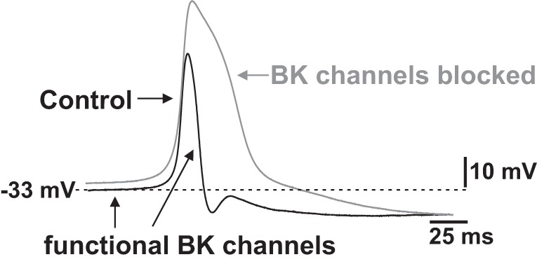
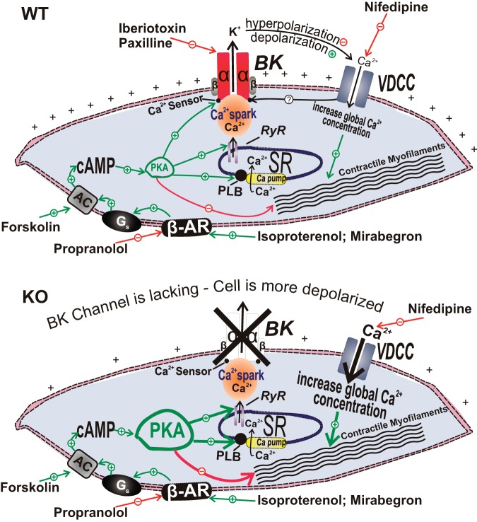
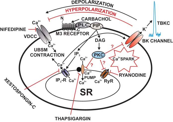
The physiological functions of the urinary bladder are to store and periodically expel urine. These tasks are facilitated by the contraction and relaxation of the urinary bladder smooth muscle (UBSM), also known as detrusor smooth muscle, which comprises the bladder wall. The large-conductance voltage- and Ca(2+)-activated K(+) (BK, BKCa, MaxiK, Slo1, or KCa1.1) channel is highly expressed in UBSM and is arguably the most important physiologically relevant K(+) channel that regulates UBSM function. Its significance arises from the fact that the BK channel is the only K(+) channel that is activated by increases in both voltage and intracellular Ca(2+). The BK channels control UBSM excitability and contractility by maintaining the resting membrane potential and shaping the repolarization phase of the spontaneous action potentials that determine UBSM spontaneous rhythmic contractility. In UBSM, these channels have complex regulatory mechanisms involving integrated intracellular Ca(2+) signals, protein kinases, phosphodiesterases, and close functional interactions with muscarinic and β-adrenergic receptors. BK channel dysfunction is implicated in some forms of bladder pathologies, such as detrusor overactivity, and related overactive bladder. This review article summarizes the current state of knowledge of the functional role of UBSM BK channels under normal and pathophysiological conditions and provides new insight toward the BK channels as targets for pharmacological or genetic control of UBSM function. Modulation of UBSM BK channels can occur by directly or indirectly targeting their regulatory mechanisms, which has the potential to provide novel therapeutic approaches for bladder dysfunction, such as overactive bladder and detrusor underactivity.

Keywords: KCa1.1 channel; detrusor; iberiotoxin; muscarinic receptors; overactive bladder; paxilline; β-adrenergic receptors.


Figures

Fig. 1.
Fig. 1.
Large-conductance voltage- and Ca2+-activated K+ (BK) channel molecular structure. Four pore-forming α-subunits and the four regulatory β1-subunits or β4-subunits form a functional BK channel in urinary bladder smooth muscle (UBSM) cells. This figure is based on information from Petkov (117, 118).
Fig. 2.
Fig. 2.
Role of BK channels in determining UBSM cell resting membrane potential and action potential repolarization. BK channels contribute to the initial repolarization phase of the UBSM spontaneous action potential and to the maintenance of the resting membrane potential in UBSM cells. This figure is based on information from Refs. , , , , and .
Fig. 3.
Fig. 3.
Illustration of the cellular mechanisms by which BK channels mediate β-adrenergic relaxation in UBSM cells with demonstration of the differential outcomes in the wild-type (WT) mouse (top) and the BK-knockout (KO) mouse (bottom), respectively. In UBSM cells from WT mice, functionally active BK channels regulate Ca2+ entry via L-type voltage-dependent Ca2+ channels (VDCC), and, thus, contractility (top). In addition, BK channels are under the local control of the so-called “Ca2+ sparks” caused by Ca2+ release from the ryanodine receptors of the sarcoplasmic reticulum, adjacent to the cell membrane. Following permanent BK channel pore forming α-subunit gene deletion in the BK channel KO mouse, an adaptive compensatory upregulation of the β-AR/cAMP/PKA signaling pathway develops. The enhanced β-AR/PKA activity compensates for the increased Ca2+ entry via L-type VDCC that occurs due to sustained membrane depolarization in the absence of the BK channels. AC, adenylyl cyclase; BK, large-conductance voltage- and Ca2+-activated K+ channels; β-AR, β-adrenergic receptors; VDCC, L-type voltage-dependent Ca2+ channels; Gs, stimulatory G protein; PKA, protein kinase-A; PLB, phospholamban; PLC, phospholipase C; RyR, ryanodine receptor; SR, sarcoplasmic reticulum. This figure is based on information from Brown et al. (21).
Fig. 4.
Fig. 4.
Illustration of the cellular mechanisms by which M3 receptors regulate BK channel function in UBSM cells. Activation of M3 receptors leads to IP3 and DAG production, via a pathway involving phospholipase-C (PLC) and PIP2. IP3 activates IP3 receptors, which releases Ca2+ from the SR. This IP3-induced Ca2+ release transiently activates the BK channels. Over time, depletion of SR Ca2+ upon activation of M3 receptors reduces Ca2+ spark activity, which leads to inhibition of TBKCs and depolarization of UBSM cell resting membrane potential causing activation of VDCC and thus increases UBSM contractility. DAG activates PKC, which may lead to direct inhibition of the SR Ca2+ pump and RyRs resulting in suppression of Ca2+ sparks and TBKCs. Pharmacological tools used in this study to inhibit cellular sources of Ca2+ are indicated. BK, large-conductance voltage- and Ca2+-activated K+ channel; DAG, 1,2-diacylglycerol; IP3, inositol triphosphate; M3 receptors, muscarinic receptors type 3; PIP2, phosphatidylinositol 4,5-bisphosphate; PKC, protein kinase-C; PLC, phospholipase-C; Ryr, ryanodine receptor; TBKCs, transient BK currents; SR, sarcoplasmic reticulum; UBSM, urinary bladder smooth muscle; VDCC, L-type voltage-dependent Ca2+ channel. This figure is based on information from (62, 112, 114).

Comment in

References

    1. Abrams P, Andersson KE, Buccafusco JJ, Chapple C, de Groat WC, Fryer AD, Kay G, Laties A, Nathanson NM, Pasricha PJ, Wein AJ. Muscarinic receptors: their distribution and function in body systems, and the implications for treating overactive bladder. Br J Pharmacol 148: 565–578, 2006 - PMC - PubMed
    1. Afeli SA, Hristov KL, Petkov GV. Do β3-adrenergic receptors play a role in guinea pig detrusor smooth muscle excitability and contractility? Am J Physiol Renal Physiol 302: F251–F263, 2012 - PMC - PubMed
    1. Afeli SA, Malysz J, Petkov GV. Molecular expression and pharmacological evidence for a functional role of Kv7 channel subtypes in Guinea pig urinary bladder smooth muscle. PLoS One 8: e75875, 2013 - PMC - PubMed
    1. Afeli SA, Petkov GV. Functional BK channels facilitate the beta3-adrenoceptor agonist-mediated relaxation of nerve-evoked contractions in rat urinary bladder smooth muscle isolated strips. Eur J Pharmacol 711: 50–56, 2013 - PMC - PubMed
    1. Afeli SA, Rovner ES, Petkov GV. BRL37344, a β3-adrenergic receptor agonist, decreases nerve-evoked contractions in human detrusor smooth muscle isolated strips: role of BK channels. Urology 82: 744 e741–747, 2013 - PMC - PubMed

Publication types

MeSH terms

Substances