. 2014 Jul 2;6(243):243ra88.
doi: 10.1126/scitranslmed.3008992.

Neutralizing antibodies to HIV-1 envelope protect more effectively in vivo than those to the CD4 receptor

Affiliations

Neutralizing antibodies to HIV-1 envelope protect more effectively in vivo than those to the CD4 receptor

Amarendra Pegu et al. Sci Transl Med. .

Abstract

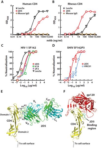
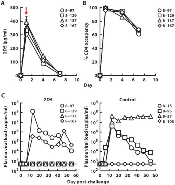
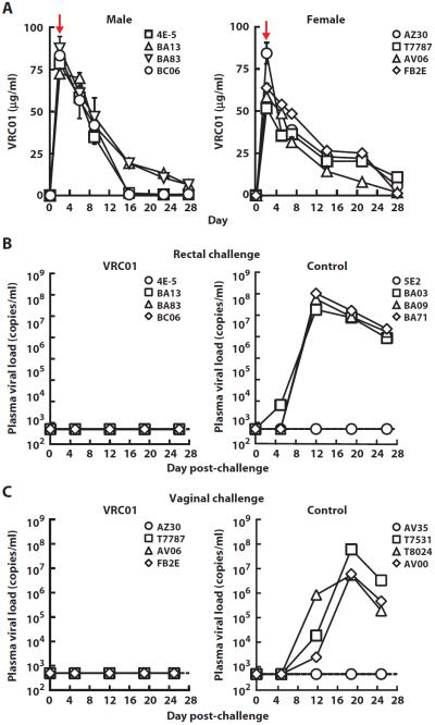
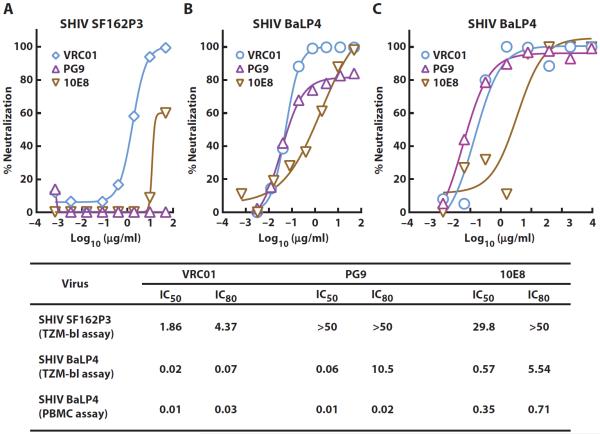
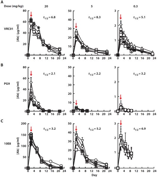
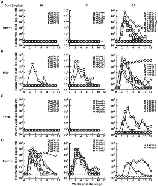
HIV-1 infection depends on effective viral entry mediated by the interaction of its envelope (Env) glycoprotein with specific cell surface receptors. Protective antiviral antibodies generated by passive or active immunization must prevent these interactions. Because the HIV-1 Env is highly variable, attention has also focused on blocking the HIV-1 primary cell receptor CD4. We therefore analyzed the in vivo protective efficacy of three potent neutralizing monoclonal antibodies (mAbs) to HIV-1 Env compared to an antibody against the CD4 receptor. Protection was assessed after mucosal challenge of rhesus macaques with simian/HIV (SHIV). Despite its comparable or greater neutralization potency in vitro, the anti-CD4 antibody did not provide effective protection in vivo, whereas the HIV-1-specific mAbs VRC01, 10E8, and PG9, targeting the CD4 binding site, membrane-proximal, and V1V2 glycan Env regions, respectively, conferred complete protection, albeit at different relative potencies. These findings demonstrate the protective efficacy of broadly neutralizing antibodies directed to the HIV-1 Env and suggest that targeting the HIV-1 Env is preferable to the cell surface receptor CD4 for the prevention of HIV-1 transmission.


Figures

Fig. 1
Fig. 1. Neutralization of HIV-1 Env by mAbs 2D5 and VRC01
(A and B) Binding of 2D5 and Leu3A anti-CD4 antibodies to either soluble human (A) or rhesus CD4 (B) as tested by enzyme-linked immunosorbent assay (ELISA). Data are representative of two independent experiments. (C) Neutralization of HIV-1 SF162 by different anti-CD4 mAbs, measured using an Env-pseudotyped lentiviral reporter assay and MAGI-CCR5 target cells that express human CD4 and CCR5. (D) Neutralization of SHIV SF162P3 by 2D5 and VRC01 using a rhesus PBMC infection assay. Means ± SEM from two independent experiments are shown. (E) Ribbon diagram showing CD4 (yellow) complexed to the 2D5 Fab (heavy chain, green; light chain, cyan). Complementarity-determining regions H1, H2, H3, and L1 of 2D5 that contact CD4 are labeled as are 2D5-contacting CD4 loops CC′ and C″D. (F) Ribbon diagram showing CD4 (yellow) complexed to the HIV-1 gp120 core (red) from Protein Data Bank (PDB) entry 1G9M. The 2D5 binding region of CD4 is shown in cyan and green.
Fig. 2
Fig. 2. Receptor occupancy, serum mAb levels, and plasma viral loads in rhesus macaques administered 2D5 followed by a single high-dose mucosal challenge with SHIV SF162P3
(A) The concentration of 2D5 was measured by ELISA in serum taken at different time points from rhesus macaques after administration of a dose (40 mg/kg) of the antibody. The red arrow indicates time of rectal SHIV challenge. (B) The occupancy of cell surface CD4 on peripheral CD4+ T cells by 2D5 was determined using flow cytometry. Day 0 indicates the time point of mAb infusion. (C) Plasma viral loads in rhesus macaques that were administered a single high dose (40 mg/kg) of 2D5 or a control human IgG and rectally challenged 1 day later with a single high dose of SHIV SF162P3 (300 TCID50).
Fig. 3
Fig. 3. Serum mAb levels and plasma viral loads in rhesus macaques administered VRC01 followed by a single high-dose mucosal challenge with SHIV SF162P3
(A) The concentration of VRC01 IgG1 was measured by an RSC3 (resurfaced stabilized gp120 core, derivative 3)–based ELISA in blood taken at different time points from male or female rhesus macaques after administration of a dose (20 mg/kg) of the antibody. The red arrows indicate time of mucosal SHIV challenge. (B) Plasma viral loads in rhesus macaques that were administered a single high dose (20 mg/kg) of VRC01 or a control human IgG and rectally challenged 2 days later with a single high dose of SHIV SF162P3 (300 TCID50). (C) Plasma viral loads in rhesus macaques that were administered a single high dose (20 mg/kg) of VRC01 or a control human IgG and vaginally challenged 2 days later with a single high dose of SHIV SF162P3 (300 TCID50).
Fig. 4
Fig. 4. Neutralization of CCR5-tropic SHIV strains by VRC01, PG9, and 10E8
(A) Neutralizing activity of VRC01 and PG9 against SHIV SF162P3 in a neutralization assay using a luciferase reporter–based TZM-bl cell line. (B) Neutralizing activity of VRC01, PG9, and 10E8 against SHIV BaLP4 in a neutralization assay using a luciferase reporter–based TZM-bl cell line. (C) Neutralizing activity of VRC01, PG9, and 10E8 against SHIV BaLP4 in a rhesus PBMC infection assay. The table lists the IC50 and IC80 values for the neutralization of SHIVs by the three anti–HIV-1 mAbs in the different assay formats. Average values from two independent experiments are shown.
Fig. 5
Fig. 5. Plasma levels of mAbs in rhesus macaques administered VRC01, PG9, and 10E8 at three different doses of each antibody
(A to C) The plasma concentrations of VRC01 (A), PG9 (B), and 10E8 (C) IgG1 were measured by ELISA at different time points after administration of the indicated doses (20, 5, and 0.3 mg/kg) of each antibody in each group. The terminal in vivo half-life (t1/2) is indicated for each antibody dose. The red arrows indicate time of mucosal SHIV challenge. Each treatment group consisted of 6 animals except for the group that received VRC01 (0.3 mg/kg), which had 10 animals.
Fig. 6
Fig. 6. Plasma viral loads in rhesus macaques administered VRC01, PG9, and 10E8 at three different doses of each antibody after a single high-dose mucosal challenge with SHIV BaLP4
(A to D) Plasma viral loads in rhesus macaques that were administered three different doses (20, 5, and 0.3 mg/kg) of VRC01 (A), PG9 (B), 10E8 (C), control IgG (D) and rectally challenged 2 days later with a single high dose of SHIV BaLP4.

References

    1. Burton DR, Desrosiers RC, Doms RW, Koff WC, Kwong PD, Moore JP, Nabel GJ, Sodroski J, Wilson IA, Wyatt RT. HIV vaccine design and the neutralizing antibody problem. Nat. Immunol. 2004;5:233–236. - PubMed
    1. Mascola JR, Montefiori DC. The role of antibodies in HIV vaccines. Annu. Rev. Immunol. 2010;28:413–444. - PubMed
    1. Baba TW, Liska V, Hofmann-Lehmann R, Vlasak J, Xu W, Ayehunie S, Cavacini LA, Posner MR, Katinger H, Stiegler G, Bernacky BJ, Rizvi TA, Schmidt R, Hill LR, Keeling ME, Lu Y, Wright JE, Chou TC, Ruprecht RM. Human neutralizing monoclonal antibodies of the IgG1 subtype protect against mucosal simian–human immunodeficiency virus infection. Nat. Med. 2000;6:200–206. - PubMed
    1. Mascola JR, Stiegler G, VanCott TC, Katinger H, Carpenter CB, Hanson CE, Beary H, Hayes D, Frankel SS, Birx DL, Lewis MG. Protection of macaques against vaginal transmission of a pathogenic HIV-1/SIV chimeric virus by passive infusion of neutralizing antibodies. Nat. Med. 2000;6:207–210. - PubMed
    1. Hessell AJ, Rakasz EG, Poignard P, Hangartner L, Landucci G, Forthal DN, Koff WC, Watkins DI, Burton DR. Broadly neutralizing human anti-HIV antibody 2G12 is effective in protection against mucosal SHIV challenge even at low serum neutralizing titers. PLOS Pathog. 2009;5:e1000433. - PMC - PubMed

Publication types

MeSH terms

Substances

Associated data