

Exercise modulates chloride homeostasis after spinal cord injury
- PMID: 24990918
- PMCID: PMC6608257
- DOI: 10.1523/JNEUROSCI.0678-14.2014
Exercise modulates chloride homeostasis after spinal cord injury
Abstract
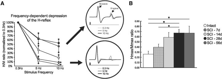
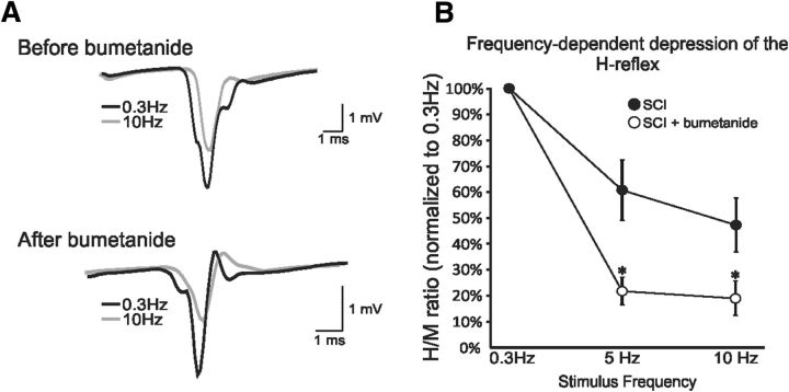
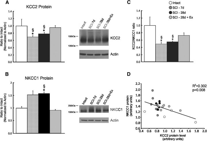
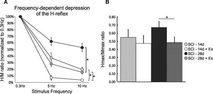
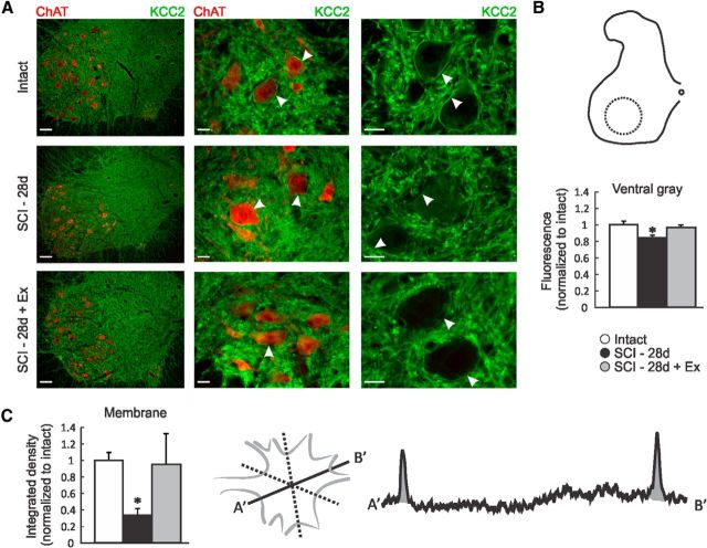
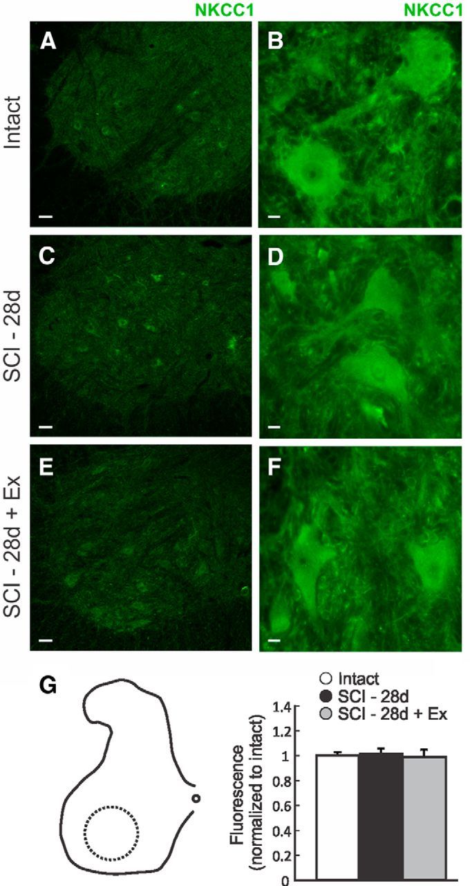
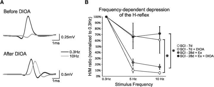
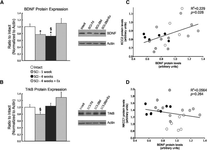
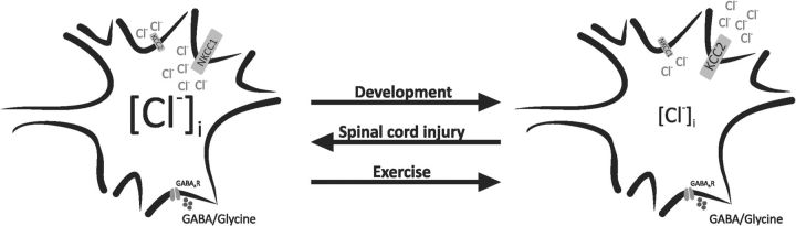
Activity-based therapies are routinely integrated in spinal cord injury (SCI) rehabilitation programs because they result in a reduction of hyperreflexia and spasticity. However, the mechanisms by which exercise regulates activity in spinal pathways to reduce spasticity and improve functional recovery are poorly understood. Persisting alterations in the action of GABA on postsynaptic targets is a signature of CNS injuries, including SCI. The action of GABA depends on the intracellular chloride concentration, which is determined largely by the expression of two cation-chloride cotransporters (CCCs), KCC2 and NKCC1, which serve as chloride exporters and importers, respectively. We hypothesized that the reduction in hyperreflexia with exercise after SCI relies on a return to chloride homeostasis. Sprague Dawley rats received a spinal cord transection at T12 and were assigned to SCI-7d, SCI-14d, SCI-14d+exercise, SCI-28d, SCI-28d+exercise, or SCI-56d groups. During a terminal experiment, H-reflexes were recorded from interosseus muscles after stimulation of the tibial nerve and the low-frequency-dependent depression (FDD) was assessed. We provide evidence that exercise returns spinal excitability and levels of KCC2 and NKCC1 toward normal levels in the lumbar spinal cord. Acutely altering chloride extrusion using the KCC2 blocker DIOA masked the effect of exercise on FDD, whereas blocking NKCC1 with bumetanide returned FDD toward intact levels after SCI. Our results indicate that exercise contributes to reflex recovery and restoration of endogenous inhibition through a return to chloride homeostasis after SCI. This lends support for CCCs as part of a pathway that could be manipulated to improve functional recovery when combined with rehabilitation programs.
Keywords: H-reflex; KCC2; NKCC1; complete transection; exercise; spinal cord injury.
Copyright © 2014 the authors 0270-6474/14/348976-12$15.00/0.
Figures
Similar articles
-
Exercise-Induced Plasticity in Signaling Pathways Involved in Motor Recovery after Spinal Cord Injury.Int J Mol Sci. 2021 May 4;22(9):4858. doi: 10.3390/ijms22094858. Int J Mol Sci. 2021. PMID: 34064332 Free PMC article. Review.
-
Rehabilitation Decreases Spasticity by Restoring Chloride Homeostasis through the Brain-Derived Neurotrophic Factor-KCC2 Pathway after Spinal Cord Injury.J Neurotrauma. 2020 Mar 15;37(6):846-859. doi: 10.1089/neu.2019.6526. Epub 2019 Nov 13. J Neurotrauma. 2020. PMID: 31578924 Free PMC article.
-
Combined use of CLP290 and bumetanide alleviates neuropathic pain and its mechanism after spinal cord injury in rats.CNS Neurosci Ther. 2024 Sep;30(9):e70045. doi: 10.1111/cns.70045. CNS Neurosci Ther. 2024. PMID: 39267289 Free PMC article.
-
Down-regulation of the potassium-chloride cotransporter KCC2 contributes to spasticity after spinal cord injury.Nat Med. 2010 Mar;16(3):302-7. doi: 10.1038/nm.2107. Epub 2010 Feb 28. Nat Med. 2010. PMID: 20190766
-
Crossing the Chloride Channel: The Current and Potential Therapeutic Value of the Neuronal K+-Cl- Cotransporter KCC2.Biomed Res Int. 2019 May 21;2019:8941046. doi: 10.1155/2019/8941046. eCollection 2019. Biomed Res Int. 2019. PMID: 31240228 Free PMC article. Review.
Cited by
-
Exercise-Induced Plasticity in Signaling Pathways Involved in Motor Recovery after Spinal Cord Injury.Int J Mol Sci. 2021 May 4;22(9):4858. doi: 10.3390/ijms22094858. Int J Mol Sci. 2021. PMID: 34064332 Free PMC article. Review.
-
Body Weight-Supported Treadmill Training Ameliorates Motoneuronal Hyperexcitability by Increasing GAD-65/67 and KCC2 Expression via TrkB Signaling in Rats with Incomplete Spinal Cord Injury.Neurochem Res. 2022 Jun;47(6):1679-1691. doi: 10.1007/s11064-022-03561-9. Epub 2022 Mar 23. Neurochem Res. 2022. PMID: 35320460 Free PMC article.
-
The Role of Ion-Transporting Proteins on Crosstalk Between the Skeletal Muscle and Central Nervous Systems Elicited by Physical Exercise.Mol Neurobiol. 2025 May;62(5):5546-5565. doi: 10.1007/s12035-024-04613-7. Epub 2024 Nov 22. Mol Neurobiol. 2025. PMID: 39578339 Review.
-
Behavioral studies of spinal conditioning: The spinal cord is smarter than you think it is.J Exp Psychol Anim Learn Cogn. 2022 Oct;48(4):435-457. doi: 10.1037/xan0000332. Epub 2022 Jul 28. J Exp Psychol Anim Learn Cogn. 2022. PMID: 35901417 Free PMC article.
-
Reactivation of Dormant Relay Pathways in Injured Spinal Cord by KCC2 Manipulations.Cell. 2018 Jul 26;174(3):521-535.e13. doi: 10.1016/j.cell.2018.06.005. Epub 2018 Jul 19. Cell. 2018. PMID: 30033363 Free PMC article.
References
Publication types
MeSH terms
Substances
LinkOut - more resources
Full Text Sources
Other Literature Sources
Medical