. 2014 Jul 2;34(27):9067-75.
doi: 10.1523/JNEUROSCI.0796-14.2014.

Development of thalamocortical connectivity during infancy and its cognitive correlations

Affiliations

Development of thalamocortical connectivity during infancy and its cognitive correlations

Sarael Alcauter et al. J Neurosci. .

Abstract

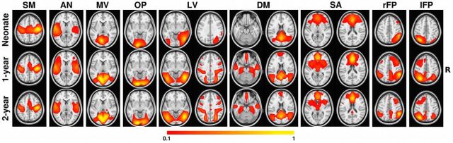
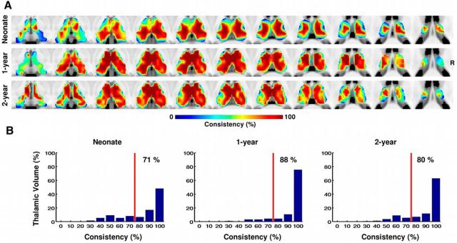
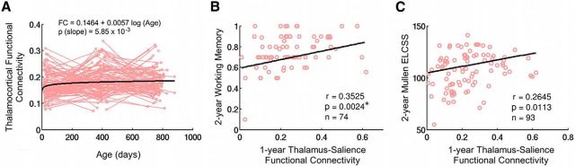
Although commonly viewed as a sensory information relay center, the thalamus has been increasingly recognized as an essential node in various higher-order cognitive circuits, and the underlying thalamocortical interaction mechanism has attracted increasing scientific interest. However, the development of thalamocortical connections and how such development relates to cognitive processes during the earliest stages of life remain largely unknown. Leveraging a large human pediatric sample (N = 143) with longitudinal resting-state fMRI scans and cognitive data collected during the first 2 years of life, we aimed to characterize the age-dependent development of thalamocortical connectivity patterns by examining the functional relationship between the thalamus and nine cortical functional networks and determine the correlation between thalamocortical connectivity and cognitive performance at ages 1 and 2 years. Our results revealed that the thalamus-sensorimotor and thalamus-salience connectivity networks were already present in neonates, whereas the thalamus-medial visual and thalamus-default mode network connectivity emerged later, at 1 year of age. More importantly, brain-behavior analyses based on the Mullen Early Learning Composite Score and visual-spatial working memory performance measured at 1 and 2 years of age highlighted significant correlations with the thalamus-salience network connectivity. These results provide new insights into the understudied early functional brain development process and shed light on the behavioral importance of the emerging thalamocortical connectivity during infancy.

Keywords: Mullen scores; development; functional connectivity; resting state; thalamus; working memory.


Figures

Figure 1.
Figure 1.
Development of the nine cortical functional networks. Color bar denotes correlation strength. The right side of the brain is in the right side of the image. SM, Sensorimotor; AN, auditory; MV, medial visual; OP, occipital poles; LV, lateral visual; DM, default mode; SA, salience; rFP/lFP, right/left frontoparietal.
Figure 2.
Figure 2.
Consistency of the thalamic parcellation based on its cortical network projections within the whole sample. A, Consistency maps across bootstrapped samples (n = 10,000) for all three age groups. The right side of the brain is on the right side of the image. B, Corresponding histograms of the consistency values for the three age groups. Red vertical lines indicate 80% consistency level. The number shown in each plot indicates the percentage of thalamic voxels with a consistency level >80%.
Figure 3.
Figure 3.
Thalamus parcellation and thalamocortical projection patterns for all three age groups. A, Thalamus parcellation scheme based on the pooled large sample. B, Cortical connectivity patterns associated with each thalamic cluster in A. The 3D rendering of each thalamic cluster is visualized alongside its cortical projection map for easier interpretation. Color bar denotes connectivity strength. The right side of the brain is on the right side of the image. SM, Sensorimotor; SA, salience; DM, default mode; MV, medial visual.
Figure 4.
Figure 4.
Thalamocortical connectivity growth and its correlation with cognitive scores. A, Longitudinal growth of thalamocortical connectivity. Light red lines denote individual growth trajectories and the bold black line represents the log-linear fit of the growth trends (significant growth at p < 0.05). B, Relationship between thalamus–salience network connectivity in 1-year-olds and working memory score in 2-year-olds. C, Relationship between thalamus–salience network connectivity in 1-year-olds and Mullen ELCSS in 2-year-olds. *Significant after FDR correction, controlling for three prediction levels and four networks.

References

    1. Alcauter S, Lin W, Keith Smith J, Gilmore JH, Gao W. Consistent anterior-posterior segregation of the insula during the first 2 years of life. Cereb Cortex. 2013 in press. - PMC - PubMed
    1. Amsterdam B. Mirror self-image reactions before age two. Dev Psychobiol. 1972;5:297–305. doi: 10.1002/dev.420050403. - DOI - PubMed
    1. Anticevic A, Cole MW, Repovs G, Murray JD, Brumbaugh MS, Winkler AM, Savic A, Krystal JH, Pearlson GD, Glahn DC. Characterizing thalamo-cortical disturbances in schizophrenia and bipolar illness. Cereb Cortex. 2013 in press. - PMC - PubMed
    1. Arichi T, Gordon-Williams R, Allievi A, Groves AM, Burdet E, Edwards AD. Computer-controlled stimulation for functional magnetic resonance imaging studies of the neonatal olfactory system. Acta Paediatr. 2013;102:868–875. doi: 10.1111/apa.12327. - DOI - PMC - PubMed
    1. Bartocci M, Bergqvist LL, Lagercrantz H, Anand KJ. Pain activates cortical areas in the preterm newborn brain. Pain. 2006;122:109–117. doi: 10.1016/j.pain.2006.01.015. - DOI - PubMed

Publication types

MeSH terms