. 2014 Jul 2;34(27):9124-33.
doi: 10.1523/JNEUROSCI.0181-14.2014.

Sleep disorders in Parkinsonian macaques: effects of L-dopa treatment and pedunculopontine nucleus lesion

Affiliations

Sleep disorders in Parkinsonian macaques: effects of L-dopa treatment and pedunculopontine nucleus lesion

Hayat Belaid et al. J Neurosci. .

Abstract

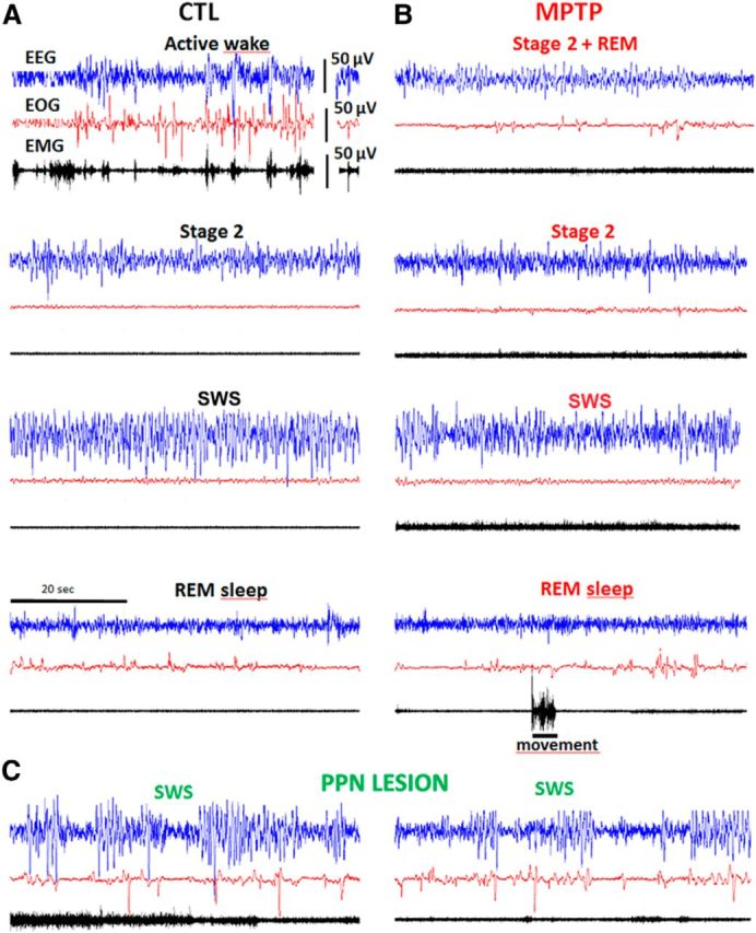
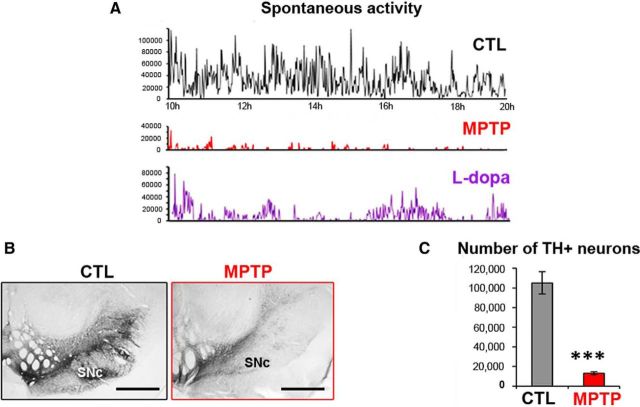
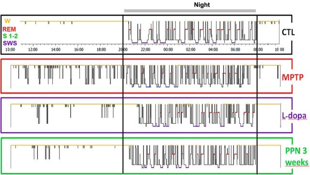
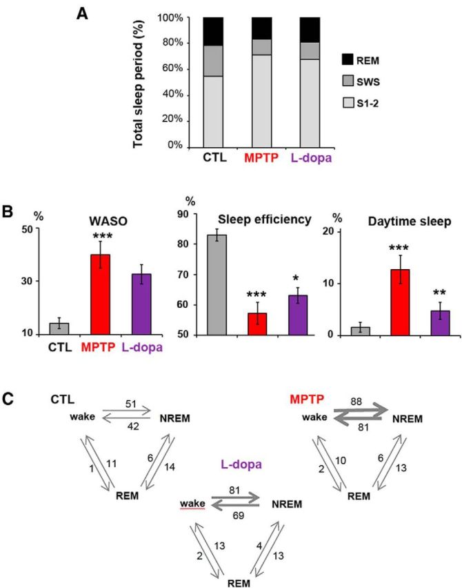
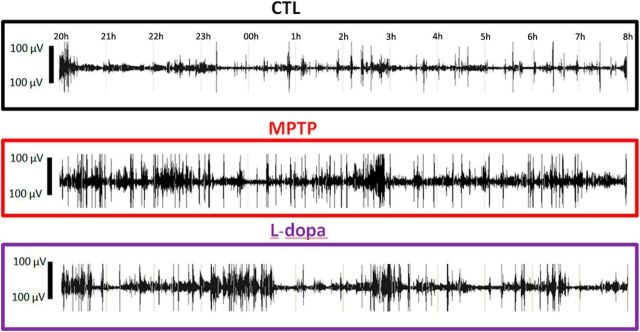
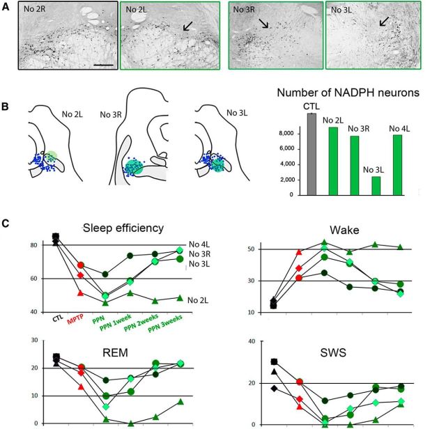
Patients with Parkinson's disease (PD) display significant sleep disturbances and daytime sleepiness. Dopaminergic treatment dramatically improves PD motor symptoms, but its action on sleep remains controversial, suggesting a causal role of nondopaminergic lesions in these symptoms. Because the pedunculopontine nucleus (PPN) regulates sleep and arousal, and in view of the loss of its cholinergic neurons in PD, the PPN could be involved in these sleep disorders. The aims of this study were as follows: (1) to characterize sleep disorders in a monkey model of PD; (2) to investigate whether l-dopa treatment alleviates sleep disorders; and (3) to determine whether a cholinergic PPN lesion would add specific sleep alterations. To this end, long-term continuous electroencephalographic monitoring of vigilance states was performed in macaques, using an implanted miniaturized telemetry device. 1-Methyl-4-phenyl-1,2,3,6-tetrahydropyridine treatment induced sleep disorders that comprised sleep episodes during daytime and sleep fragmentation and a reduction of sleep efficiency at nighttime. It also induced a reduction in time spent in rapid eye movement (REM) sleep and slow-wave sleep and an increase in muscle tone during REM and non-REM sleep episodes and in the number of awakenings and movements. l-Dopa treatment resulted in a partial but significant improvement of almost all sleep parameters. PPN lesion induced a transient decrease in REM sleep and in slow-wave sleep followed by a slight improvement of sleep quality. Our data demonstrate the efficacy of l-dopa treatment in improving sleep disorders in parkinsonian monkeys, and that adding a cholinergic PPN lesion improves sleep quality after transient sleep impairment.

Keywords: Parkinson's disease; cholinergic neurons; l-dopa; macaques; pedunculopontine nucleus; sleep disorders.


Figures

Figure 1.
Figure 1.
Examples of 1 min polysomnographic recordings. A, At baseline (control state, CTL). B, After MPTP intoxication. C, At 1 week after further PPN lesion. Blue represents EEG; red represents EOG; black represents EMG. Note the existence, after MPTP, of eye movements during Stage 2 (top right: Stage 2 + REM), and of movements during Stage 2 and REM sleep, and the occurrence, after PPN lesion, of ample δ frequencies on EEG and increased muscle tone.
Figure 2.
Figure 2.
Measurement of motor disability and assessment of DA lesion within the mesencephalon of MPTP-treated macaques. A, Examples of 10 h daytime recording of spontaneous activity of MPTP-treated Macaque 3 during the control (CTL) state, after MPTP intoxication, then after l-dopa medication. l-Dopa treatment (bottom graph) was given at 10 h and at 14 h. B, Example of TH immunostaining in the mesencephalon of a control macaque and an MPTP-treated macaque. Scale bar, 2 mm. C, Graphic representation of the number of TH+ neurons in the substantia nigra pars compacta (SNc) in the 4 MPTP-treated macaques and 5 controls. ***p < 0.001 (Mann–Whitney rank-sum test).
Figure 3.
Figure 3.
Examples of hypnograms of 24 h recordings in Macaque 3 at baseline (CTL), 1 month after MPTP intoxication, after 1 week of l-dopa treatment, and 3 weeks after subsequent PPN lesion. MPTP intoxication induced sleep episodes during the day and the impairment of sleep organization with marked sleep fragmentation and awakenings during the night. l-Dopa had a beneficial effect on daytime and nighttime sleep. A subsequent PPN lesion resulted in a better sleep quality 3 weeks after the lesion than after MPTP intoxication alone.
Figure 4.
Figure 4.
Sleep disorders after MPTP intoxication and partial improvement after l-dopa treatment. A, Comparison of the contribution of REM, Stages 1 and 2 and SWS, expressed as a percentage of the total sleep period in the 4 macaques in the control state (CTL), after MPTP intoxication, and after l-dopa treatment. B, Histograms showing increase in duration of WASO, reduction of sleep efficiency (ratio of total sleep time to the 12 h of nighttime), and development of daytime sleepiness (expressed as percentage of time spent asleep during the 12 h of daytime), and their improvement after l-dopa treatment. C, Transitions between wake, NREM, and REM periods. The mean number of transitions is given and is represented by the thickness of the arrows. The number of transitions between wake and NREM stages was increased after MPTP intoxication and improved slightly after l-dopa treatment.
Figure 5.
Figure 5.
Examples of EMG recordings during the 12 h of nighttime sleep, at baseline (CTL) (first line), after MPTP intoxication (second line), and after l-dopa treatment in Macaque 2. The increase in muscle tone and in the number of movements that developed in the MPTP state was slightly improved after l-dopa treatment.
Figure 6.
Figure 6.
Effect of PPN lesion on sleep in three MPTP-treated macaques. A, Photomicrographs of PPN sections labeled for NADPH histochemistry, showing the toxin injection site into the PPN (arrow) in the left (L) side of Macaque 2 and in both sides of Macaque 3 compared with the right (R) unlesioned side of Macaque 2. B, Left, NADPH+ neurons (blue) were mapped in two lesioned animals. Each dot represents an NADPH+ neuron. Right, Quantification of the total number of NADPH+ neurons in the PPN showing the neuronal loss for unilaterally lesioned macaques (Macaques 2 and 4), and for bilaterally lesioned macaque (Macaque 3). C, Values of relevant sleep parameters in the three macaques 1–3 weeks after PPN lesion compared with control (CTL) and MPTP values. Note a decrease of sleep efficiency, REM sleep and SWS, and an increase of nighttime awakenings (wake). These disorders were much more severe in Macaque 2 than in Macaques 3 and 4. These parameters progressively returned to values observed after MPTP treatment, except for Macaque 2, which was therefore killed 3 weeks after PPN lesion. Scale bar: A, 1 mm.

Similar articles

Cited by

References

    1. Abbott RD, Ross GW, White LR, Tanner CM, Masaki KH, Nelson JS, Curb JD, Petrovitch H. Excessive daytime sleepiness and subsequent development of Parkinson disease. Neurology. 2005;65:1442–1446. doi: 10.1212/01.wnl.0000183056.89590.0d. - DOI - PubMed
    1. Almirall H, Pigarev I, de la Calzada MD, Pigareva M, Herrero MT, Sagales T. Nocturnal sleep structure and temperature slope in MPTP treated monkeys. J Neural Transm. 1999;106:1125–1134. doi: 10.1007/s007020050228. - DOI - PubMed
    1. Arnulf I. Excessive daytime sleepiness in parkinsonism. Sleep Med Rev. 2005;9:185–200. doi: 10.1016/j.smrv.2005.01.001. - DOI - PubMed
    1. Arnulf I. REM sleep behavior disorder: motor manifestations and pathophysiology. Mov Disord. 2012;27:677–689. doi: 10.1002/mds.24957. - DOI - PubMed
    1. Arnulf I, Konofal E, Merino-Andreu M, Houeto JL, Mesnage V, Welter ML, Lacomblez L, Golmard JL, Derenne JP, Agid Y. Parkinson's disease and sleepiness: an integral part of PD. Neurology. 2002;58:1019–1024. doi: 10.1212/WNL.58.7.1019. - DOI - PubMed

Publication types

MeSH terms