. 2014 Aug 22;289(34):23917-27.
doi: 10.1074/jbc.M114.583385. Epub 2014 Jul 2.

Direct evidence of an elongation factor-Tu/Ts·GTP·Aminoacyl-tRNA quaternary complex

Affiliations

Direct evidence of an elongation factor-Tu/Ts·GTP·Aminoacyl-tRNA quaternary complex

Benjamin J Burnett et al. J Biol Chem. .

Abstract

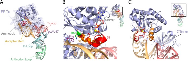
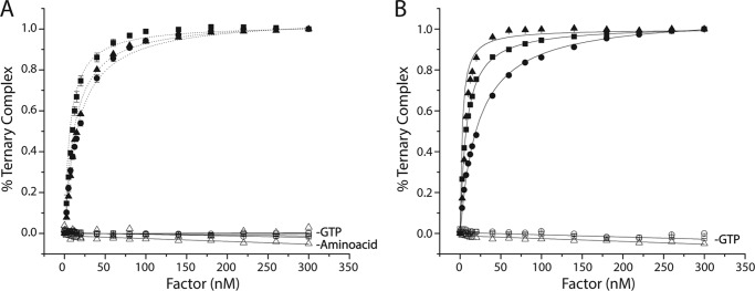
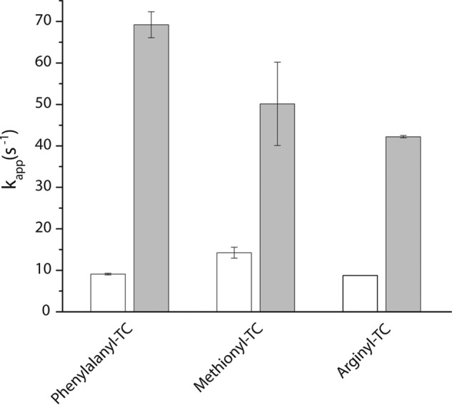
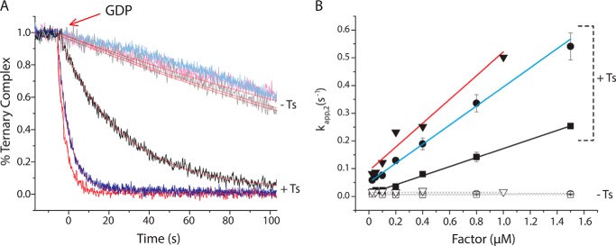
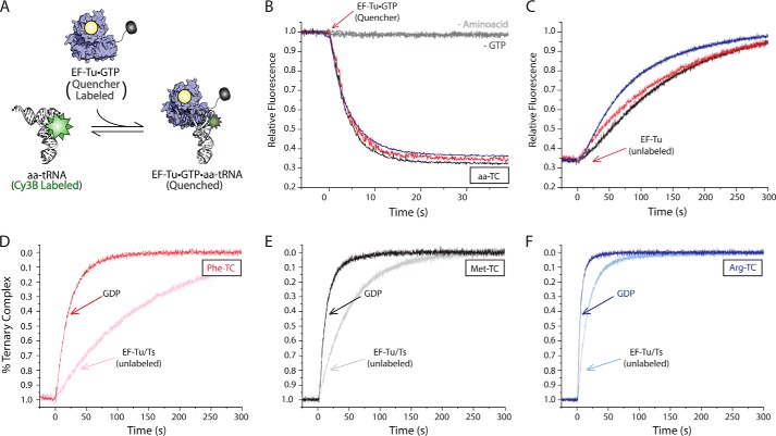
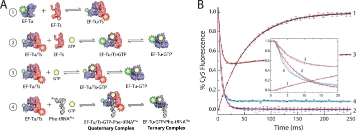
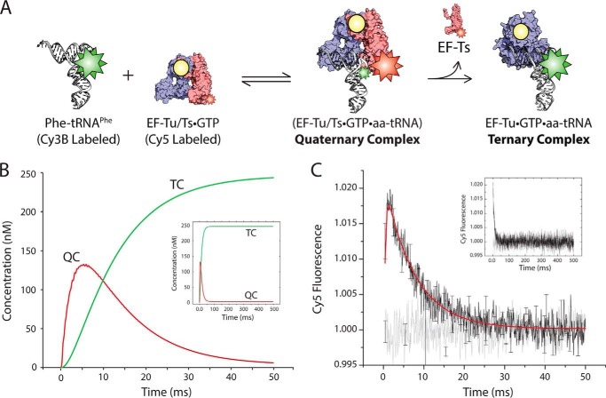
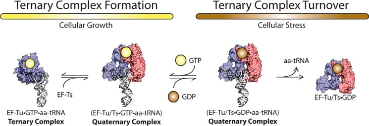
During protein synthesis, elongation factor-Tu (EF-Tu) bound to GTP chaperones the entry of aminoacyl-tRNA (aa-tRNA) into actively translating ribosomes. In so doing, EF-Tu increases the rate and fidelity of the translation mechanism. Recent evidence suggests that EF-Ts, the guanosine nucleotide exchange factor for EF-Tu, directly accelerates both the formation and dissociation of the EF-Tu-GTP-Phe-tRNA(Phe) ternary complex (Burnett, B. J., Altman, R. B., Ferrao, R., Alejo, J. L., Kaur, N., Kanji, J., and Blanchard, S. C. (2013) J. Biol. Chem. 288, 13917-13928). A central feature of this model is the existence of a quaternary complex of EF-Tu/Ts·GTP·aa-tRNA(aa). Here, through comparative investigations of phenylalanyl, methionyl, and arginyl ternary complexes, and the development of a strategy to monitor their formation and decay using fluorescence resonance energy transfer, we reveal the generality of this newly described EF-Ts function and the first direct evidence of the transient quaternary complex species. These findings suggest that EF-Ts may regulate ternary complex abundance in the cell through mechanisms that are distinct from its guanosine nucleotide exchange factor functions.

Keywords: Elongation Factor Ts; Elongation Factor Tu; G Protein; Guanine Nucleotide Exchange Factor (GEF); Guanosine Nucleotide Exchange Factor; Protein Synthesis; Ternary Complex; Translation; Translation Elongation Factor.


Figures

FIGURE 1.
FIGURE 1.
EF-Tu binds both the aminoacyl moiety and the tRNA body. A, E. coli EF-Tu (blue) bound in a ternary complex with GDPNP and Saccharomyces cerevisiae Phe-tRNAPhe (Protein Data Bank code 1OB2). The following functional domains of tRNA are highlighted: acceptor stem (orange), T-loop (red), D-loop (blue), and the anticodon loop (green) as well as GDPNP (carbon atoms in gray, nitrogen in blue, oxygen in red, and phosphorus in orange) and the acp3U47 residue, the site of fluorophore labeling used in this report. B, the aminoacyl binding pocket is stabilized by the GTP-dependent coordination of switch 1 (S1; red) and switch 2 (S2; orange) regions. C, conserved contacts between the acceptor stem of aa-tRNA and all three domains of EF-Tu. The approximate position of the C terminus (CTerm) of EF-Tu is shown.
FIGURE 2.
FIGURE 2.
EF-Tu binds ternary complex with nanomolar affinity. Titration of EF-Tu·GTP (A) or EF-Tu/Ts·GTP (B) into a solution of Cy3-labeled Phe-tRNAPhe (solid triangles), Met-tRNAMet (solid squares), or Arginyl-tRNAArg (solid circles) in the presence of 10 μm GTP. The apparent dissociation constant (KD) for each tRNA species was estimated by fitting (see “Experimental Procedures”). Identical experiments performed in the absence of GTP or the tRNA synthetase (open symbols). Error bars represent the S.D. of three separate experiments.
FIGURE 3.
FIGURE 3.
EF-Ts directly facilitates ternary complex formation. The estimated rate of ternary complex formation (kapp,1) measured by a change in fluorescence intensity upon stopped-flow delivery of 5 μm EF-Tu·GTP (open bars) or 5 μm EF-Tu/Ts (solid bars) to 100 nm Cy3-labeled phenylalanyl-tRNAPhe, arginyl-tRNAArg, or methionyl-tRNAMet. Error bars represent the S.D. of three separate experiments.
FIGURE 4.
FIGURE 4.
EF-Ts accelerates ternary complex decay in response to GDP. A, addition of a 10-fold molar excess of GDP to Arg-TC (blue) or Met-TC (black) preformed with EF-Tu (light colored curves) or EF-Tu/Ts (dark colored curves) in the presence of 10 μm GTP. B, the apparent rate of ternary complex dissociation (kapp,2) was estimated by fitting each decay process to a single exponential function. The relationship between kapp,2 and the concentration of EF-Tu (open symbols) or EF-Tu/Ts (solid symbols) is shown for both Met-TC (squares) and Arg-TC (circles). Error bars represent the S.D. of three separate experiments. For comparison, kapp,2 (acquired previously) is shown for Phe-TC (red triangles) (1).
FIGURE 5.
FIGURE 5.
Measuring ternary complex formation and dissociation via FRET. A, schematic illustration of the TC quenching assay showing Cy5Q-labeled EF-Tu (blue) bound to GTP (yellow) quenches the fluorescence of Cy3B-labeled aa-tRNA (white) upon ternary complex formation. B, addition of 0.4 μm EF-Tu·GTP (Cy5Q) to 5 nm Cy3B-labeled Phe-tRNAPhe (red), Met-tRNAMet (black), or Arg-tRNAArg (blue) in the presence of 10 μm GTP. Omission of the tRNA synthetase (−Amino acid) or GTP (−GTP) from the ternary complex formation assay resulted in no change in Cy3B fluorescence intensity. C, addition of excess unlabeled EF-Tu to the ternary complexes formed in B. Addition of excess GDP (bold colors) or unlabeled EF-Tu/Ts (light colors) to Phe-TC (D), Met-TC (E), and Arg-TC (F) preformed with EF-Tu/Ts (Cy5Q). Apparent rates of ternary complex decay were estimated by fitting to a single exponential function.
FIGURE 6.
FIGURE 6.
Significant amounts of EF-Tu/Ts remain bound to GTP under physiological nucleotide concentrations. A, schematic illustrating the EF-Tu/Ts FRET assay. 1, Cy3-labeled EF-Tu (blue) is added to Cy5-labeled EF-Ts (red). 2, EF-Tu(Cy3)/Ts(Cy5) mixed with unlabeled EF-Ts and GTP. 3, EF-Tu(Cy3)/Ts(Cy5) added to GTP. 4, EF-Tu(Cy3)/Ts(Cy5) mixed with unlabeled Phe-tRNAPhe and GTP. B, 2 μm EF-Tu(Cy3) mixed with 2 μm EF-Ts(Cy5) while monitoring Cy5 fluorescence (curve 1), 2 μm EF-Tu(Cy3)/Ts(Cy5) added to 20 μm unlabeled EF-Ts and 1 mm GTP (curve 2), 2 μm EF-Tu(Cy3)/Ts(Cy5) mixed with 1 mm GTP (curve 3), and 2 μm EF-Tu(Cy3)/Ts(Cy5) mixed with 0.5 μm Phe-tRNAPhe and 1 mm GTP (curve 4). A plot focusing on the early time points in the reactions is shown for clarity (inset). Apparent rates were estimated by fitting to either a single exponential function (curves 1, 2, and 4) or a double exponential function (curve 3). Error bars represent the S.D. of three separate experiments.
FIGURE 7.
FIGURE 7.
Direct evidence of the transient quaternary complex. A, schematic illustrating the FRET assay used to detect quaternary complex. Cy3B-labeled Phe-tRNAPhe (white) is mixed with EF-Tu/Ts·GTP harboring a Cy5 fluorophore linked to EF-Ts. B, simulation of the time-dependent formation of quaternary complex (red curve; QC) and ternary complex (green curve; TC) based on the schematic presented in A and outlined under “Experimental Procedures.” C, transient FRET observed upon mixing 2 μm EF-Tu/Ts(Cy5) and 250 nm Phe-tRNAPhe (Cy3B) in the presence of 1 mm GTP (black). These data were fit to a double exponential function (red). This experiment was repeated with deacylated tRNAPhe (Cy3B) (gray). Error bars represent the S.D. of three separate experiments.
FIGURE 8.
FIGURE 8.
EF-Ts regulates ternary complex abundance. During growth-promoting conditions, EF-Ts (red) facilitates the formation of GTP (yellow)-bound EF-Tu (blue) to aa-tRNA (white) by accelerating rate-determining conformational processes in EF-Tu. The guanosine nucleotide exchange function of EF-Ts also serves to preferentially load GDP during conditions of cellular stress.

Similar articles

Cited by

References

    1. Burnett B. J., Altman R. B., Ferrao R., Alejo J. L., Kaur N., Kanji J., Blanchard S. C. (2013) Elongation factor Ts directly facilitates the formation and disassembly of the Escherichia coli elongation factor Tu·GTP·aminoacyl-tRNA ternary complex. J. Biol. Chem. 288, 13917–13928 - PMC - PubMed
    1. Vetter I. R., Wittinghofer A. (2001) The guanine nucleotide-binding switch in three dimensions. Science 294, 1299–1304 - PubMed
    1. Leipe D. D., Wolf Y. I., Koonin E. V., Aravind L. (2002) Classification and evolution of P-loop GTPases and related ATPases. J. Mol. Biol. 317, 41–72 - PubMed
    1. Thomposon R. C., Dix D. B. (1982) Accuracy of protein biosynthesis. A kinetic study of the reaction of poly(U)-programmed ribosomes with a leucyl-tRNA2-elongation factor Tu-GTP complex. J. Biol. Chem. 257, 6677–6682 - PubMed
    1. Rodnina M. V., Pape T., Fricke R., Wintermeyer W. (1995) Elongation factor Tu, a GTPase triggered by codon recognition on the ribosome: mechanism and GTP consumption. Biochem. Cell Biol. 73, 1221–1227 - PubMed

Publication types

MeSH terms

Associated data

LinkOut - more resources