. 2014 Sep 5;289(36):25227-40.
doi: 10.1074/jbc.M114.554881. Epub 2014 Jul 2.

Akt-dependent activation of mTORC1 complex involves phosphorylation of mTOR (mammalian target of rapamycin) by IκB kinase α (IKKα)

Affiliations

Akt-dependent activation of mTORC1 complex involves phosphorylation of mTOR (mammalian target of rapamycin) by IκB kinase α (IKKα)

Han C Dan et al. J Biol Chem. .

Abstract

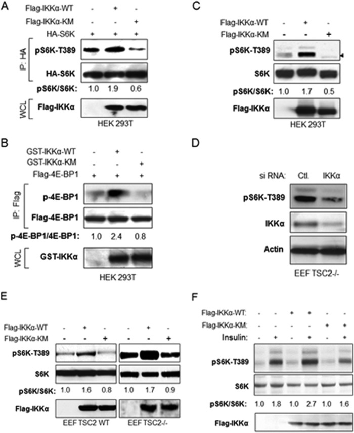
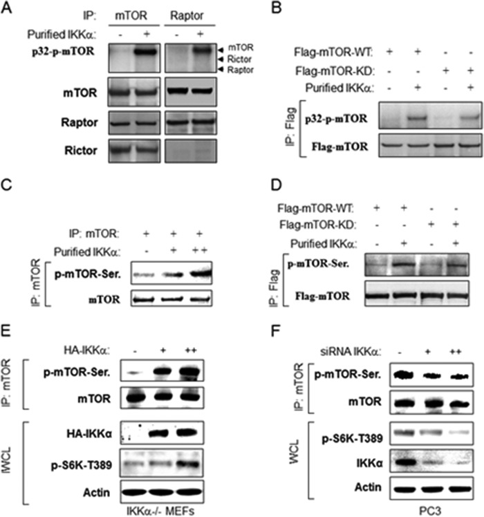
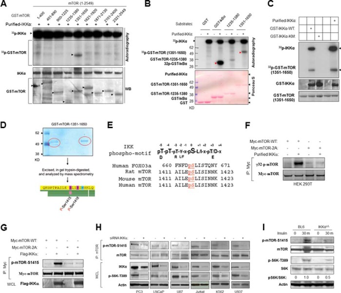
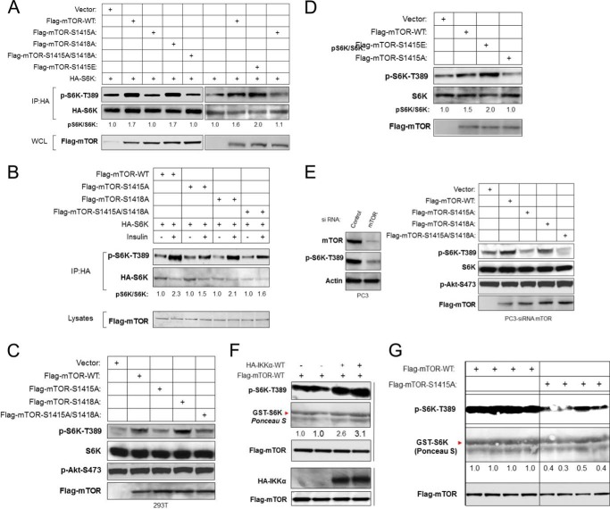
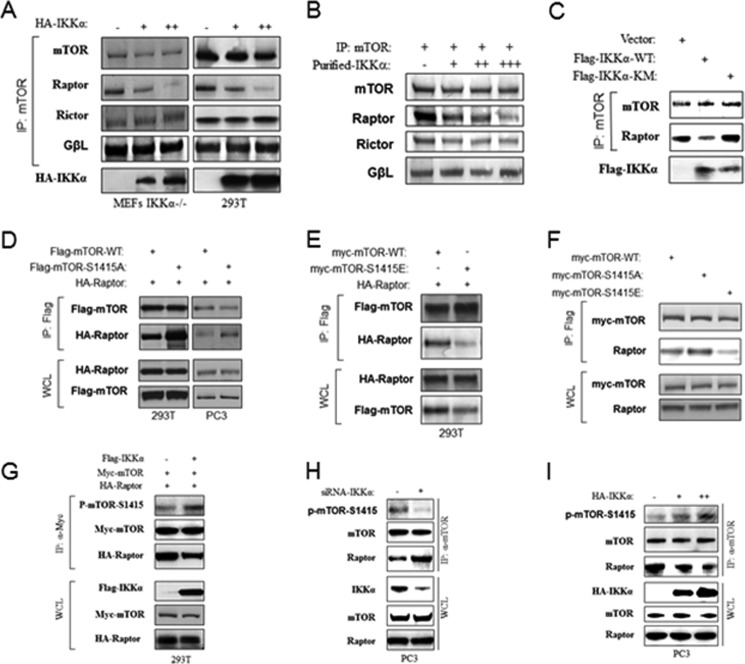
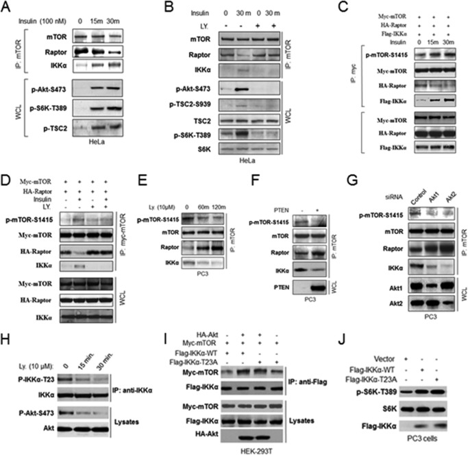
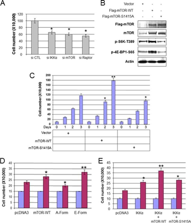
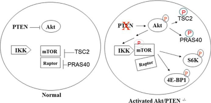
The serine/threonine protein kinase Akt promotes cell survival, growth, and proliferation through phosphorylation of different downstream substrates. A key effector of Akt is the mammalian target of rapamycin (mTOR). Akt is known to stimulate mTORC1 activity through phosphorylation of tuberous sclerosis complex 2 (TSC2) and PRAS40, both negative regulators of mTOR activity. We previously reported that IκB kinase α (IKKα), a component of the kinase complex that leads to NF-κB activation, plays an important role in promoting mTORC1 activity downstream of activated Akt. Here, we demonstrate IKKα-dependent regulation of mTORC1 using multiple PTEN null cancer cell lines and an animal model with deletion of IKKα. Importantly, IKKα is shown to phosphorylate mTOR at serine 1415 in a manner dependent on Akt to promote mTORC1 activity. These results demonstrate that IKKα is an effector of Akt in promoting mTORC1 activity.

Keywords: Akt; Cell Proliferation; IKK; Mammalian Target of Rapamycin (mTOR); Phosphatase and Tensin Homolog (PTEN); Phosphorylation; Raptor.


Figures

FIGURE 1.
FIGURE 1.
IKKα activates mTORC1 in a kinase-dependent manner independent of TSC2. A, effect of wild type and mutant IKKα on S6K phosphorylation. HEK 293T cells were transfected with HA-S6K, FLAG-IKKα wild type, or IKKα mutants, and HA immunoprecipitates (IP) and whole cell lysates (WCL) were analyzed with the indicated antibodies. The bands of phospho-S6K and S6K were quantified, and the ratio of pS6K/S6K was measured as indicated. The experiments were carried out on three separate occasions. B, effect of wild type and mutant IKKα on 4E-BP1 phosphorylation. HEK 293T cells were transfected with FLAG-4E-BP1, GST-IKKα wild type, or IKKα mutants, and FLAG immunoprecipitates and whole cell lysates were analyzed with the indicated antibody. The bands phospho-4E-BP1 and FLAG-4E-BP1 were quantified, and the ratio of p-4E-BP1/4E-BP1 was measured as described in A. Results are representative of three experimental repetitions. C, effect of wild type and mutant IKKα on endogenous S6K phosphorylation. HEK 293 cells were transfected with FLAG-IKKα wild type or IKKα mutants, and endogenous phospho-S6K, S6K, and expression of FLAG-IKKα were detected. The bands phospho-S6K and S6K were quantified, and the ratio of pS6K/S6K was measured as described in A. The results are representative of three experimental repetitions. D, Eker rat embryo fibroblast TSC2−/− cells were transfected with siRNA control or siIKKα, and endogenous phospho-S6K and expression of IKKα and Actin were detected. E, Eker rat embryo fibroblast TSC2+/+ and TSC2−/− cells were transfected with FLAG-IKKα wild type or IKKα mutants, and endogenous phospho-S6K, S6K, and expression of FLAG-IKKα was detected. The results are representative of three experimental experiments. F, HeLa cells were transfected with siRNA IKKα for 48 h and then expressed with FLAG-IKKα wild type or IKKα mutants, the cells were serum-deprived (16 h), incubated in the absence or presence of insulin (100 nm) for 15–30 min, and whole cell lysates were analyzed with indicated antibodies. The results are representative of three experimental repetitions.
FIGURE 2.
FIGURE 2.
IKKα phosphorylates mTOR in vitro and in vivo. A, IKKα phosphorylates mTOR in vitro. Endogenous mTOR or Raptor was immunoprecipitated (IP) with anti-mTOR or anti-Raptor from 293T cells and incubated with recombinant active IKKα and [γ-32P]ATP. Autoradiography was performed followed by immunoblotting with anti-mTOR, Raptor, and Rictor. B, FLAG-mTOR wild type and kinase dead (KD) were immunoprecipitated by anti-FLAG from 293T cells transfected and incubated with recombinant active IKKα and [γ-32P]ATP. Autoradiography was performed followed by immunoblotting with indicated antibodies. C, nonradioactive in vitro kinase assay. Endogenous mTOR was immunoprecipitated by anti-mTOR from 293T cells and probed with anti-phosphoserine antibody followed by anti-mTOR. D, nonradioactive in vitro kinase assay. FLAG-mTOR wild type and kinase dead were immunoprecipitated by anti-FLAG from 293T cells and blotted with anti-phospho-serine antibody followed by anti-FLAG. E, IKKα induces mTOR phosphorylation. IKKα−/− MEFs were transfected with HA-IKKα and lysed, and the endogenous mTOR was immunoprecipitated by anti-mTOR and blotted with anti-phosphoserine antibody followed by anti-mTOR. The whole cell lysates (WCL) also were tested by indicated antibodies. F, knockdown of IKKα decreases mTOR phosphorylation. PC3 cells were transfected with siRNA against IKKα, lysed, and immunoprecipitated with anti-mTOR. mTOR immunoprecipitate and whole cell lysates were blotted with the indicated antibodies.
FIGURE 3.
FIGURE 3.
IKKα phosphorylates mTOR at serine 1415 in vitro and in vivo. A, IKKα phosphorylates mTOR in vitro. Recombinant IKKα was incubated with GST-mTOR fragments for in vitro, radioactive IKKα kinase assays. Proteins from this assay were blotted with the indicated antibodies. WB, Western blot. B, IKKα phosphorylates GST-mTOR fragment and GST-IκBα in vitro. Recombinant IKKα was incubated with the indicated GST-mTOR fragments or GST-IκBα for in vitro IKKα kinase assays. C, purified IKKα used in A and B, and GST-IKKα wild type and kinase mutant were analyzed relative to their ability to phosphorylate GST-mTOR-(1351–1650). D, Ser-1415 and Ser-1418 are the primary direct IKKα phosphorylation sites in vitro. Purified GST-mTOR-1351–1650 was incubated with active IKKα for phosphorylation, and phosphorylation site mapping was determined by mass spectrometry. E, consensus IKKα phosphorylation sites on human mTOR and alignment with conserved sites on mouse and rat mTOR compared with human FOXO3a and IKK phosphorylation motif. F, IKKα phosphorylates mTOR in serine 1415 in vitro. Myc-tagged mTOR wild type and S1415A/S1418A mutants were immunoprecipitated (IP) by anti-Myc from 293T cells and used as substrates for in vitro kinase assay using recombinant active IKKα. Autoradiography was performed followed by immunoblotting with anti-Myc. G, HEK293T cells were cotransfected with Myc-mTOR (WT or S1415A) and FLAG-IKKα (WT) as indicated. The immunoprecipitates of Myc were analyzed with the phospho-mTOR-Ser-1415 antibody followed by FLAG antibody. WCL, whole cell lysates. H, PC3 and other PTEN-mutated cancer cell lines were transfected with siRNA against IKKα, and mTOR immunoprecipitates and whole cell lysates were analyzed with the indicated antibodies. I, wild type and IKKαLoxP/LoxP PB-Cre+ mice of 15 weeks of age were injected with insulin (intraperitoneally, 1 unit/kg) for 30 min, and protein lysates obtained from prostates were analyzed with the indicated antibodies.
FIGURE 4.
FIGURE 4.
IKKα regulates mTORC1 activity through phosphorylation of mTOR at serine 1415. A, mutation of IKKα phosphorylation site (S1415A) decreases mTOR activity. HA-S6K was cotransfected with wild type or various mTOR mutants in HEK293T cells as indicated. Phosphorylation of S6K-Thr-389 was determined in conjunction with expression levels of S6K, mTOR, and IKKα. IP, immunoprecipitated; WCL, whole cell lysates. B, IKKα phosphorylation of mTOR is involved in insulin-induced mTOR activation. HeLa cells were cotransfected with HA-S6K and FLAG-tagged wild type or various mTOR mutants as indicated, serum-starved overnight, stimulated with insulin, lysed, and analyzed with phospho-S6K-Thr-389 and other antibodies. C, mutation of IKKα phosphorylation sites with alanine substitution blocks endogenous mTOR activity. HEK 293T cells were transfected with wild type or various mTOR mutants as indicated. Endogenous phosphorylation of S6K-Thr-389 and Akt-Ser-473 was determined in conjunction with expression levels of S6K and FLAG-mTOR. D, HEK 293T cells were transfected with wild type or various mTOR mutants as indicated. Endogenous phosphorylation of S6K-Thr-389 was determined in conjunction with expression levels of S6K and FLAG-mTOR. E, PC3 cells were transfected with siRNA against mTOR and then wild type or various mTOR mutants as indicated 48 h after siRNA transfection, lysed, and analyzed with the indicated antibodies. F, expression of IKKα enhances in vitro mTOR kinase activity. HEK293T cells were cotransfected with HA-IKKα and FLAG-mTOR. FLAG-mTOR was IP with antibody, and mTOR kinase activity toward GST-S6K was determined in the immunoprecipitates. G, mutation of IKKα Ser-1415 by alanine substitution decreases mTOR kinase activity. FLAG-mTOR (WT and S1415A) was transfected in HEK293T cells and lysed. Kinase activity of FLAG immunoprecipitates toward GST-S6K were measured by phospho-S6K-Thr-389 antibody.
FIGURE 5.
FIGURE 5.
IKKα phosphorylation of mTOR at serine 1415 modulates association with Raptor downstream of Akt. A, IKKα activity modulates mTOR-Raptor interaction. MEFs IKKα−/− and 293 cells were transfected with different amounts of HA-IKKα in full serum. Lysates were immunoprecipitated (IP) with anti-mTOR and blotted with mTOR, raptor, Rictor, and GβL antibodies. B, IKKα weakens mTOR-Raptor interaction in vitro. mTOR immunoprecipitates from HEK293T cells were incubated with recombinant IKKα and unlabeled ATP in IKK kinase buffer for 30 min and washed with lysis buffer three times, blotted with mTOR, Raptor, Rictor, and GβL antibodies, respectively. C, HEK293 cells were transfected with FLAG-IKKα (WT or mutant), lysed, and immunoprecipitated with anti-mTOR and blotted with mTOR and Raptor antibodies. D, 293 cells and PC3 cells were cotransfected with FLAG-mTOR WT or the S1415A mutant with HA-Raptor, immunoprecipitated with anti-FLAG, and blotted with FLAG and HA antibodies, respectively. WCL, whole cell lysates. E, 293 cells were cotransfected with FLAG-mTOR WT or S1415E with HA-Raptor, immunoprecipitated with anti-FLAG, and blotted with FLAG and HA antibodies, respectively. F, 293 cells were cotransfected with FLAG-mTOR WT or S1415E with HA-Raptor, immunoprecipitated with anti-FLAG, and blotted with FLAG and HA antibodies, respectively. G, IKKα mediates mTOR phosphorylation and mTOR-Raptor interaction in PC3 cells. siRNA to IKKα was transfected. Lysates were immunoprecipitated with anti-mTOR and blotted with phospho-mTOR-Ser-1415 antibody and followed by other antibodies. Additionally, whole cell lysates were analyzed with the indicated antibodies. H, IKKα mediates mTOR phosphorylation and mTOR-Raptor interaction in PC3 cells. siRNA to IKKα was transfected. Lysates were immunoprecipitated with anti-mTOR and blotted with phospho-mTOR-Ser-1415 antibody and followed by other antibodies. Additionally, whole cell lysates were analyzed with indicated antibodies. I, IKKα mediates mTOR phosphorylation and mTOR-Raptor interaction in PC3 cells. HA-IKKα was transfected in PC3 cells. Lysates were immunoprecipitated with anti-mTOR and blotted with phospho-mTOR-Ser-1415 antibody and followed by other antibodies. Additionally, whole cell lysates were analyzed with the indicated antibodies.
FIGURE 6.
FIGURE 6.
IKKα-mediated mTOR-Raptor interaction is regulated by Akt. A, HeLa cells were serum-deprived (16 h), incubated in the absence or presence of insulin (100 nm) for 15–30 min, and lysed. mTOR immunoprecipitates (IP) and whole cell lysates (WCL) were immunoblotted as indicated. B, HeLa cells were serum-deprived (16 h), pretreated with or without LY 294000 (LY), incubated in the absence or presence of insulin for 30 min, and lysed. mTOR immunoprecipitates and whole cell lysates were immunoblotted as indicated antibodies. C and D, HeLa cells were cotransfected with Myc-mTOR, HA-Raptor, and FLAG-IKKα and then serum-deprived (16 h), pretreated with or without LY 294000, incubated in the absence or presence of insulin for 30 min as indicated, and mTOR immunoprecipitates and whole cell lysates were immunoblotted as the indicated antibodies. E, PI3K inhibitor, LY 294000, blocks IKKα regulation of mTORC1. PC3 cells were treated with LY 294000 for the indicated times, lysed, immunoprecipitated with mTOR antibody, and immunoblotted with antibodies as indicated. F and G, PC3 cells were transfected with PTEN (F) and siRNA against Akt1 or Akt2 (G), and mTOR immunoprecipitates and whole cell lysates were analyzed with the indicated antibodies. H, PC3 cells were treated with PI3K inhibitor, LY 294000, at the indicated times, lysed, immunoprecipitated by IKKα antibodies, and immunoblotted with the indicated antibodies. I, 293T cells were co-transfected as indicated and immunoprecipitated with FLAG antibody and immunoblotted with the indicated antibodies. J, PC3 cells were transfected with FLAG-IKKα wild type or mutant, and whole cell lysates were analyzed with antibodies as indicated.
FIGURE 7.
FIGURE 7.
IKKα phosphorylation of mTOR promotes PTEN null and Akt active PC3 prostate cancer cell proliferation. A, knockdown of IKKα and mTOR decreases cell proliferation. PC3 cells were transfected with siRNA against IKKα, mTOR, and Raptor and plated in 6-well plates (2.0 × 10⁴) 48 h posttransfection, and cell numbers were counted using a hemocytometer after 3 days. The numbers were calculated and presented as the mean ± S.D. from triplicates. B, stably transfected PC3 cells (mTOR-WT or mTOR-S1415A) were lysed and analyzed with antibodies as indicated. C, the stably transfected cells (pcDNA3, mTOR-WT, and mTOR-S1415A) were plated in 6 well plates (10 × 104), and cell numbers were counted using a hemocytometer every day for 3 days. The numbers were calculated and are presented as the mean ± S.D. from triplicates. The single asterisk indicates statistical significance compared with controls (t test, p < 0.05), and the double asterisks indicate p < 0.01. D, PC3 cells were transfected with mTOR wild type and its mutants, plated in 6-well plates (15 × 104) 48 h posttransfection, and cell numbers were counted using a hemocytometer in 3 days. Statistical analysis was performed as described in A and C. E, PC3 cells were transfected with IKKα and mTOR wild type and its mutants as indicated, and plated in 6-well plates (10 × 104) 48 h posttransfection, and cell numbers were counted using a hemocytometer after 3 days followed by statistical analysis. The single asterisk indicates statistical significance compared with controls (t test, p < 0.05), and the double asterisks indicate p < 0.01.
FIGURE 8.
FIGURE 8.
A proposed model of IKKα regulation of mTORC1 downstream of activated Akt.

Similar articles

Cited by

References

    1. Zoncu R., Efeyan A., Sabatini D. M. (2011) mTOR: from growth signal integration to cancer, diabetes, and ageing. Nat. Rev. Mol. Cell Biol. 12, 21–35 - PMC - PubMed
    1. Howell J. J., Manning B. D. (2011) mTOR couples cellular nutrient sensing to organismal metabolic homeostasis. Trends Endocrinol. Metab. 22, 94–102 - PMC - PubMed
    1. Inoki K., Kim J., Guan K.-L. (2012) AMPK and mTOR in cellular energy homeostasis and drug targets. Annu. Rev. Pharmacol. Toxicol. 52, 381–400 - PubMed
    1. Jewell J. L., Guan K. L. (2013) Nutrient signaling to mTOR and cell growth. Trends Biochem. Sci. 38, 233–242 - PMC - PubMed
    1. Ma X. M., Blenis J. (2009) Molecular mechanisms of mTOR-mediated translational control. Nat. Rev. Mol. Cell Biol. 10, 307–318 - PubMed

Publication types

MeSH terms

LinkOut - more resources