Review
. 2014 Aug 15;289(33):22583-22588.
doi: 10.1074/jbc.R114.566091. Epub 2014 Jul 2.

Phospholipase D and the maintenance of phosphatidic acid levels for regulation of mammalian target of rapamycin (mTOR)

Affiliations
Review

Phospholipase D and the maintenance of phosphatidic acid levels for regulation of mammalian target of rapamycin (mTOR)

David A Foster et al. J Biol Chem. .

Abstract

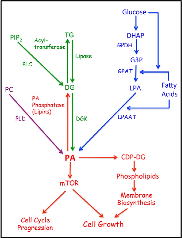
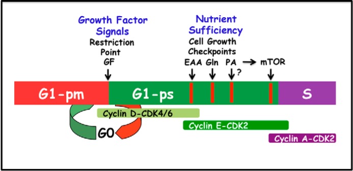
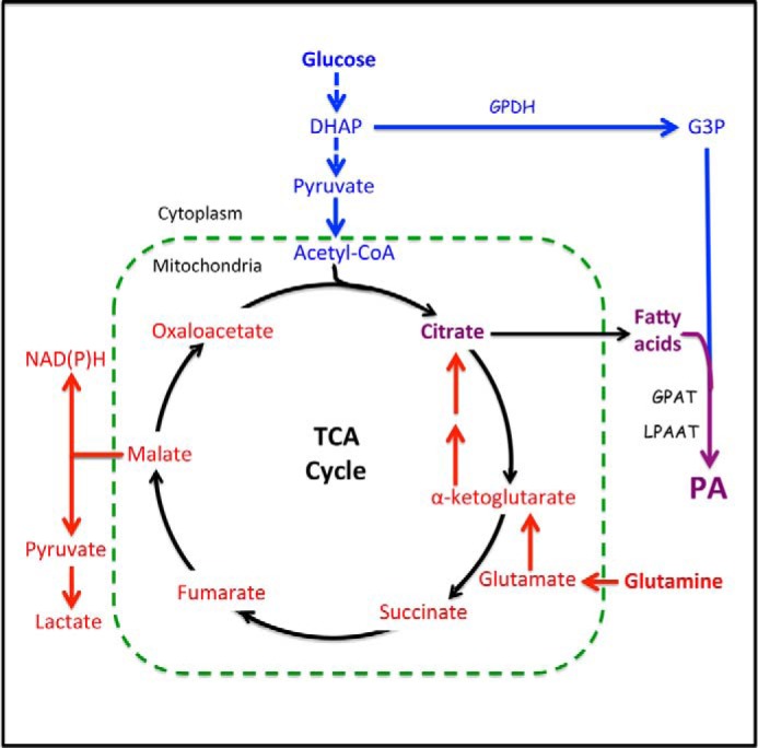
Phosphatidic acid (PA) is a critical metabolite at the heart of membrane phospholipid biosynthesis. However, PA also serves as a critical lipid second messenger that regulates several proteins implicated in the control of cell cycle progression and cell growth. Three major metabolic pathways generate PA: phospholipase D (PLD), diacylglycerol kinase (DGK), and lysophosphatidic acid acyltransferase (LPAAT). The LPAAT pathway is integral to de novo membrane phospholipid biosynthesis, whereas the PLD and DGK pathways are activated in response to growth factors and stress. The PLD pathway is also responsive to nutrients. A key target for the lipid second messenger function of PA is mTOR, the mammalian/mechanistic target of rapamycin, which integrates both nutrient and growth factor signals to control cell growth and proliferation. Although PLD has been widely implicated in the generation of PA needed for mTOR activation, it is becoming clear that PA generated via the LPAAT and DGK pathways is also involved in the regulation of mTOR. In this minireview, we highlight the coordinated maintenance of intracellular PA levels that regulate mTOR signals stimulated by growth factors and nutrients, including amino acids, lipids, glucose, and Gln. Emerging evidence indicates compensatory increases in one source of PA when another source is compromised, highlighting the importance of being able to adapt to stressful conditions that interfere with PA production. The regulation of PA levels has important implications for cancer cells that depend on PA and mTOR activity for survival.

Keywords: Diacylglycerol Kinase; Lipid Metabolism; Lysophosphatidic Acid Acyltransferase; Mammalian Target of Rapamycin (mTOR); Phosphatidic Acid; Phospholipase D; glycolysis.


Figures

FIGURE 1.
FIGURE 1.
Metabolic pathways for PA production. There are three main pathways leading to the production of PA. For de novo synthesis of membrane phospholipids is the LPAAT pathway where G3P, derived largely from the glycolytic intermediate DHAP, is doubly acylated with a fatty acid, first by G3P acyltransferase (GPAT) to generate LPA, and then by LPAAT to generate PA. The DGK pathway involves the phosphorylation of DG to generate PA. DG can be generated from stored triglycerides (TG) by a lipase, or from phosphatidylinositol 4,5-bisphosphate (PIP2) via growth factor-stimulated phospholipase C. The third mechanism is the hydrolysis of phosphatidylcholine (PC) by PLD. Like PLC, the PLD reaction is commonly stimulated by growth factors. The balance between PA and DG is carefully controlled by both DGK and PA phosphatases that convert PA to DG. Both PA and DG are important intermediates in phospholipid biosynthesis. It is hypothesized that the PA input to mTOR is an indicator of sufficient lipid precursors for cell growth and a signal to promote cell cycle progression. GPDH, G3P dehydrogenase.
FIGURE 2.
FIGURE 2.
Regulation of G1 cell cycle progression by growth factors and nutrients. G1 can be separated into two phases referred to as G1-pm (post-mitotic) and G1-ps (pre-S) by a growth factor (GF)-dependent restriction point (23). At the restriction point, the cell receives signals signifying that it is appropriate to divide. Later in G1-ps there is a series of metabolic checkpoints that evaluate whether there are sufficient nutrients for the cell to double in mass and divide. There are distinct checkpoints for essential amino acids (EAA), the conditionally essential amino acid Gln, and a later checkpoint mediated by mTOR. The schematic shows the relative order of the checkpoints, but does not reflect an accurate time frame. Because mTOR requires PA for stability of the mTOR complexes (30), this late mTOR checkpoint also requires PA. It is not clear whether there is a separate checkpoint for PA like there is for the essential amino acids (EAA), which are also required for mTOR activity.
FIGURE 3.
FIGURE 3.
Metabolic pathways from glucose and Gln to PA. Glucose is converted into lipids via two pathways. The first pathway is the conversion of the glycolytic intermediate DHAP to G3P by G3P dehydrogenase (GPDH). G3P is then fatty acylated, first to LPA by G3P acyltransferase (GPAT) and then to PA by LPAAT. The second pathway utilizes the end product of glycolysis, pyruvate. Pyruvate is converted to acetyl-CoA, which condenses with oxaloacetate to form citrate. Citrate leaves the mitochondria and is then converted back to oxaloacetate and acetyl-CoA, which is then used to synthesize the fatty acids that will be used to acylate G3P and generate PA. With the exit of citrate from the TCA cycle, there is a need for anaplerotic replenishment of the carbon provided by citrate. This is provided by the conditionally essential amino acid Gln, which enters the TCA cycle by being deaminated to glutamate and then to α-ketoglutarate by transamination. Via the TCA cycle, most of the Gln is converted to malate and then to pyruvate to generate NADPH for fatty acid synthesis. Gln can also go from malate to oxaloacetate where it can then condense with acetyl-CoA derived from glucose to form citrate and then fatty acids as above. Gln can also be reductively carboxylated to isocitrate and then converted to citrate in a reverse TCA cycle reaction of isocitrate dehydrogenase. In the absence of Gln, glucose cannot be converted to fatty acid synthesis.

Similar articles

Cited by

References

    1. Athenstaedt K., Daum G. (1999) Phosphatidic acid, a key intermediate in lipid metabolism. Eur. J. Biochem. 266, 1–16 - PubMed
    1. Pascual F., Carman G. M. (2013) Phosphatidate phosphatase, a key regulator of lipid homeostasis. Biochim. Biophys. Acta 1831, 514–522 - PMC - PubMed
    1. Foster D. A. (2007) Regulation of mTOR by phosphatidic acid? Cancer Res. 67, 1–4 - PubMed
    1. Kraft C. A., Garrido J. L., Fluharty E., Leiva-Vega L., Romero G. (2008) Role of phosphatidic acid in the coupling of the ERK cascade. J. Biol. Chem. 283, 36636–36645 - PMC - PubMed
    1. Foster D. A. (2009) Phosphatidic acid signaling to mTOR: signals for the survival of human cancer cells. Biochim. Biophys. Acta 1791, 949–955 - PMC - PubMed

Publication types

MeSH terms

LinkOut - more resources