. 2014 Jul 1;28(13):1429-44.
doi: 10.1101/gad.242131.114.

Prolyl hydroxylation by EglN2 destabilizes FOXO3a by blocking its interaction with the USP9x deubiquitinase

Affiliations

Prolyl hydroxylation by EglN2 destabilizes FOXO3a by blocking its interaction with the USP9x deubiquitinase

Xingnan Zheng et al. Genes Dev. .

Abstract

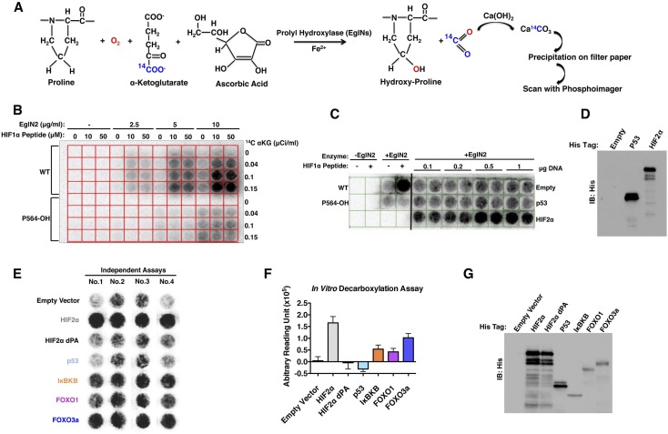
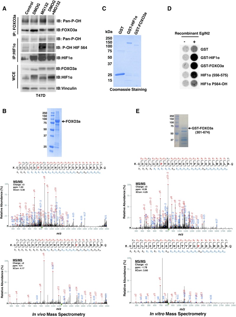
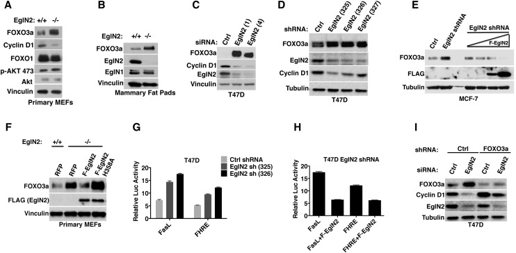
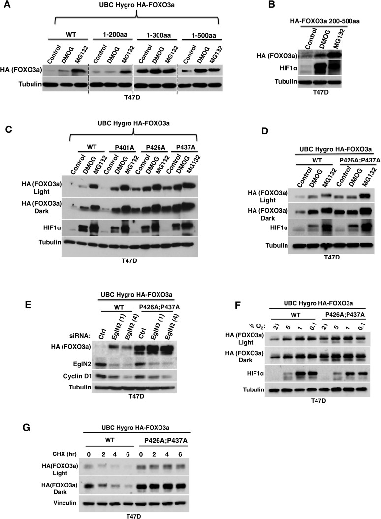
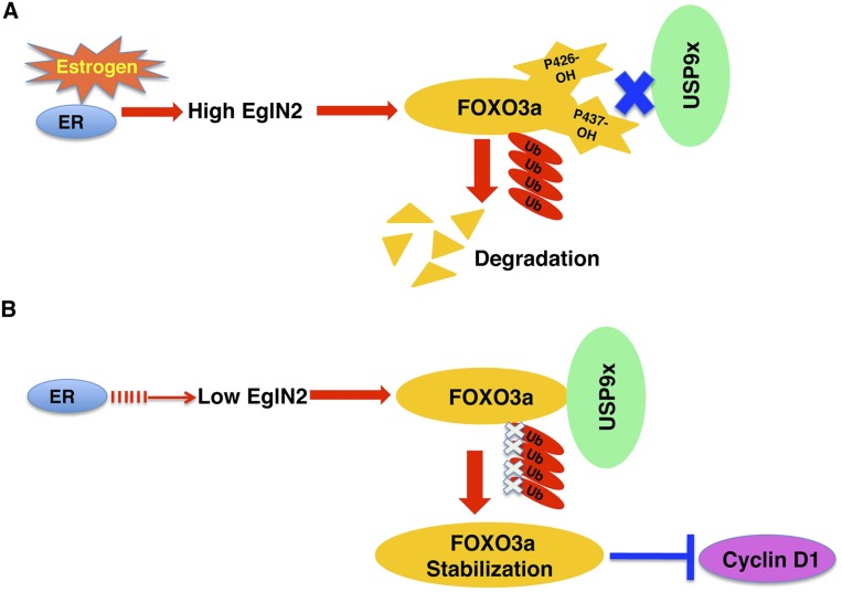
The three EglN prolyl hydroxylases (EglN1, EglN2, and EglN3) regulate the stability of the HIF transcription factor. We recently showed that loss of EglN2, however, also leads to down-regulation of Cyclin D1 and decreased cell proliferation in a HIF-independent manner. Here we report that EglN2 can hydroxylate FOXO3a on two specific prolyl residues in vitro and in vivo. Hydroxylation of these sites prevents the binding of USP9x deubiquitinase, thereby promoting the proteasomal degradation of FOXO3a. FOXO transcription factors can repress Cyclin D1 transcription. Failure to hydroxylate FOXO3a promotes its accumulation in cells, which in turn suppresses Cyclin D1 expression. These findings provide new insights into post-transcriptional control of FOXO3a and provide a new avenue for pharmacologically altering Cyclin D1 activity.

Keywords: Cyclin D1; EglN2; FOXO3a; USP9x; breast cancer; prolyl hydroxylation.


Figures

Figure 1.
Figure 1.
Screen for EglN2 substrates. (A) Biochemical basis for screen. Hydroxylation of a substrate by EglN2 is coupled to release and capture of 14C-labeled CO2. (B) Hydroxylation assays performed with varying amounts of recombinant EglN2, 14C-αKG, and synthetic HIF1α (556–575) peptides (wild-type or containing hydroxyproline at 564 position). (C) Hydroxylation assays using unprogrammed wheat germ lysate (Empty) or wheat germ lysate programmed to produce His-tagged p53 or HIF2α. Hydroxylation assays with no EglN2 (first two columns) or with EglN2 in the absence or presence of synthetic HIF1α peptides (columns 3,4) were included as controls. Increasing amounts of plasmid DNA were used for in vitro transcription/translation, as indicated. (D) Immunoblot analysis of in vitro translation products used in C. (E) Representative images from four independent hydroxylation assays. (F) Quantification of signal intensity in E (arbitrary units) after subtracting signal intensity from the empty vector well. (G) Immunoblot analysis of in vitro translation products as in E.
Figure 2.
Figure 2.
EglN2 prolyl hydroxylates FOXO3a in vivo and in vitro. (A) Immunoblot (IB) assays of whole-cell extracts (WCEs) and immunoprecipitates (IPs) of T47D cells treated with either vehicle control, 1 mM DMOG, 10 μM MG132, or both 1 mM DMOG and 10 μM MG132. (B) 293FT cells were cotransfected to produce HA-FOXO3a and Flag-EglN2 and then treated with MG132. (Inset) HA-FOXO3a was purified by anti-HA immunoprecipitation followed by SDS–polyacrylamide gel electrophoresis and Coomassie blue staining. Shown are liquid chromatography-tandem mass spectrometry (LC-MS/MS) data for the excised FOXO3a band corresponding to FOXO3a peptides hydroxylated at Pro426 or Pro437. The red “P” indicates a hydroxylated proline residue. (C,D) Coomassie blue staining (C) and in vitro hydroxylation assays (D) with the indicated GST fusion proteins. Synthetic HIF1α peptides corresponding to residues 556–575 were included as controls in D. P564-OH indicates 556–575 peptide hydroxylated at Pro564. (E) GST-FOXO3a (301–674) was incubated with recombinant EglN2, resolved by SDS–polyacrylamide gel electrophoresis, and stained with Coomassie blue. Shown are MS/MS data for the excised FOXO3a band corresponding to FOXO3a peptides hydroxylated at Pro426 or Pro437. The red “P” indicates a hydroxylated proline residue. The peak heights are the relative abundances of the corresponding fragment ions, with the annotation of the identified matched N terminus-containing ions (b ions) in blue and C terminus-containing ions (y ions) in red.
Figure 3.
Figure 3.
FOXO3a protein abundance is regulated by EglN2 and links EglN2 to Cyclin D1. (A,B) Immunoblot analysis of primary MEFs (A) and mammary fat pads (B) derived from EglN2+/+ and EglN2−/− littermates. (C,D) Immunoblot analysis of T47D cells after transfection with EglN2 siRNA (C) or infection with lentiviruses encoding EglN2 shRNAs. Unrelated nontargeting siRNA and shRNA sequences were used as controls (Ctrl). (E) Immunoblot analysis of MCF-7 cells that were infected with a lentivirus encoding EglN2 shRNA or Ctrl shRNA and, after drug selection, superinfected with a lentivirus encoding Flag-EglN2 (F-EglN2) under the control of a doxycycline-inducible promoter. Increasing amounts of doxycycline were added to the cells 48 h before lysis as indicated by the triangle. (F) Immunoblot analysis of primary MEF cells that were infected with a lentivirus encoding red fluorescent protein (RFP) or Flag-tagged EglN2 (wild-type or H358A). (G,H) Luciferase reporter assay of T47D cells that were infected with a lentivirus encoding EglN2 shRNA (325 or 326) or Control (Ctrl) shRNA followed by transfection with either FasL or FHRE reporter plasmid in the absence or presence of shRNA-resistant Flag-EglN2 (F-EglN2) with TK-Renilla as an internal control. (I) Immunoblot analysis of T47D cells that were infected with a lentivirus encoding FOXO3a shRNA or control (Ctrl) shRNA and then transfected with either EglN2 siRNA or Ctrl siRNA.
Figure 4.
Figure 4.
Regulation of FOXO3a by EglN2 is post-transcriptional and HIF-independent. (A) Immunoblot analysis of T47D cells that were infected with a lentivirus encoding EglN2 shRNA or control shRNA (Ctrl) and then treated with 200 μM CoCl2, 1 mM DMOG, 200 μM DFO, 10 μM MG132, or DMSO (Control) for 16 h. (B) Immunoblot analysis of T47D cells that were infected with a lentivirus encoding either ARNT shRNA (3146) or Ctrl shRNA and, after drug selection, transfected with either EglN2 siRNA or Ctrl siRNA or treated with 1 mM DMOG. (C) Immunoblot analysis of T47D cells that were infected with a lentivirus encoding either EglN2 shRNA or Ctrl shRNA and then treated with 100 μg/mL cycloheximide (CHX) for the indicated amount of time. (D) Quantitative RT–PCR (qRT–PCR) analysis of mRNA from T47D cells that were transfected with the indicated siRNAs. Error bars represent one SEM. (E,F) Immunoblot analysis of MCF-7 cells that were infected with a lentivirus encoding HA-FOXO3a and, after hygromycin selection, treated with either 10 μM MG132, 200 μM CoCl2, 200 μM DFO, 1 mM DMOG, or DMSO control for 16 h. (F) The cells were superinfected with a lentivirus encoding EglN2 shRNA (327) or control shRNA after the initial hygromycin selection. The cells were then placed under both hygromycin and puromycin selection and treated with DMOG, MG132, or vehicle control.
Figure 5.
Figure 5.
FOXO3a prolyl hydroxylation sites govern FOXO3a stability. (AE) Immunoblot analysis of T47D cells that were infected with lentiviruses encoding the indicated HA-FOXO3a variants and then treated with either 1 mM DMOG, 10 μM MG132, or DMSO control for 16 h (A–D) or transfected with EglN2 siRNA (1 or 4) or control siRNA (E). (WT) Wild-type. (C,D) Light or dark refers to different exposure times. (F,G) Immunoblot analysis of T47D cells that were infected with a lentivirus encoding either wild-type FOXO3a or P426A;P437A followed by hygromycin selection and treatment with either hypoxia for 16 h (F) or 100 μg/mL cycloheximide (CHX) for the indicated amount of time (G). Light or dark refers to different exposure times.
Figure 6.
Figure 6.
Differential binding of USP9x underlies control of FOXO3a stability by EglN2. (A) Immunoblot analysis of T47D cells that were infected with a lentivirus encoding the indicated USP9x shRNAs or Ctrl shRNA. (B) Immunoblot analysis of T47D cells that were infected with a lentivirus encoding HA-FOXO3a and then superinfected with a lentivirus encoding USP9x shRNA (364 or 064) or Ctrl shRNA. (C) Immunoblot analysis of 293T cells that were infected with a lentivirus encoding USP9x shRNA (364) or Ctrl shRNA and then, after drug selection, transfected with a plasmid encoding V5-tagged mouse full-length USP9x or empty vector (Ctrl). (D) Immunoblot (IB) assays of whole-cell extracts (WCE) and immunoprecipitates (IP) of T47D cells that were infected with a lentivirus encoding USP9x shRNA (sh128 or sh364) or Ctrl shRNA and then, after drug selection, transiently transfected with plasmids encoding Flag-ubiquitin and HA-FOXO3a followed by treatment with 10 μM MG132 for 16 h. (E) Immunoblot analysis of MCF-7 cells that were infected with a lentivirus encoding shRNA against either USP9x (364) or Ctrl shRNA followed by 100 μg/mL cycloheximide for the indicated duration. (F) Immunoblot analysis of 293T cells that were cotransfected with plasmids encoding V5-tagged USP9x (wild-type or C1556S) and HA-FOXO3a. (G) Immunoblot (IB) assays of whole-cell extracts (WCE) and immunoprecipitates (IP) of T47D cells that were transfected with a plasmid encoding HA-FOXO3a (wild-type or P426A;P437A) followed by treatment with 10 μM MG132 for 16 h. (H) Immunoblot analysis of bound USP9x recovered from lysed T47D cells that were incubated with 20 μL of Neutravidin-Sepharose preloaded with the indicated FOXO3a peptides. (I,J) Immunoblot analysis of T47D cells that were infected with a lentivirus encoding USP9x shRNA (364) or Ctrl shRNA and then, after drug selection, treated with the indicated drugs or siRNAs.
Figure 7.
Figure 7.
Proposed models for FOXO3a regulation by EglN2. (A) Estrogen binding with ER transcriptionally induces EglN2, which triggers FOXO3a hydroxylation on prolyl residue 426 and 327 sites. Hydroxylation of these sites dissociates FOXO3a from USP9x deubiquitinase, thereby facilitating FOXO3a ubiquitylation and degradation. (B) Depletion of EglN2 by either lack of ER activity or EglN2 siRNAs/shRNAs prevents FOXO3a from being hydroxylated. As a result, USP9x binds with FOXO3a and prevents FOXO3a from being ubiquitylated and degraded. Stabilization of FOXO3a decreases Cyclin D1 expression.

References

    1. Appelhoff RJ, Tian YM, Raval RR, Turley H, Harris AL, Pugh CW, Ratcliffe PJ, Gleadle JM 2004. Differential function of the prolyl hydroxylases PHD1, PHD2, and PHD3 in the regulation of hypoxia-inducible factor. J Biol Chem 279: 38458–38465 - PubMed
    1. Aragones J, Schneider M, Van Geyte K, Fraisl P, Dresselaers T, Mazzone M, Dirkx R, Zacchigna S, Lemieux H, Jeoung NH, et al. 2008. Deficiency or inhibition of oxygen sensor Phd1 induces hypoxia tolerance by reprogramming basal metabolism. Nat Genet 40: 170–180 - PubMed
    1. Bakker WJ, Harris IS, Mak TW 2007. FOXO3a is activated in response to hypoxic stress and inhibits HIF1-induced apoptosis via regulation of CITED2. Mol Cell 28: 941–953 - PubMed
    1. Berra E, Benizri E, Ginouves A, Volmat V, Roux D, Pouyssegur J 2003. HIF prolyl-hydroxylase 2 is the key oxygen sensor setting low steady-state levels of HIF-1α in normoxia. EMBO J 22: 4082–4090 - PMC - PubMed
    1. Beuck S, Schanzer W, Thevis M 2012. Hypoxia-inducible factor stabilizers and other small-molecule erythropoiesis-stimulating agents in current and preventive doping analysis. Drug Test Anal 4: 830–845 - PubMed

Publication types

LinkOut - more resources