. 2014 Jul 1;28(13):1445-60.
doi: 10.1101/gad.241950.114.

Heterochromatin-associated interactions of Drosophila HP1a with dADD1, HIPP1, and repetitive RNAs

Affiliations

Heterochromatin-associated interactions of Drosophila HP1a with dADD1, HIPP1, and repetitive RNAs

Artyom A Alekseyenko et al. Genes Dev. .

Abstract

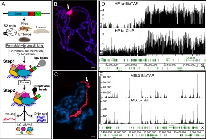
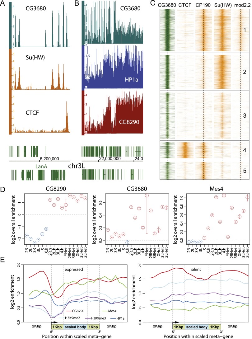
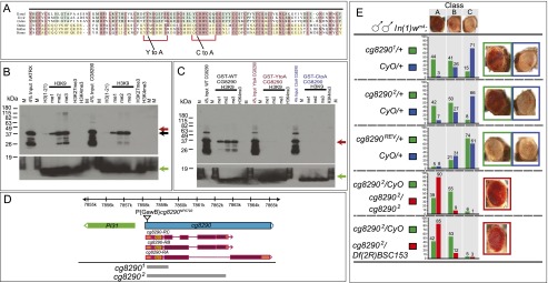
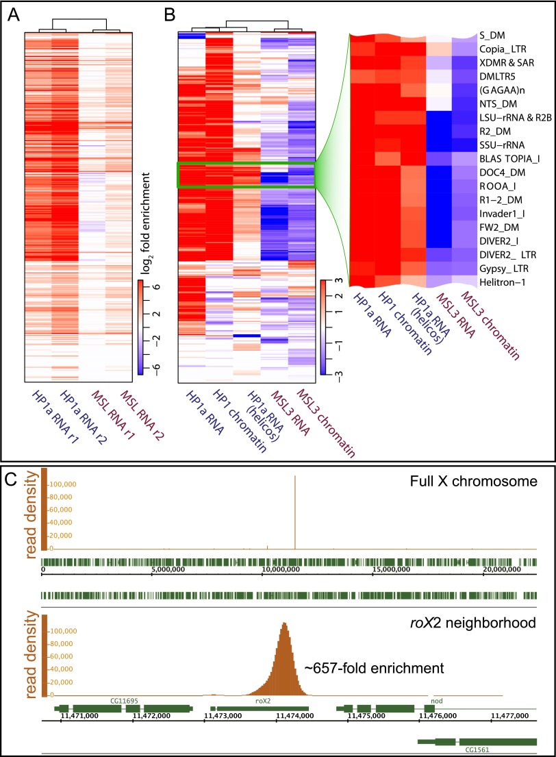
Heterochromatin protein 1 (HP1a) has conserved roles in gene silencing and heterochromatin and is also implicated in transcription, DNA replication, and repair. Here we identify chromatin-associated protein and RNA interactions of HP1a by BioTAP-XL mass spectrometry and sequencing from Drosophila S2 cells, embryos, larvae, and adults. Our results reveal an extensive list of known and novel HP1a-interacting proteins, of which we selected three for validation. A strong novel interactor, dADD1 (Drosophila ADD1) (CG8290), is highly enriched in heterochromatin, harbors an ADD domain similar to human ATRX, displays selective binding to H3K9me2 and H3K9me3, and is a classic genetic suppressor of position-effect variegation. Unexpectedly, a second hit, HIPP1 (HP1 and insulator partner protein-1) (CG3680), is strongly connected to CP190-related complexes localized at putative insulator sequences throughout the genome in addition to its colocalization with HP1a in heterochromatin. A third interactor, the histone methyltransferase MES-4, is also enriched in heterochromatin. In addition to these protein-protein interactions, we found that HP1a selectively associated with a broad set of RNAs transcribed from repetitive regions. We propose that this rich network of previously undiscovered interactions will define how HP1a complexes perform their diverse functions in cells and developing organisms.

Keywords: Bayesian analysis; ChIP; HP1a; LC-MS/MS; formaldehyde cross-linking.


Figures

Figure 1.
Figure 1.
Overview of the BioTAP-XL purification strategy and validation of MSL3-BioTAP- and HP1a-BioTAP-tagged proteins. (A) The BioTAP tag includes two epitopes: protein A and Bio, a 75-amino-acid sequence that is biotinylated in vivo. To preserve endogenous expression patterns, genomic BioTAP-tagged transgenes (generated by BAC recombineering) were introduced into flies or S2 cells. Crude nuclear extracts from cells and from different life stages of flies were cross-linked using formaldehyde, sonicated, and subjected to TAP, first with rabbit IgG agarose beads eluted under denaturing conditions and subsequently using streptavidin agarose beads. The resulting DNA and RNA were analyzed by high-throughput sequencing. Peptides from the protein fraction were released by direct on-bead trypsin digestion and then identified by LC-MS/MS. Immunostaining detected the specific signal on pericentric heterochromatin for HP1a-BioTAP (B) and on the male X chromosome for MSL3-BioTAP with PAP antibody (red) (C). Hoechst staining of DNA is shown in blue. Bars, 10 μm. (D) Representative ChIP-seq profiles of HP1a-BioTAP on the heterochromatin–euchromatin border of chromosome 3L correspond well with ChIP-seq data (HP1a-ChIP) obtained from the modENCODE consortium (Kharchenko et al. 2011). (E) Representative ChIP-seq profiles of MSL3-BioTAP in S2 cells on the X chromosome. The results correlated well with previous MSL3-TAP ChIP-seq (Alekseyenko et al. 2008).
Figure 2.
Figure 2.
Bamse. (A) Our analysis method aims to identify proteins exhibiting statistically significant enrichment in target pull-downs relative to controls based on an agglomeration of data from different cell types. The peptide counts observed for CG3680 in different mass spectrometry samples (top table) are modeled as variables distributed according to a negative binomial distribution, with the mean rate described by a log-linear model (middle formula). The log-linear model separates contributions of base-level protein abundance in the cell (αt), enrichment in pull-downs due to nonspecific effects (β), and enrichment due to specific association with the target protein (γ). (Bottom plots) A Bayesian approach was used to infer the distributions of likely values for these parameters (i.e., posterior distributions), and the range of statistically likely values of the target-specific fold enrichment magnitude γ was used to evaluate the significance of the association with the target. For CG3680, the 95% confidence interval of the target-specific enrichment magnitude γ lies within the range of 5.6–8.0 (log2 scale). (B) The posterior distributions are shown for the ISWI protein, which is detected in the HP1a BioTAP pull-downs but shows only small enrichment magnitude (<1.7 on log2 scale), partly due to nonspecific pull-down abundance β (with MSL3 control).
Figure 3.
Figure 3.
Genomic distribution of CG3680, CG8290, and MES-4 proteins. (A) Within the euchromatic portion of the Drosophila genome, CG3680 exhibits prominent binding positions coinciding with those of several insulator proteins, including CTCF and Su(Hw). The plots show enrichment of the DNA fraction of the CG3680 BioTAP pull-down for a euchromatic region of chromosome 3L along with ChIP-seq enrichment for the insulator proteins in S2 cells. (B) A region of chromosome 3L in S2 cells containing the euchromatic–heterochromatic boundary is shown. Both CG3680 and CG8290 show a broad pattern of enrichment in the heterochromatin, resembling that of HP1a. However, the enrichment magnitude of CG3680 is lower, particularly in contrast to its insulator-associated euchromatic binding positions. (C) CG3680 euchromatic binding positions coincide with several different classes of insulators. The heat maps show ChIP-seq enrichments of Drosophila insulator proteins around all euchromatic CG3680 positions in S2 cells (from top to bottom). Most of the euchromatic CG3680 sites can be classified into those associated with gypsy-like insulator combinations [Su(Hw) + CP190 + mod2.2; cluster 1], standalone Su(Hw) (clusters 2 and 3), or CTCF + CP190 (cluster 4 insulators). (D) CG3680, CG8290, and MES-4 show statistically significant enrichment in the heterochromatic portions of the Drosophila genome. The maximum likelihood estimate (circles) over the overall enrichment levels ([red] enriched; [blue] depleted) and the 95% confidence intervals (whiskers) are shown for the euchromatic portions of the assembled chromosome arms (e.g., 2L), assembled pericentromeric heterochromatin regions (e.g., 2L.h), and unassembled heterochromatic contigs (e.g,. 2LHet). (E) Metagene profiles of CG8290 and MES-4 in S2 cells with comparison with HP1 and H3K9me2/3 modification (Kharchenko et al. 2011) around expressed and silent heterochromatic genes.
Figure 4.
Figure 4.
CG8290 recognizes H3K9me2/3 through its ADD domain and functions in heterochromatin silencing. (A) Alignment of the ADD domains from dipteran CG8290 (Drosophila melanogaster, Drosophila virilis, and Culex pipiens) and vertebrate ATRX (Danio rerio, Gallus gallus, and Homo sapiens). Residues conserved among dipterans or vertebrates are shaded blue and yellow, respectively. Residues shared by at least five of the six species shown here are highlighted in pink. Mutations Y101A and C117A at conserved positions implicated in H3K9me recognition and used as controls in H3K9 peptide pull-downs are depicted below. Red brackets denote the residues forming a composite H3K9me3 pocket (Iwase et al. 2011). (B) Anti-GST Western blot analysis showing recovery of the CG8290 GST-ADD domain after pull-down with biotinylated histone H3 tail peptides methylated at H3K9 but not at H3K4 or H3K27 residues, consistent with the published human ATRX ADD data (positive control). Mobilities of the GST-tagged CG8290 and ATRX ADD domains are slightly different and are marked by red and black arrows. A green arrow indicates biotinylated histone H3 peptides used in pull-downs, detected with streptavidin-HRP. (C) Western blot analysis of the CG8290 GST-ADD domain showing that binding to methylated histone H3K9 peptides is abrogated by point mutations Y101A and C117A. Positions of wild-type and mutant CG8290 ADD are indicated by the red arrow. The green arrow indicates the signal from biotinylated histone H3 peptides used in pull-downs. (D) Organization of the cg8290 locus. cg8290 (blue) produces three types of transcripts encoding three CG8290 isoforms. Coding sequences and UTRs are colored purple and orange, respectively, with the position of the ADD domain indicated. Insertion of P{GawB}cg8290NP0793 is indicated by a triangle. Two null alleles, cg82901 and cg82902, are deletions of the cg8290 coding sequence, not affecting the upstream PI31 gene. The deletions are indicated by gray boxes. (E) cg8290 alleles display a dose-dependent PEV phenotype and act as Su(var)s in the white-mottled genetic background. A dominant Su(var) phenotype is observed in wm4h/Y; cg82901 or 2/+ flies, which is further enhanced in homozygotes (wm4h/Y; cg82902/cg82902) and trans-heterozygotes (wm4h/Y; cg82902/Df(2R)BSC153). cg8290REV is a precise excision of P{GawB} and serves as a genetic background control. Df(2R)BSC153 is a 341-kb deletion removing cg8290 and other genes; it was obtained on a background distinct from that of cg82902, so its use in combination with cg8290 helps exclude the possible contribution of linked PEV modifiers on the cg82902 chromosome. Distribution of eye color classes (A, B, or C, depicted above) is distinct between cg82901 or 2/+ and cg8290REV/+ or CyO/+. Cg8290-null animals (cg82902/cg82902 and cg82902/Df(2R)BSC153) display a further increase in eye pigmentation.
Figure 5.
Figure 5.
RNA-seq analysis of BioTAP-XL pull-downs. (A) Enrichment of repeat-derived RNA in HP1a-BioTAP cross-linked complexes from S2 cells compared with MSL3-BioTAP complexes from S2 cells detected using a random-priming approach for cDNA synthesis and Illumina RNA-seq. The plot shows log2 average enrichment across all annotated repeat types in Drosophila (each row corresponds to a repeat type), estimated relative to a corresponding input sample (total chromatin-associated RNA). Only statistically significant enrichment/depletion levels are shown in color. (B) Enrichment of the same spectrum of repeat-derived RNAs (rows are ordered as in A) assessed using HP1a-BioTAP embryos and either an on-bead ligation method for cDNA priming (HP1a RNA) or direct RNA-seq (HP1a RNA; Helicos). The enrichment was calculated compared with unrelated BioTAP-XL pull-down samples from embryos (see the Supplemental Material). The enrichment levels are also shown for the DNA fraction of the BioTAP pull-downs, illustrating the abundance of HP1a binding to the repetitive regions of the genome (HP1a chromatin) as well as the lack of such binding in the case of the control MSL3 complex (MSL3 chromatin). The zoomed-in version shows selected repeat details, with the full list in Supplemental Data File 1. Repeat enrichment was not observed for MSL3-associated RNA from S2 cells using the on-bead ligation approach (MSL3 RNA). (C) Analysis of the RNA recovered from the MSL3-BioTAP pull-down (on-bead method, Illumina platform) confirms the presence of roX2 RNA as the main RNA component of the MSL complex in S2 cells.

Similar articles

Cited by

References

    1. Ainsztein AM, Kandels-Lewis SE, Mackay AM, Earnshaw WC 1998. INCENP centromere and spindle targeting: identification of essential conserved motifs and involvement of heterochromatin protein HP1. J Cell Biol 143: 1763–1774 - PMC - PubMed
    1. Alekseyenko AA, Peng S, Larschan E, Gorchakov AA, Lee OK, Kharchenko P, McGrath SD, Wang CI, Mardis ER, Park PJ, et al. 2008. A sequence motif within chromatin entry sites directs MSL establishment on the Drosophila X chromosome. Cell 134: 599–609 - PMC - PubMed
    1. Alekseyenko AA, Gorchakov AA, Kharchenko PV, Kuroda MI 2014. Reciprocal interactions of human C10orf12 and C17orf96 with PRC2 revealed by BioTAP-XL cross-linking and affinity purification. Proc Natl Acad Sci 111: 2488–2493 - PMC - PubMed
    1. Amrein H, Axel R 1997. Genes expressed in neurons of adult male Drosophila. Cell 88: 459–469 - PubMed
    1. Ashburner M (1989). Drosophila, A Laboratory Manual. Cold Spring Harbor Laboratory Press, Cold Spring Harbor, NY

Publication types

MeSH terms

Associated data