. 2014 Jul 1;28(13):1498-514.
doi: 10.1101/gad.241133.114.

A fly trap mechanism provides sequence-specific RNA recognition by CPEB proteins

Affiliations

A fly trap mechanism provides sequence-specific RNA recognition by CPEB proteins

Tariq Afroz et al. Genes Dev. .

Abstract

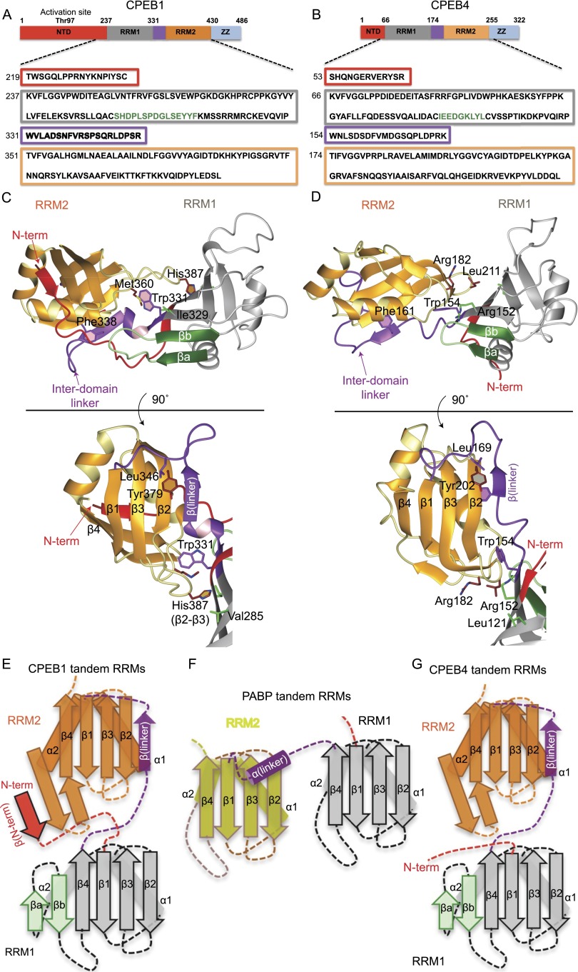
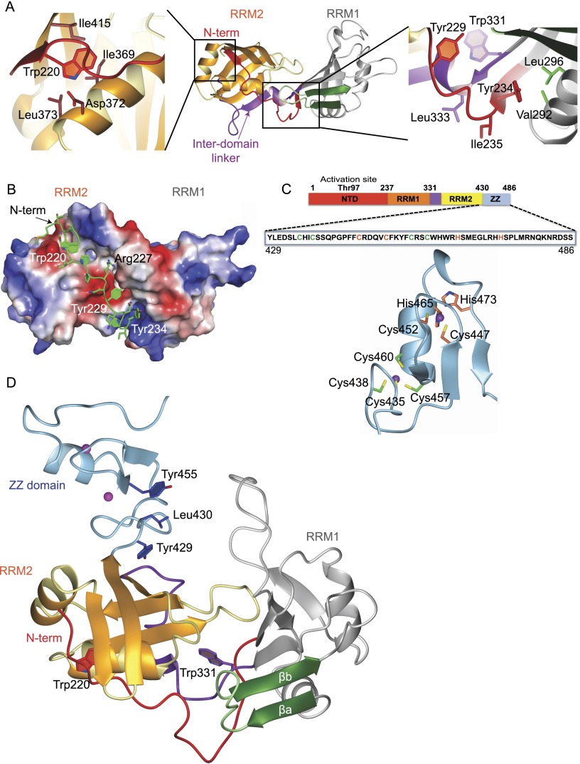
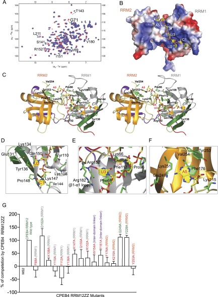
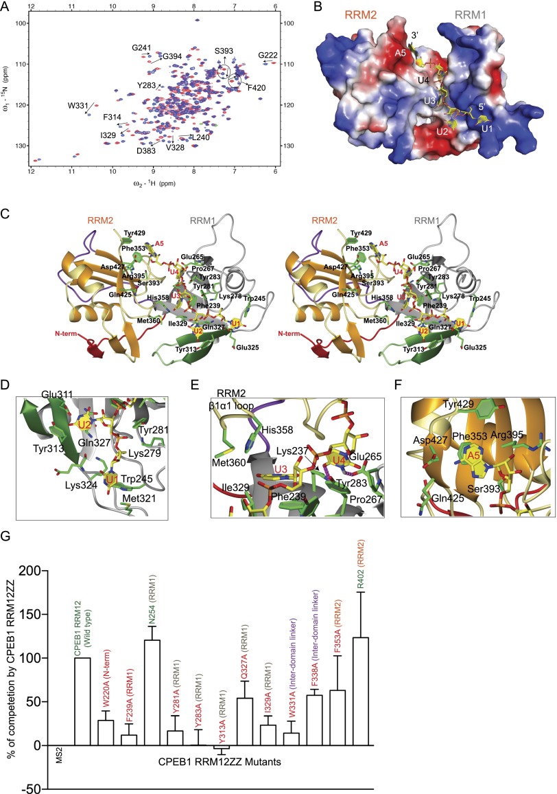
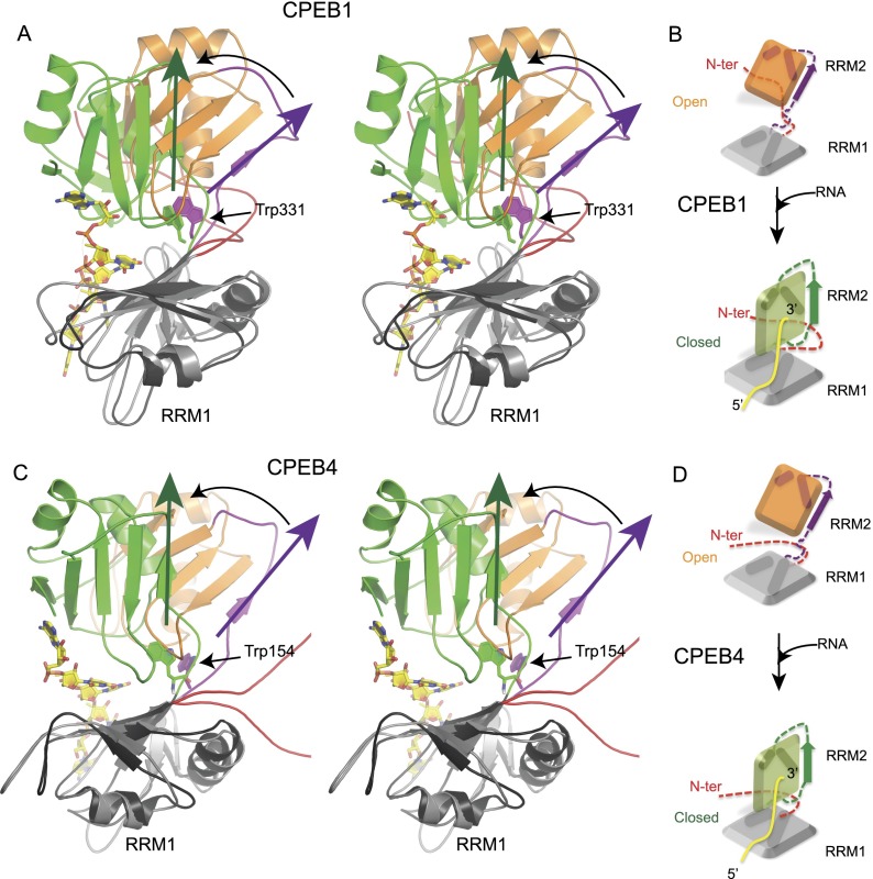
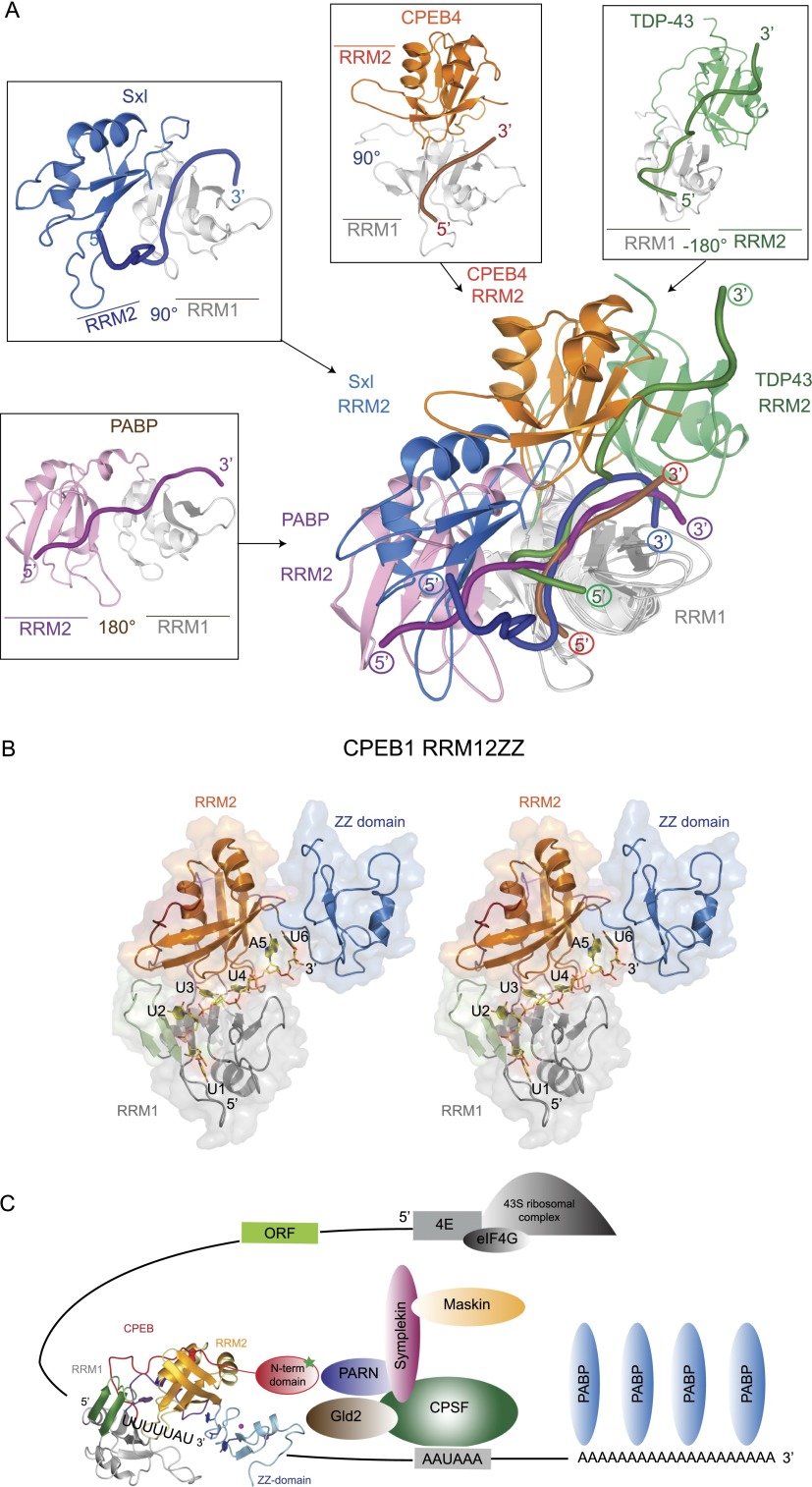
Cytoplasmic changes in polyA tail length is a key mechanism of translational control and is implicated in germline development, synaptic plasticity, cellular proliferation, senescence, and cancer progression. The presence of a U-rich cytoplasmic polyadenylation element (CPE) in the 3' untranslated regions (UTRs) of the responding mRNAs gives them the selectivity to be regulated by the CPE-binding (CPEB) family of proteins, which recognizes RNA via the tandem RNA recognition motifs (RRMs). Here we report the solution structures of the tandem RRMs of two human paralogs (CPEB1 and CPEB4) in their free and RNA-bound states. The structures reveal an unprecedented arrangement of RRMs in the free state that undergo an original closure motion upon RNA binding that ensures high fidelity. Structural and functional characterization of the ZZ domain (zinc-binding domain) of CPEB1 suggests a role in both protein-protein and protein-RNA interactions. Together with functional studies, the structures reveal how RNA binding by CPEB proteins leads to an optimal positioning of the N-terminal and ZZ domains at the 3' UTR, which favors the nucleation of the functional ribonucleoprotein complexes for translation regulation.

Keywords: CPEB1; CPEB4; binuclear zinc-binding domain; cytoplasmic polyadenylation; protein–RNA interactions; translational regulation.


Figures

Figure 1.
Figure 1.
Solution structures of CPEB1 and CPEB4 tandem RRMs in the free state (see also Supplemental Fig. S1). (A,B) Schematic representation of full-length CPEB1 and CPEB4 proteins. The regions corresponding to the N-terminal domain, RRM1, the inter-domain linker, RRM2, and the ZZ domain are shown by red, gray, purple, orange, and blue boxes, respectively. The same color-coding is also used for the structures. The protein is shown with the amino acid sequence of the RRMs used in these studies. (C,D) Representative structure of CPEB1 and CPEB4 tandem RRMs in ribbon representation. The structures shown below were obtained by a 90° rotation. (E–G) Two-dimensional (2D) schematic representation of CPEB1 (E), PABP (F), and CPEB4 (G) tandem RRMs. CPEB1/4 RRM1 has an insertion of two anti-parallel β strands in the β sheet (βa and βb, shown in green) compared with the canonical RRM of PABP. (E) In CPEB1, the N-terminal extension (in red) forms a parallel β strand with the β hairpin between the α2 helix and the β4 strand of RRM2. (E,G) In both CPEB1 and CPEB4, the interdomain linker (purple) forms an anti-parallel β strand with the β2 strand of RRM2.
Figure 2.
Figure 2.
Intramolecular interactions of the N-terminal extension with RRM2 and structural model of CPEB1 RRM12ZZ (see also Supplemental Fig. S2). (A) Ribbon representation of a representative structure from the ensemble of CPEB1 RRM12 structures. (Left and right panels) Two enlarged views show the details from the part of the structure marked in black boxes. (B) Surface charge of CPEB1 RRM12. Negative charge is represented in red, and positive charge is represented in blue. A similar color code is also used for surface charges in other structures. The N-terminal extension with side chains in stick representation is shown in green. (C) The CPEB1 protein is shown with the amino acid sequence of the ZZ domain used in these studies. Shown below is one representative structure from the ensemble of the CPEB1 ZZ domain in light blue. The two zinc ions are shown as purple spheres. The two clusters of zinc-coordinating residues are shown in green and orange (also colored in the sequence). (D) Structural model of CPEB1 tandem RRMs and the ZZ domain.
Figure 3.
Figure 3.
Structure of CPEB4 tandem RRMs in complex with the RNA (see also Supplemental Fig. S3). (A) Overlay of 1H–15N HSQC spectra of CPEB4 RRM12 in the free state (red) and bound to 5′-CUUUA-3′ (blue) at 30°C. Peaks undergoing CSPs upon RNA binding are labeled. (B) Surface charge representation of CPEB4 RRM12 in complex with RNA (yellow). (C) Stereo view of the representative structure of CPEB4 tandem RRMs in complex with RNA. Protein is shown in ribbon, while the RNA is shown in yellow stick representation. (D–F) Close-up view showing the interactions between amino acids involved in RNA interaction in CPEB4 RRM12 in complex with RNA. Protein side chains (green) involved in interactions with the RNA (yellow) are shown in stick representation. (G) In vivo functional assay to validate CPEB4 residues involved in RNA binding. The extent of RNA probe polyadenylation is plotted as a percentage of competition, quantified from three independent experiments (100% competition was assigned to the wild-type protein, and 0% competition was assigned to the MS2-negative control). Positive controls and wild-type protein are labeled in green, while mutants are labeled in red. The plotted values were obtained from the gels from Supplemental Figure S3, H and I.
Figure 4.
Figure 4.
Structural model of CPEB1 RRMs in complex with RNA (see also Supplemental Fig. S4). (A) Overlay of 1H–15N HSQC spectra of CPEB1 RRM12 in the free state (red) and in complex with 5′-UUUUA-3′ (blue) measured at 40°C. Peaks undergoing CSPs upon RNA binding are labeled. (B) Surface charge representation of CPEB1 RRM12 in complex with RNA (yellow). (C) Stereo view of the structural model of CPEB1 RRM12 in complex with 5′-UUUUA-3′. The protein is shown in ribbon representation. The RNA is shown in stick representation in yellow, and the bases are labeled. (DF) Close-up view showing the interactions between amino acids involved in RNA interaction in CPEB1 RRM12 in complex with 5′-UUUUA-3′. Protein side chains (green) involved in interactions with the RNA (yellow) are shown in stick representation. (G) In vivo functional assay to validate CPEB1 residues involved in RNA binding. The extent of RNA probe polyadenylation is plotted as a percentage of competition, quantified from three independent experiments (100% competition was assigned to the wild-type protein, and 0% competition was assigned to the MS2-negative control). Positive controls and wild-type protein are labeled in green, while mutants are labeled in red. The plotted values were obtained from the gels from Supplemental Figure S4, G–I.
Figure 5.
Figure 5.
Conformational changes upon RNA binding (see also Supplemental Fig. S5). (A,C) Stereo view of overlay of CPEB1 (A) and CPEB4 (C) RRM12 in the free state and in complex with RNA. The structures have been overlaid on RRM1. The structure of the complex is shown in ribbon with RRM1 in black, RRM2 and the interdomain linker in green, and RNA as yellow sticks. In the free state, RRM1 is shown in gray, the interdomain linker is in purple, and RRM2 is in orange. The conformational change is depicted with the help of colored arrows. For clarity, the loops in the structures have been smoothened. (B,D) Schematic diagram depicting the conformational change upon RNA binding in CPEB1 (B) and CPEB4 (D) tandem RRMs.
Figure 6.
Figure 6.
Molecular mechanism of assembly of translation regulatory complexes by CPEB1 (see also Supplemental Fig. S6). (A) Overlay of CPEB4 RRM12 in complex with three previously characterized tandem RRM–RNA complexes: PABP (1CVJ), Sxl (1B7F), and TDP-43 (4BS2). All structures were overlaid on the RRM1 domain (shown in gray for all structures). The RRM2 domain is shown in pink, blue, orange, and green for PABP, Sxl, CPEB4, and TDP43, respectively. RNA is shown in tube representation in the corresponding RRM2 colors, with the 5′ and 3′ ends labeled in colored circles. An individual structure is shown around the overlay in the same orientation. For clarity, the loops in the structures have been smoothened. (B) Structural model of CPEB1 RRM12ZZ in complex with the RNA in stereo view. (C) A model depicting the assembly of a translational regulatory complex by CPEB1. mRNA is shown in black, with the 5′ and 3′ ends labeled in blue. A model of full-length CPEB1 is shown in ribbon representation. (Red) N-terminal domain; (gray) RRM1; (orange) RRM2; (cyan) ZZ domain. The activation phosphorylation site is shown in the N-terminal domain of CPEB1 as a green star. Other protein factors of this complex are schematically depicted, with ovals in different colors and labeled inside.

Similar articles

Cited by

References

    1. Alexandrov IM, Ivshina M, Jung DY, Friedline R, Ko HJ, Xu M, O’Sullivan-Murphy B, Bortell R, Huang YT, Urano F, et al. 2012. Cytoplasmic polyadenylation element binding protein deficiency stimulates PTEN and Stat3 mRNA translation and induces hepatic insulin resistance. PLoS Genet 8: e1002457. - PMC - PubMed
    1. Allain FH, Gilbert DE, Bouvet P, Feigon J 2000. Solution structure of the two N-terminal RNA-binding domains of nucleolin and NMR study of the interaction with its RNA target. J Mol Biol 303: 227–241 - PubMed
    1. Bae E, Reiter NJ, Bingman CA, Kwan SS, Lee D, Phillips GN Jr, Butcher SE, Brow DA 2007. Structure and interactions of the first three RNA recognition motifs of splicing factor prp24. J Mol Biol 367: 1447–1458 - PMC - PubMed
    1. Barraud P, Allain FH 2013. Solution structure of the two RNA recognition motifs of hnRNP A1 using segmental isotope labeling: how the relative orientation between RRMs influences the nucleic acid binding topology. J Biol NMR 55: 119–138 - PubMed
    1. Bava FA, Eliscovich C, Ferreira PG, Minana B, Ben-Dov C, Guigo R, Valcarcel J, Mendez R 2013. CPEB1 coordinates alternative 3′-UTR formation with translational regulation. Nature 495: 121–125 - PubMed

Publication types

MeSH terms

LinkOut - more resources