

The phosphatidylinositol 3-kinase (PI3K) isoform dependence of tumor formation is determined by the genetic mode of PI3K pathway activation rather than by tissue type
- PMID: 24991009
- PMCID: PMC4178865
- DOI: 10.1128/JVI.01409-14
The phosphatidylinositol 3-kinase (PI3K) isoform dependence of tumor formation is determined by the genetic mode of PI3K pathway activation rather than by tissue type
Abstract
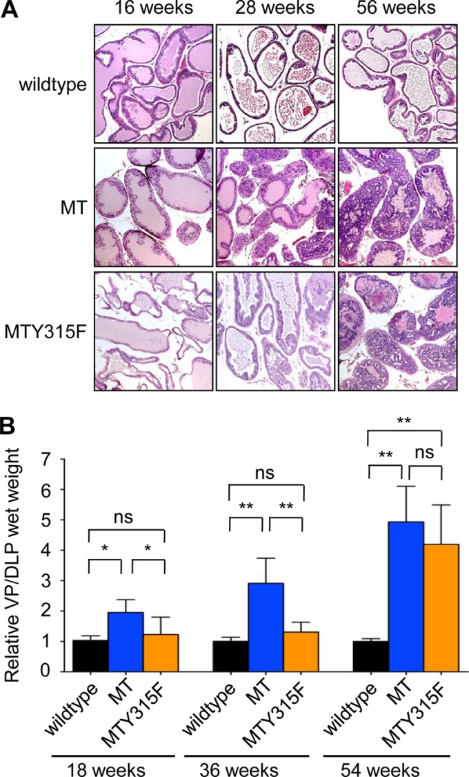
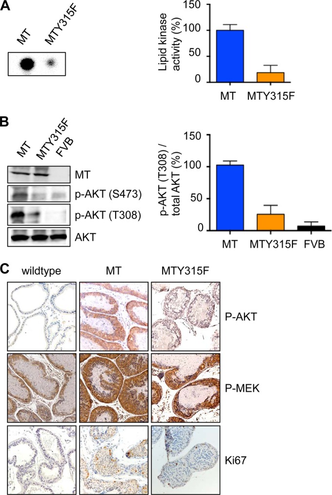
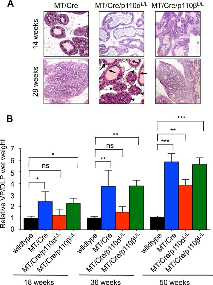
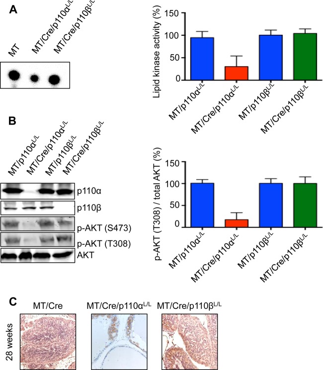
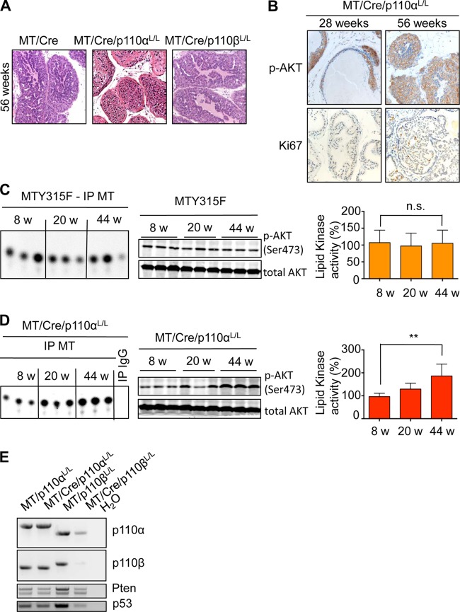
Previous work has shown that prostate cancer in a Pten-null murine model is dependent on the p110β isoform of phosphatidylinositol 3-kinase (PI3K), while breast cancer driven by either polyoma middle T antigen (MT) or HER2 is p110α dependent. Whether these differences in isoform dependence arise from tissue specificity or from the nature of the oncogenic signal activating the PI3K pathway is important, given increasing interest in using isoform-specific PI3K inhibitors in cancer therapy. To approach this question, we studied the PI3K isoform dependence of our recently constructed prostate cancer model driven by MT. Since MT activates a number of signaling pathways, we first confirmed that the MT-driven prostate cancer model was actually dependent on PI3K. A newly generated transgenic prostate line expressing an MT allele (Y315F) known to be defective for PI3K binding displayed a markedly reduced ability to drive tumor formation. We next selectively ablated expression of either p110α or p110β in mice in which wild-type MT was expressed in the prostate. We found that tumor formation driven by MT was significantly delayed by the loss of p110α expression, while ablation of p110β had no effect. Since the tumor formation driven by MT is p110α dependent in the prostate as well as in the mammary gland, our data suggest that PI3K isoform dependence is driven by the mode of PI3K pathway activation rather than by tissue type.
Importance: Middle T antigen (MT), the oncogene of polyomavirus, can drive tumor formation in a variety of cell types and tissues. Interestingly, MT has no intrinsic enzymatic activity but instead functions by binding and activating cellular signaling proteins. One of the most important of these is the lipid kinase PI3K, which was first studied in MT immunoprecipitates. Ubiquitously expressed PI3K comes in two major isoforms: p110α and p110β. Previous work in animal models showed that p110α was the key isoform in breast tumors driven by oncogenes, including MT and HER2, while p110β was key in prostate tumors driven by Pten loss. We asked the simple question of whether a prostate tumor driven by MT depends on p110α, which would suggest that the mode of activation determines p110 isoform dependence, or p110β, which would suggest that tissue type determines isoform dependence. The clear answer is that MT depends on p110α in both the prostate and breast.
Copyright © 2014, American Society for Microbiology. All Rights Reserved.
Figures
Similar articles
-
The p110alpha isoform of phosphatidylinositol 3-kinase is essential for polyomavirus middle T antigen-mediated transformation.J Virol. 2007 Jul;81(13):7069-76. doi: 10.1128/JVI.00115-07. Epub 2007 Apr 18. J Virol. 2007. PMID: 17442716 Free PMC article.
-
PI3K-p110α mediates resistance to HER2-targeted therapy in HER2+, PTEN-deficient breast cancers.Oncogene. 2016 Jul 7;35(27):3607-12. doi: 10.1038/onc.2015.406. Epub 2015 Oct 26. Oncogene. 2016. PMID: 26500061 Free PMC article.
-
The p110α and p110β isoforms of PI3K play divergent roles in mammary gland development and tumorigenesis.Genes Dev. 2012 Jul 15;26(14):1573-86. doi: 10.1101/gad.191973.112. Genes Dev. 2012. PMID: 22802530 Free PMC article.
-
Class I Phosphoinositide 3-Kinase PIK3CA/p110α and PIK3CB/p110β Isoforms in Endometrial Cancer.Int J Mol Sci. 2018 Dec 7;19(12):3931. doi: 10.3390/ijms19123931. Int J Mol Sci. 2018. PMID: 30544563 Free PMC article. Review.
-
Should individual PI3 kinase isoforms be targeted in cancer?Curr Opin Cell Biol. 2009 Apr;21(2):199-208. doi: 10.1016/j.ceb.2008.12.007. Epub 2009 Feb 4. Curr Opin Cell Biol. 2009. PMID: 19200708 Free PMC article. Review.
Cited by
-
Divergent Roles of PI3K Isoforms in PTEN-Deficient Glioblastomas.Cell Rep. 2020 Sep 29;32(13):108196. doi: 10.1016/j.celrep.2020.108196. Cell Rep. 2020. PMID: 32997991 Free PMC article.
-
Addition of the p110α inhibitor BYL719 overcomes targeted therapy resistance in cells from Her2-positive-PTEN-loss breast cancer.Tumour Biol. 2016 Nov;37(11):14831-14839. doi: 10.1007/s13277-016-5381-7. Epub 2016 Sep 17. Tumour Biol. 2016. PMID: 27639383
-
PTEN-regulated PI3K-p110 and AKT isoform plasticity controls metastatic prostate cancer progression.Oncogene. 2024 Jan;43(1):22-34. doi: 10.1038/s41388-023-02875-4. Epub 2023 Oct 24. Oncogene. 2024. PMID: 37875657 Free PMC article.
-
Isoform-specific activities of the regulatory subunits of phosphatidylinositol 3-kinases - potentially novel therapeutic targets.Expert Opin Ther Targets. 2018 Oct;22(10):869-877. doi: 10.1080/14728222.2018.1522302. Epub 2018 Sep 24. Expert Opin Ther Targets. 2018. PMID: 30205700 Free PMC article. Review.
-
PI3Kβ-A Versatile Transducer for GPCR, RTK, and Small GTPase Signaling.Endocrinology. 2019 Mar 1;160(3):536-555. doi: 10.1210/en.2018-00843. Endocrinology. 2019. PMID: 30601996 Free PMC article. Review.
References
Publication types
MeSH terms
Substances
Grants and funding
LinkOut - more resources
Full Text Sources
Other Literature Sources
Medical
Molecular Biology Databases
Research Materials
Miscellaneous
