. 2014 Sep;88(18):10714-24.
doi: 10.1128/JVI.01331-14. Epub 2014 Jul 2.

Homeostatic cytokines induce CD4 downregulation in African green monkeys independently of antigen exposure to generate simian immunodeficiency virus-resistant CD8αα T cells

Affiliations

Homeostatic cytokines induce CD4 downregulation in African green monkeys independently of antigen exposure to generate simian immunodeficiency virus-resistant CD8αα T cells

Molly R Perkins et al. J Virol. 2014 Sep.

Abstract

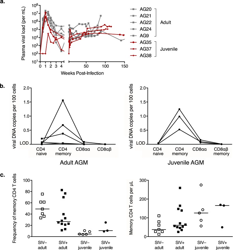
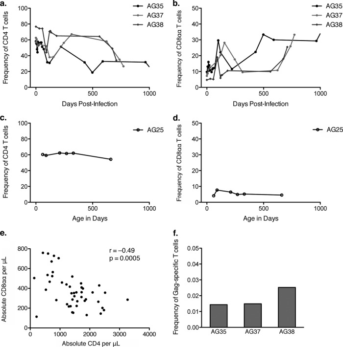
African green monkeys (AGMs; genus Chlorocebus) are a natural host of simian immunodeficiency virus (SIVAGM). As they do not develop simian AIDS, there is great interest in understanding how this species has evolved to avoid immunodeficiency. Adult African green monkeys naturally have low numbers of CD4 T cells and a large population of major histocompatibility complex class II-restricted CD8α(dim) T cells that are generated through CD4 downregulation in CD4(+) T cells. Mechanisms that drive this process of CD4 downregulation are unknown. Here, we show that juvenile AGMs accelerate CD4-to-CD8αα conversion upon SIV infection and avoid progression to AIDS. The CD4 downregulation induced by SIV infection is not limited to SIV-specific T cells, and vaccination of an adult AGM who had a negligible number of CD4 T cells demonstrated that CD4 downregulation can occur without antigenic exposure. Finally, we show that the T cell homeostatic cytokines interleukin-2 (IL-2), IL-7, and IL-15 can induce CD4 downregulation in vitro. These data identify a mechanism that allows AGMs to generate a large, diverse population of T cells that perform CD4 T cell functions but are resistant to SIV infection. A better understanding of this mechanism may allow the development of treatments to induce protective CD4 downregulation in humans.

Importance: Many African primate species are naturally infected with SIV. African green monkeys, one natural host species, avoid simian AIDS by creating a population of T cells that lack CD4, the human immunodeficiency virus/SIV receptor; therefore, they are resistant to infection. However, these T cells maintain properties of CD4(+) T cells even after receptor downregulation and preserve immune function. Here, we show that juvenile AGMs, who have not undergone extensive CD4 downregulation, accelerate this process upon SIV infection. Furthermore, we show that in vivo, CD4 downregulation does not occur exclusively in antigen-experienced T cells. Finally, we show that the cytokines IL-2, IL-7, and IL-15, which induce homeostatic T cell proliferation, lead to CD4 downregulation in vitro; therefore, they can provide signals that lead to antigen-independent CD4 downregulation. These results suggest that if a similar process of CD4 downregulation could be induced in humans, it could provide a cure for AIDS.


Figures

FIG 1
FIG 1
CD4+-to-CD8αα conversion in AGMs is driven by immunologic experience. The frequency of a cell population out of the total population of CD3+ T cells (a and c) and absolute number of cells per microliter of blood (b and d) of T cell populations were measured by flow cytometry, and absolute numbers were calculated from complete blood counts. (a and b) CD4+ T cells gated by CD3+ CD4+ expression. (c and d) CD8αα T cells gated by CD3+ CD4 CD8αdim expression. The Mann-Whitney test was used for panels a to d. For panels a and b, P < 0.0015 for comparisons to all juveniles. (e) Change in CD4+ T cell count over 5 years (Wilcoxon matched-pair signed-rank test). Adult animals are denoted by squares, juvenile animals by circles, SIV+ animals by filled symbols, and SIV animals by open symbols. Median values for each population are shown.
FIG 2
FIG 2
Viral replication dynamics are similar in juvenile and adult AGMs. (a) Plasma viral load in adult (gray squares) and juvenile (red circles) AGMs at time points measured in weeks after inoculation with SIVAGM. (b) Infection frequencies of sorted lymphocyte subsets from SIVAGM-infected adult (left) and juvenile (right) AGMs, as determined by quantitative PCR for viral DNA. Naive and memory subsets were gated based on expression of CD28 and CD95. LOD denotes samples at or below the assay limit of detection. (c) Frequency of memory CD4+ T cells out of total CD4+ T cells (left) and absolute number of memory CD4+ T cells per microliter of blood (right) measured by flow cytometry and absolute number calculated from complete blood count. Memory CD4+ T cells were gated by CD3+ CD4+ CD95+ and CD28hi or CD28low expression. Adult animals are denoted by squares, juvenile animals by circles, SIV+ animals by filled symbols, and SIV animals by open symbols. Median values for each population are shown.
FIG 3
FIG 3
Accelerated CD4+-to-CD8αα conversion in SIV-infected juvenile AGMs. (a) Frequency of CD4+ T cells out of total T cells in 3 SIV-infected juvenile AGMs relative to time postinfection. (b) Frequency of CD8αα T cells out of total T cells in 3 SIV-infected juvenile AGMs relative to time postinfection. (c) Frequency of CD4+ T cells out of total T cells in an SIV-uninfected juvenile AGM relative to time postbirth. (d) Frequency of CD8αα T cells out of total T cells in an SIV-uninfected juvenile AGM relative to time postbirth. (e) CD4+ T cell count versus CD8αα T cell count in SIV-infected juvenile AGMs. All time points from panels a and b are shown as individual data points (n = 46). The Spearman correlation was calculated; r and P values are shown. (f) SIV Gag-specific T cell response magnitude measured by flow cytometry and intracellular cytokine staining. PBMC were stimulated with the SIV Gag peptide pool or media control overnight in the presence of brefeldin A and CD28 and then stained for intracellular cytokines. The background-subtracted frequency of Gag-specific T cells out of total T cells is shown for 3 SIV-infected juvenile AGMs.
FIG 4
FIG 4
MML vaccination of AGMs elicits an MHC class II (MHC-II)-restricted T cell response. (a) Flow cytometry intracellular cytokine staining data showing live CD3+ T cells from vaccinated SIV+ juvenile AG37 at week 8. PBMC were stimulated for 14 h, with brefeldin A added after 2 h. The control tube was incubated with CD28 only. The MML tube was incubated with CD28 and MML protein. The MHC-II tube was preincubated with blocking antibody to MHC class II, and then CD28 and MML protein were added. The MHC-I tube was preincubated with blocking antibody to MHC class I, and then CD28 and MML protein were added. The frequency of cells expressing IL-2 (y axis) and TNF (x axis) is shown. (b) Density plot showing CD4 (y axis) and CD8α expression (x axis) of live CD3+ T cells with MML-specific T cells overlaid as a dot plot. SIV-uninfected juvenile AG36 (left), shown in red, and SIV+ juvenile AG37 (right), shown in blue, from the week 12 time point. (c) Percent MML-specific T cells that were CD4+ at each time point. Vaccine dose timing is indicated by arrows. (d) Density plot showing CD4 (y axis) and CD8α expression (x axis) of live CD3+ T cells with MML-specific T cells overlaid as a dot plot at week 2 for adult AG346 who had consistently low CD4+ T cell counts. (e) Flow cytometry intracellular cytokine staining data showing live CD3+ T cells from vaccinated SIV+ adult AG346 at week 2. The frequency of cells expressing IL-2 (y axis) and TNF (x axis) is shown.
FIG 5
FIG 5
CD4 downregulation induced by homeostatic cytokines. AGM and rhesus macaque PBMC were CFSE labeled and sorted for CD4+ CD3+ T cells and CD3 NKG2A HLA-DR+ antigen-presenting cells. These were cultured with IL-2 for 6 days or IL-7 or IL-15 for 7 days and analyzed by flow cytometry. (a) Plots gated on live CD3+ T cells showing CD4 (y axis) and CFSE (x axis). (b to d, left) Median fluorescence intensity of CD4 plotted for AGM cells that have divided the indicated number of times based on CFSE dilution. MFI of CD4 in undivided cells (zero divisions) compared to cells that had divided six times using a paired t test. (b to d, right) Percentage of AGM T cells in each population that expressed a memory phenotype by CD28 and CD95 staining. Undivided CD4+ T cells were compared to each of the other populations by paired t test (P < 0.004 in all cases). Adult animals are denoted by squares, juvenile animals by circles, SIV+ animals by filled symbols, and SIV animals by open symbols. Median values for each population are shown.

Similar articles

Cited by

References

    1. VandeWoude S, Apetrei C. 2006. Going wild: lessons from naturally occurring T-lymphotropic lentiviruses. Clin. Microbiol. Rev. 19:728–762. 10.1128/CMR.00009-06 - DOI - PMC - PubMed
    1. Goldstein S, Ourmanov I, Brown CR, Plishka R, Buckler-White A, Byrum R, Hirsch VM. 2005. Plateau levels of viremia correlate with the degree of CD4+-T-cell loss in simian immunodeficiency virus SIVagm-infected pigtailed macaques: variable pathogenicity of natural SIVagm isolates. J. Virol. 79:5153–5162. 10.1128/JVI.79.8.5153-5162.2005 - DOI - PMC - PubMed
    1. Hirsch VM, Dapolito G, Johnson PR, Elkins WR, London WT, Montali RJ, Goldstein S, Brown C. 1995. Induction of AIDS by simian immunodeficiency virus from an African green monkey: species-specific variation in pathogenicity correlates with the extent of in vivo replication. J. Virol. 69:955–967 - PMC - PubMed
    1. Beaumier CM, Harris LD, Goldstein S, Klatt NR, Whitted S, McGinty J, Apetrei C, Pandrea I, Hirsch VM, Brenchley JM. 2009. CD4 downregulation by memory CD4+ T cells in vivo renders African green monkeys resistant to progressive SIVagm infection. Nat. Med. 15:879–885. 10.1038/nm.1970 - DOI - PMC - PubMed
    1. Pandrea IV, Gautam R, Ribeiro RM, Brenchley JM, Butler IF, Pattison M, Rasmussen T, Marx PA, Silvestri G, Lackner AA, Perelson AS, Douek DC, Veazey RS, Apetrei C. 2007. Acute loss of intestinal CD4+ T cells is not predictive of simian immunodeficiency virus virulence. J. Immunol. 179:3035–3046. 10.4049/jimmunol.179.5.3035 - DOI - PMC - PubMed

Publication types

MeSH terms

LinkOut - more resources