. 2014 Sep;88(18):10303-11.
doi: 10.1128/JVI.01341-14. Epub 2014 Jul 2.

Interferon-induced protein Ifit2 protects mice from infection of the peripheral nervous system by vesicular stomatitis virus

Affiliations

Interferon-induced protein Ifit2 protects mice from infection of the peripheral nervous system by vesicular stomatitis virus

Volker Fensterl et al. J Virol. 2014 Sep.

Abstract

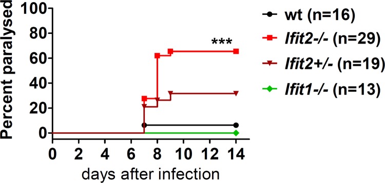
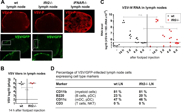
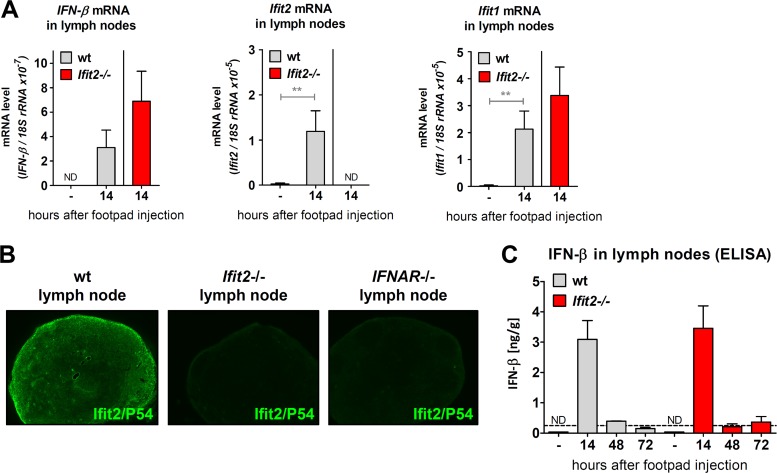
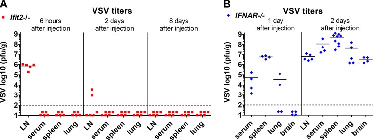
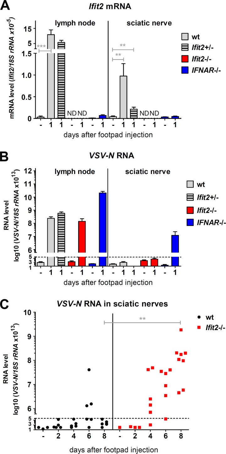
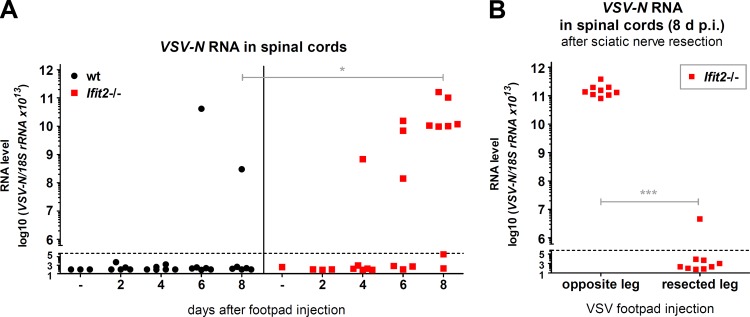
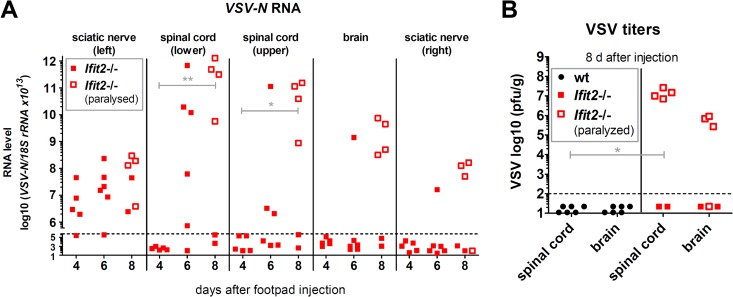
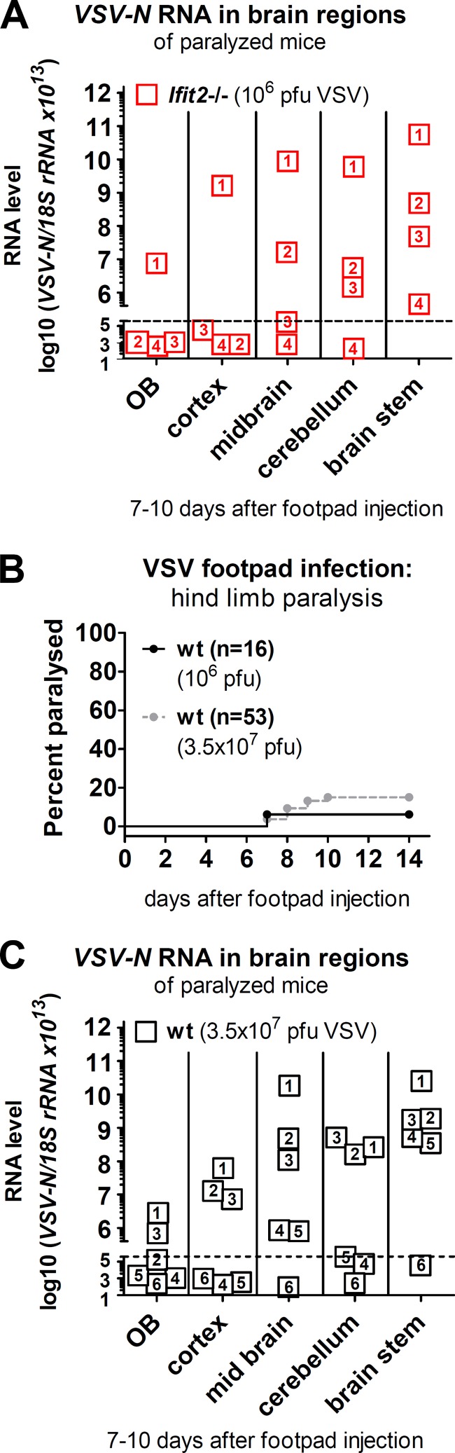
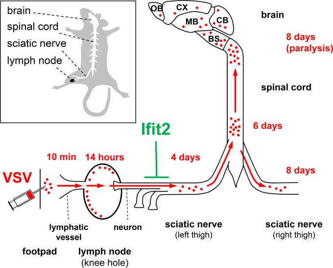
The interferon system provides the first line of host defense against virus infection. Mouse pathogenesis studies have revealed the importance of specific interferon-induced proteins in providing protection against specific viruses. We have previously reported that one such protein, Ifit2, protects neurons of the central nervous system from intranasal infection by the neurotropic rhabdovirus, vesicular stomatitis virus (VSV). Here, we demonstrate that Ifit2 protects the peripheral nervous system from VSV infection as well. In Ifit2(-/-) mice, VSV, injected subcutaneously into the footpad, entered the proximal lymph node, where it replicated and infected the nodal nerve endings. The infection spread to the sciatic nerve, the spinal cord, and the brain, causing paralysis. In contrast, in the wild-type mice, although VSV replicated equally well in the lymph node, infection of the sciatic nerve and the rest of the nervous system was impaired, thus preventing paralysis. Ifit2 protected only the nervous system from VSV infection; other tissues were well protected even in Ifit2(-/-) mice. These results indicate that Ifit2 is the interferon-induced protein that prevents VSV infection of neurons of both the peripheral and the central nervous systems, thus inhibiting the consequent neuropathy, but it is dispensable for protecting the cells of other tissues from VSV infection.

Importance: Although viral infection is quite common, the immune system effectively protects us from viral diseases. A major part of this protection is mediated by interferon, the antiviral cytokine secreted by virus-infected cells. To empower the neighboring uninfected cells in combating the oncoming infection, interferon induces the synthesis of more than 200 new proteins, many of which have antiviral activities. The virus studied here, vesicular stomatitis virus (VSV), like its relative, rabies virus, can cause neuropathy in mice if it enters the peripheral nervous system through skin lesions; however, interferon can protect neurons from VSV infection. We have identified a specific interferon-induced protein, Ifit2, as the protein that protects neurons from VSV infection. Surprisingly, Ifit2 was not needed to protect other cell types from VSV. Our results indicate that the effector antiviral proteins of the interferon system have highly specialized functions.


Figures

FIG 1
FIG 1
Ifit2 protects mice from paralytic VSV neuropathogenesis after footpad infection. Hind limb paralysis of wt, Ifit2−/−, Ifit2+/−, and Ifit1−/− mice after subcutaneous injection of 106 PFU of VSV was examined (n = number of animals). The asterisks (***) indicate statistical significance (wt versus Ifit2−/−; P < 0.0005).
FIG 2
FIG 2
Ifit2 does not restrict VSV replication in lymph nodes. (A) The top panels show the localization of VSV P protein in wt, Ifit2−/−, and IFNAR−/− popliteal lymph node sections 14 h after VSV footpad injection by immunohistofluorescence microscopy. The bottom panels show the localization of active viral gene expression in wt and Ifit2−/− popliteal lymph node sections by fluorescence microscopy of VSV-encoded GFP (arrowheads) 14 h after VSV/GFP footpad injection. All photos were taken at ×50 magnification. (B) Infectious VSV titers in wt and Ifit2−/− popliteal lymph nodes at 14 h after infection, plotted on a log scale. (C) VSV-N RNA levels in wt and Ifit2−/− popliteal lymph nodes, measured by real-time RT-PCR, plotted on a log scale. (D) Expression of cell type markers on VSV/GFP-infected wt and Ifit2−/− lymph node cells at 14 h after infection, measured by flow cytometry. Only GFP-positive cells from eight pooled lymph nodes per experiment were subjected to analysis. Dashed lines indicate the threshold of detection. Abbreviations: LN, lymph nodes; pDC, plasmacytoid dendritic cell; mDC, myeloid dendritic cell; n.s., not significant.
FIG 3
FIG 3
Induction of IFN-β and IFN-stimulated genes in lymph nodes after VSV footpad infection does not require Ifit2. (A) mRNA levels of IFN-β and the IFN-stimulated genes Ifit2 and Ifit1 in the popliteal lymph nodes of wt and Ifit2−/− mice, 14 h after footpad infection, as measured by real-time RT-PCR (three to four infected mice per group). (B) Ifit2 (= P54) protein expression in popliteal lymph nodes of wt, Ifit2−/−, and IFNAR−/− mice at 14 h after footpad infection, detected by immunohistofluorescence microscopy on tissue sections at ×50 magnification. Sections shown are adjacent sections from lymph nodes in Fig. 2A. (C) IFN-β protein production in popliteal lymph nodes after VSV infection of wt and Ifit2−/− mice, as measured by ELISA (three infected mice per group). Asterisks indicate statistical significance (**, P < 0.005). Dashed lines indicate the threshold of detection.
FIG 4
FIG 4
Interferon protects non-neuronal tissues from peripheral VSV infection even in the absence of Ifit2. (A and B) Infectious VSV titers in tissues at different times after infection of Ifit2−/− (A) and IFNAR−/− (B) mice, plotted on log scale. Dashed lines indicate the threshold of detection.
FIG 5
FIG 5
Interferon-induced Ifit2 suppresses VSV replication in sciatic nerves after footpad infection. (A and B) Ifit2 mRNA (A) and VSV-N RNA (B) levels in popliteal lymph nodes and sciatic nerves of wt, Ifit2−/−, and IFNAR−/− mice, 1 day after infection, as measured by real-time RT-PCR (four to six infected mice per group). (C) VSV-N RNA levels in wt and Ifit2−/− sciatic nerves, measured by real-time RT-PCR. All VSV RNA levels were plotted on log scale. Asterisks indicate statistical significance (***, P = 0.0007; **, P < 0.003). Dashed lines indicate the threshold of detection.
FIG 6
FIG 6
The sciatic nerve is the exclusive entry route of VSV into the spinal cord after footpad infection. (A) VSV-N RNA levels in wt and Ifit2−/− spinal cords at different times after footpad infection, measured by real-time RT-PCR. (B) VSV-N RNA levels in Ifit2−/− spinal cords of mice, after surgical removal of the midthigh sciatic nerve segment (i.e., resection), followed by VSV infection of the same or the opposite leg's footpad. RNA was measured at 8 days after infection by real-time RT-PCR. All VSV RNA levels were plotted on a log scale. Asterisks indicate statistical significance (*, P = 0.02; ***, P = 0.0002). Dashed lines indicate the threshold of detection.
FIG 7
FIG 7
VSV spread into the CNS precedes hind-limb paralysis of Ifit2−/− mice after footpad infection. (A) VSV-N RNA levels in left and right leg sciatic nerves, in the lower and upper segments of spinal cord, and in brains of Ifit2−/− mice at different times after footpad infection, as measured by real-time RT-PCR and plotted on a log scale. (B) Infectious VSV titers in tissues 8 days after footpad infection of wt and Ifit2−/− mice, plotted on a log scale. The tissues of mice showing leg paralysis at the time of harvest are labeled with empty squares. Asterisks indicate statistical significance (**, P = 0.004; *, P = 0.04). Dashed lines indicate the threshold of detection.
FIG 8
FIG 8
VSV infection spreads to all regions of the brain. (A and C) VSV-N RNA levels in different regions of the brains of paralyzed mice. (A) Four Ifit2−/− mice, numbered 1 to 4, after infection with 106 PFU of VSV; (C) six wt mice, numbered 1 to 6, after infection with 3.5 × 10⁷ PFU of VSV. (B) Hind-limb paralysis of wt mice after footpad injection of 106 PFU (from Fig. 1) or 3.5 × 10⁷ PFU of VSV (n = number of animals). No significant difference in RNA levels was found between respective wt and Ifit2−/− brain regions in panels A and C. The dashed lines in panels A and B indicate the threshold of detection.
FIG 9
FIG 9
Route of spread of VSV infection from the footpad to the brain, blocked by Ifit2 at the sciatic nerve. Within minutes after subcutaneous deposition of VSV in the left footpad, virus drains to the local lymph node in the knee hole, replicates in its periphery, and induces IFN-β, which acts on sciatic nerve neurons innervating the lymph node to induce Ifit2 within less than 24 h after infection. Ifit2 blocks slow VSV spread via the sciatic nerve into spinal cord and brain, thereby preventing limb paralysis. Abbreviations: OB, olfactory bulbs; CX, cortex; MB, “midbrain”; CB, cerebellum; BS, brain stem.

Similar articles

Cited by

References

    1. Broz P, Monack DM. 2013. Newly described pattern recognition receptors team up against intracellular pathogens. Nat. Rev. Immunol. 13:551–565. 10.1038/nri3479 - DOI - PubMed
    1. Der SD, Zhou A, Williams BR, Silverman RH. 1998. Identification of genes differentially regulated by interferon alpha, beta, or gamma using oligonucleotide arrays. Proc. Natl. Acad. Sci. U. S. A. 95:15623–15628. 10.1073/pnas.95.26.15623 - DOI - PMC - PubMed
    1. Schoggins JW, Rice CM. 2011. Interferon-stimulated genes and their antiviral effector functions. Curr. Opin. Virol. 1:519–525. 10.1016/j.coviro.2011.10.008 - DOI - PMC - PubMed
    1. Ivashkiv LB, Donlin LT. 2014. Regulation of type I interferon responses. Nat. Rev. Immunol. 14:36–49. 10.1038/nri3581 - DOI - PMC - PubMed
    1. Lopez CB, Hermesh T. 2011. Systemic responses during local viral infections: type I IFNs sound the alarm. Curr. Opin. Immunol. 23:495–499. 10.1016/j.coi.2011.06.003 - DOI - PMC - PubMed

Publication types

MeSH terms

LinkOut - more resources