Review
. 2014 May 12:5:610-21.
doi: 10.3762/bjnano.5.72. eCollection 2014.

Biocalcite, a multifunctional inorganic polymer: Building block for calcareous sponge spicules and bioseed for the synthesis of calcium phosphate-based bone

Affiliations
Review

Biocalcite, a multifunctional inorganic polymer: Building block for calcareous sponge spicules and bioseed for the synthesis of calcium phosphate-based bone

Xiaohong Wang et al. Beilstein J Nanotechnol. .

Abstract

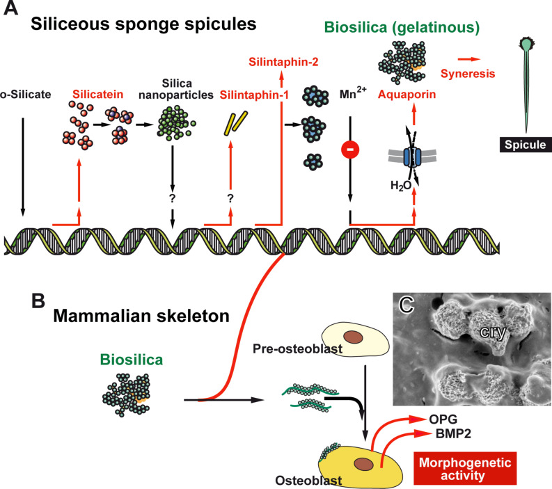
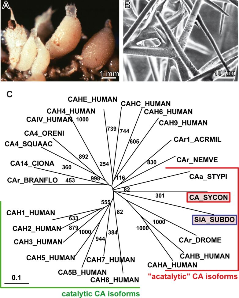
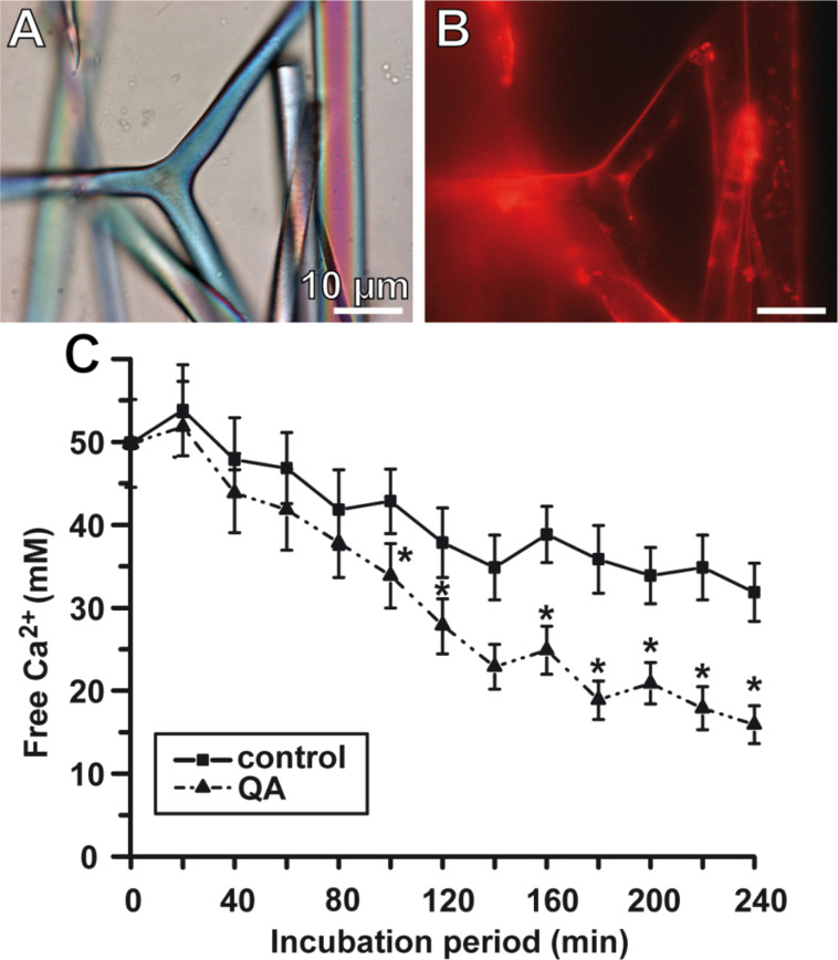
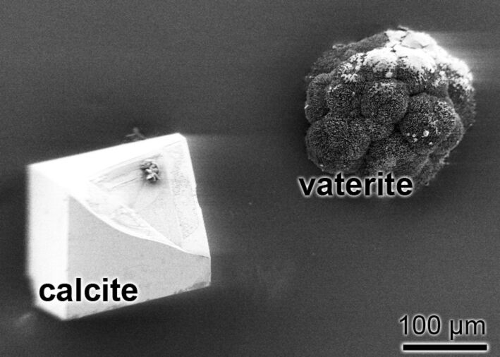
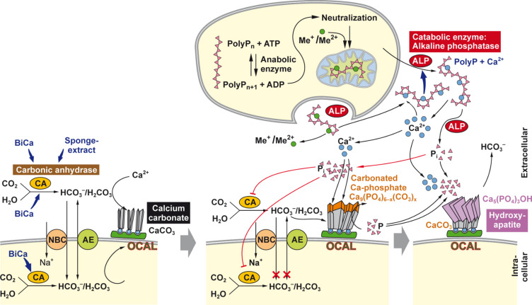
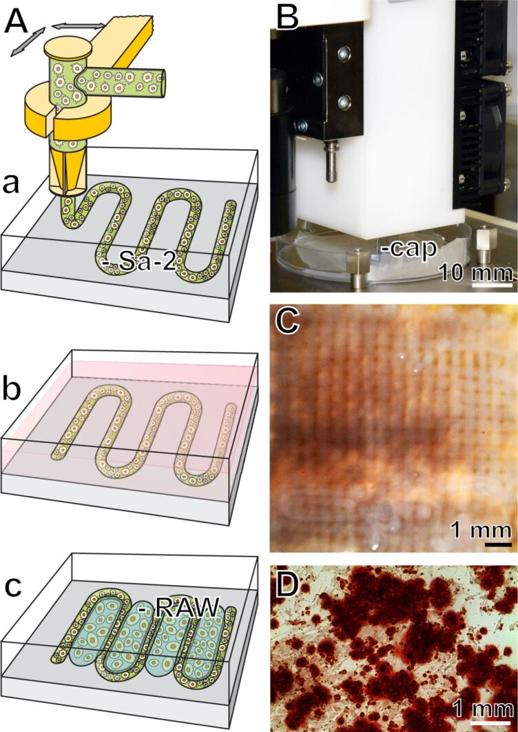
Calcium carbonate is the material that builds up the spicules of the calcareous sponges. Recent results revealed that the calcium carbonate/biocalcite-based spicular skeleton of these animals is formed through an enzymatic mechanism, such as the skeleton of the siliceous sponges, evolutionarily the oldest animals that consist of biosilica. The enzyme that mediates the calcium carbonate deposition has been identified as a carbonic anhydrase (CA) and has been cloned from the calcareous sponge species Sycon raphanus. Calcium carbonate deposits are also found in vertebrate bones besides the main constituent, calcium phosphate/hydroxyapatite (HA). Evidence has been presented that during the initial phase of HA synthesis poorly crystalline carbonated apatite is deposited. Recent data summarized here indicate that during early bone formation calcium carbonate deposits enzymatically formed by CA, act as potential bioseeds for the precipitation of calcium phosphate mineral onto bone-forming osteoblasts. Two different calcium carbonate phases have been found during CA-driven enzymatic calcium carbonate deposition in in vitro assays: calcite crystals and round-shaped vaterite deposits. The CA provides a new target of potential anabolic agents for treatment of bone diseases; a first CA activator stimulating the CA-driven calcium carbonate deposition has been identified. In addition, the CA-driven calcium carbonate crystal formation can be frozen at the vaterite state in the presence of silintaphin-2, an aspartic acid/glutamic acid-rich sponge-specific protein. The discovery that calcium carbonate crystals act as bioseeds in human bone formation may allow the development of novel biomimetic scaffolds for bone tissue engineering. Na-alginate hydrogels, enriched with biosilica, have recently been demonstrated as a suitable matrix to embed bone forming cells for rapid prototyping bioprinting/3D cell printing applications.

Keywords: biocalcite; bioprinting; bone; bone formation; calcareous spicules; sponge.


Figures

Figure 1
Figure 1
Function of biosilica during (A) the formation of siliceous sponge spicules and (B) mammalian bone mineral formation; scheme. (A) The different key molecules involved in spicule formation are sequentially expressed [11]. o-Silicate induces the expression of silicatein, followed by the expression of silintaphin-1 and silintaphin-2, a process that is most likely induced by biosilica nanoparticles. These two classes of molecules (silicateins and silintaphins) form mesoscopic gelatinous flocs that harden through syneresis to spicules [15]. The latter process is driven by water extrusion via aquaporin channels. (B) Effect of biosilica on mammalian bone-forming cells (SaOS-2 cells) in vitro. After incubation with biosilica those cells differentiate and express osteoprotegerin (OPG) and the bone morphogenetic protein 2 (BMP2), two components of their newly acquired morphogenetic activities. (C) SaOS-2 cells that have formed calcium-based carbonate and phosphate crystallites (cry).
Figure 2
Figure 2
Sycon raphanus, its spicules and its CA. (A) Specimens of S. raphanus; (B) the calcareous spicules. (C) Phylogenetic, radial tree computed with the putative calcareous sponge S. raphanus carbonic anhydrase (CA_SYCON; accession number CCE46072) and the demosponge S. domuncula silicase (SIA_SUBDO; DD298191), as well as the carbonic anhydrase isoforms from human: I (CA-I) (CAH1_HUMAN; P00915); II (CA-II) (CAH2_HUMAN; P00918); II-2 (CA II) (CAHB_HUMAN; O75493), III (CA-III) (CAH3_HUMAN; P07451); IV (CAIV_HUMAN; AAA35625.1); IV (CA-IV) (CAH4_HUMAN; P22748); VA (CAH5_HUMAN; P35218); VB (CA5B_HUMAN; O75493); VI (CA-VI) (CAH6_HUMAN; P23280); VII (CA-VII) (CAH7_HUMAN; P43166); VIII (CA-VIII) (CAH8_HUMAN; P35219); IX (CA-IX) (CAH9_HUMAN; Q16790); X (CA-RP X) (CAHA_HUMAN; Q9NS85); XII (CA-XII) (CAHC_HUMAN; O43570); XIV (CA-XIV) (CAHE_HUMAN; Q9ULX7). In addition, the related sequences from the scleractinian Acropora millepora-1 (CAr1_ACRMIL; ACJ64662.1), the stony coral Stylophora pistillata (CAa_STYPI; ACA53457.1, EU159467.1), the anthozoan Nematostella vectensis (CAr_NEMVE; XP_001627923.1), the tunicate Ciona intestinalis (CA14_CIONA; XP_002123314.1), the lancelet Branchiostoma floridae (CAr_BRANFLO; XP_002601262.1), the shark Squalus acanthias (CA4_SQUAAC; AAZ03744.1), the fish Oreochromis niloticus (CA4_ORENI; XP_003456174.1), and the insect enzyme from Drosophila melanogaster (CAr_DROME; NP_572407.3) are included. The CAs, belonging to the "acatalytic" CA isoforms and of the catalytic CA isoforms, are surrounded. Partially taken from [38] with permission.
Figure 3
Figure 3
Sycon CA, its localization and in vitro function. Reacting of Sycon spicule with antibodies, raised against the homologous CA. (A) Light microscopic image of the spicules (in the center is a large triactine). (B) The spicules have been reacted with polyclonal antibodies, raised against the Sycon CA. The immunocomplexes were stained with Cy5-labelled anti-rabbit IgG. (C) Formation of CaCO3 in the ammonium carbonate diffusion assay in the presence of CA. For this series of experiments the recombinant human CA2 enzyme, expressed in Escherichia coli (C6624, Sigma), with a specific activity of about 5,000 units/mg, was added at a concentration of 35 W-A units (10 μg)/500 μL of CaCl2 to the assays. The formation of calcium carbonate was determined quantitatively on the basis of the consumption of free Ca2+ ions using the EDTA titration procedure [31]. The assays either remained free of additional compound(s) (filled square) or were supplemented with 10 μM quinolinic acid (QA, filled triangle). Samples of six parallel determinations were quantitated; means ± SD are given. *p < 0.05.
Figure 4
Figure 4
Calcium carbonate crystals formed in vitro (ammonium carbonate diffusion assay) by using Sycon CA. Left: a calcite crystal formed; at the right a vaterite crystal that has been formed.
Figure 5
Figure 5
Sketch proposing the sequential deposition of calcium carbonate and Ca phosphate on the surface of bone-forming cells (SaOS-2 cells). The CA drives/accelerates the formation of bicarbonate, which reacts to carbonic acid and finally undergoes precipitation to calcium carbonate. Bicarbonate is provided to the CA via the chloride/bicarbonate anion exchanger (AE), or by the sodium bicarbonate co-transporter (NBC). It is proposed that the calcium carbonate crystallites are formed in the vicinity of the plasma membrane, perhaps under the participation of osteocalcin (OCAL). Sponge extracts (as well as their components, e.g., quinolinic acid) and bicarbonate (abbreviated as BiCa) stimulate the CA. In the second step calcium phosphate precipitates in the extracellular space onto the calcium carbonate bioseeds under formation of calcium carbonated apatite. Finally, orthophosphate, released from polyphosphate (polyP), downregulates the activity of the CA. In the third step it is outlined that the Ca2+-salt of polyphosphate (polyP) undergoes hydrolysis through the alkaline phosphatase (ALP), resulting in the liberation of both orthophosphate (pink filled triangles) and Ca2+, both components are required for the synthesis of HA [(Ca5(PO4)3(OH)]. Partially taken from [58] with permission.
Figure 6
Figure 6
Computer-aided rapid prototyping bioprinting. (A-a) A sketch outlining the computer-guided extrusion of Na alginate hydrogel (supplemented with biosilica or bicarbonate) through a capillary in a meander-like pattern. This matrix contained SaOS-2 cells (Sa-2). (A-b and A-c) The blocks formed were incubated in medium into which RAW 264.7 cells (RAW) had been suspended. (B) Part of the bioplotter showing the capillary (cap) through which the alginate/cell matrix is extruded. (C) Completed 4 mm high blocks into which the cells have been embedded into the alginate. (D) The cells retain the capacity to form crystallites, which can be visualized after staining with Alizarin Red S.

Similar articles

Cited by

References

    1. Müller W E G, Wiens M, Adell T, Gamulin V, Schröder H C, Müller I M. Int Rev Cytol. 2004;235:53–92. doi: 10.1016/S0074-7696(04)35002-3. - DOI - PubMed
    1. Wang X, Schröder H C, Müller W E G. Int Rev Cell Mol Biol. 2009;273:69–115. doi: 10.1016/S1937-6448(08)01803-0. - DOI - PubMed
    1. Morse D E. Trends Biotechnol. 1999;17:230–232. doi: 10.1016/S0167-7799(99)01309-8. - DOI
    1. Wang X, Schröder H C, Wang K, Kaandorp J A, Müller W E G. Soft Matter. 2012;8:9501–9518. doi: 10.1039/c2sm25889g. - DOI
    1. Knoll A H. Rev Mineral Geochem. 2003;54:329–356. doi: 10.2113/0540329. - DOI

LinkOut - more resources