

Injection of ligand-free gold and silver nanoparticles into murine embryos does not impact pre-implantation development
- PMID: 24991505
- PMCID: PMC4077524
- DOI: 10.3762/bjnano.5.80
Injection of ligand-free gold and silver nanoparticles into murine embryos does not impact pre-implantation development
Abstract
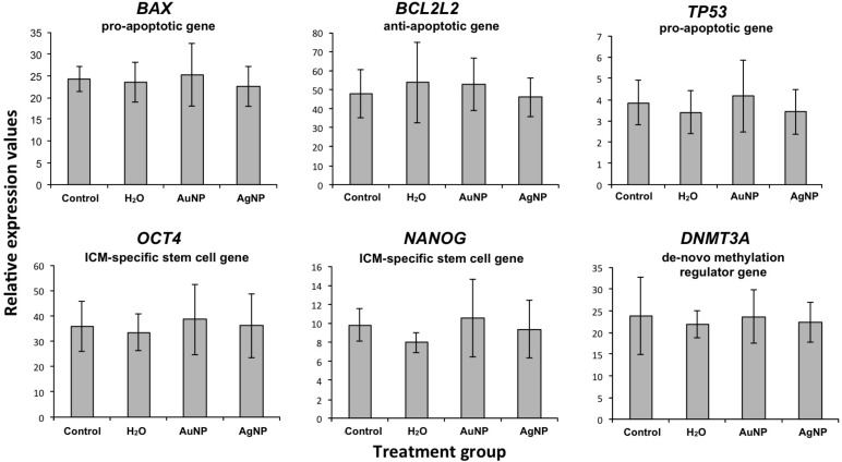
Intended exposure to gold and silver nanoparticles has increased exponentially over the last decade and will continue to rise due to their use in biomedical applications. In particular, reprotoxicological aspects of these particles still need to be addressed so that the potential impacts of this development on human health can be reliably estimated. Therefore, in this study the toxicity of gold and silver nanoparticles on mammalian preimplantation development was assessed by injecting nanoparticles into one blastomere of murine 2 cell-embryos, while the sister blastomere served as an internal control. After treatment, embryos were cultured and embryo development up to the blastocyst stage was assessed. Development rates did not differ between microinjected and control groups (gold nanoparticles: 67.3%, silver nanoparticles: 61.5%, sham: 66.2%, handling control: 79.4%). Real-time PCR analysis of six developmentally important genes (BAX, BCL2L2, TP53, OCT4, NANOG, DNMT3A) did not reveal an influence on gene expression in blastocysts. Contrary to silver nanoparticles, exposure to comparable Ag(+)-ion concentrations resulted in an immediate arrest of embryo development. In conclusion, the results do not indicate any detrimental effect of colloidal gold or silver nanoparticles on the development of murine embryos.
Keywords: biomedical application; confocal microscopy; gene expression; protein corona; toxicity.
Figures


Similar articles
-
Influence of gold, silver and gold-silver alloy nanoparticles on germ cell function and embryo development.Beilstein J Nanotechnol. 2015 Mar 5;6:651-664. doi: 10.3762/bjnano.6.66. eCollection 2015. Beilstein J Nanotechnol. 2015. PMID: 25821705 Free PMC article.
-
TEAD4 regulates trophectoderm differentiation upstream of CDX2 in a GATA3-independent manner in the human preimplantation embryo.Hum Reprod. 2022 Jul 30;37(8):1760-1773. doi: 10.1093/humrep/deac138. Hum Reprod. 2022. PMID: 35700449
-
Comparative analysis of mouse and human preimplantation development following POU5F1 CRISPR/Cas9 targeting reveals interspecies differences.Hum Reprod. 2021 Apr 20;36(5):1242-1252. doi: 10.1093/humrep/deab027. Hum Reprod. 2021. PMID: 33609360
-
Potential toxicity of engineered nanoparticles in mammalian germ cells and developing embryos: treatment strategies and anticipated applications of nanoparticles in gene delivery.Hum Reprod Update. 2016 Sep;22(5):588-619. doi: 10.1093/humupd/dmw020. Epub 2016 Jul 6. Hum Reprod Update. 2016. PMID: 27385359 Review.
-
The cytogenetic constitution of human blastocysts: insights from comprehensive chromosome screening strategies.Hum Reprod Update. 2019 Jan 1;25(1):15-33. doi: 10.1093/humupd/dmy036. Hum Reprod Update. 2019. PMID: 30395265 Review.
Cited by
-
Bone Regeneration, Reconstruction and Use of Osteogenic Cells; from Basic Knowledge, Animal Models to Clinical Trials.J Clin Med. 2020 Jan 4;9(1):139. doi: 10.3390/jcm9010139. J Clin Med. 2020. PMID: 31947922 Free PMC article. Review.
-
Influence of gold, silver and gold-silver alloy nanoparticles on germ cell function and embryo development.Beilstein J Nanotechnol. 2015 Mar 5;6:651-664. doi: 10.3762/bjnano.6.66. eCollection 2015. Beilstein J Nanotechnol. 2015. PMID: 25821705 Free PMC article.
-
Current state of laser synthesis of metal and alloy nanoparticles as ligand-free reference materials for nano-toxicological assays.Beilstein J Nanotechnol. 2014 Sep 12;5:1523-41. doi: 10.3762/bjnano.5.165. eCollection 2014. Beilstein J Nanotechnol. 2014. PMID: 25247135 Free PMC article. Review.
-
Polyamide Nanogels from Generally Recognized as Safe Components and Their Toxicity in Mouse Preimplantation Embryos.Biomacromolecules. 2015 Nov 9;16(11):3491-8. doi: 10.1021/acs.biomac.5b00900. Epub 2015 Oct 6. Biomacromolecules. 2015. PMID: 26367020 Free PMC article.
References
-
- [Oct 23;2013 ];Woodrow Wilson Database. Available from: http://www.nanotechproject.org/
-
- Jain P K, El-Sayed I H, El-Sayed M A. Nano Today. 2007;2:18–29. doi: 10.1016/S1748-0132(07)70016-6. - DOI
-
- Challier J C, Panigel M, Meyer E. Int J Nucl Med Biol. 1973;1:103–106. - PubMed
LinkOut - more resources
Full Text Sources
Other Literature Sources
Research Materials
Miscellaneous