. 2014 Jul 3;5(7):e1311.
doi: 10.1038/cddis.2014.275.

Ketamine alters cortical integration of GABAergic interneurons and induces long-term sex-dependent impairments in transgenic Gad67-GFP mice

Affiliations

Ketamine alters cortical integration of GABAergic interneurons and induces long-term sex-dependent impairments in transgenic Gad67-GFP mice

C Aligny et al. Cell Death Dis. .

Abstract

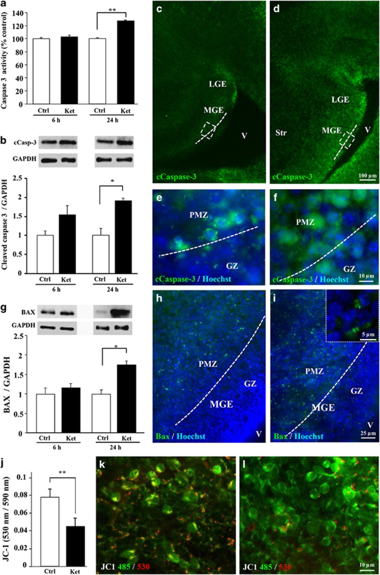
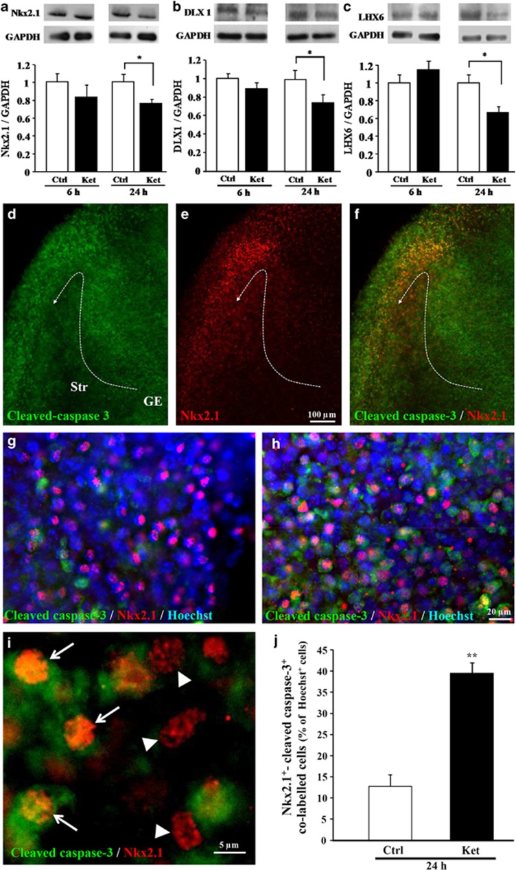
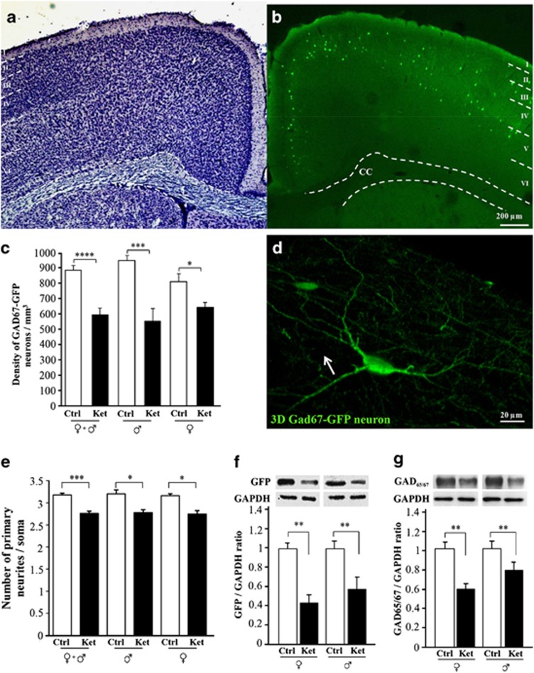
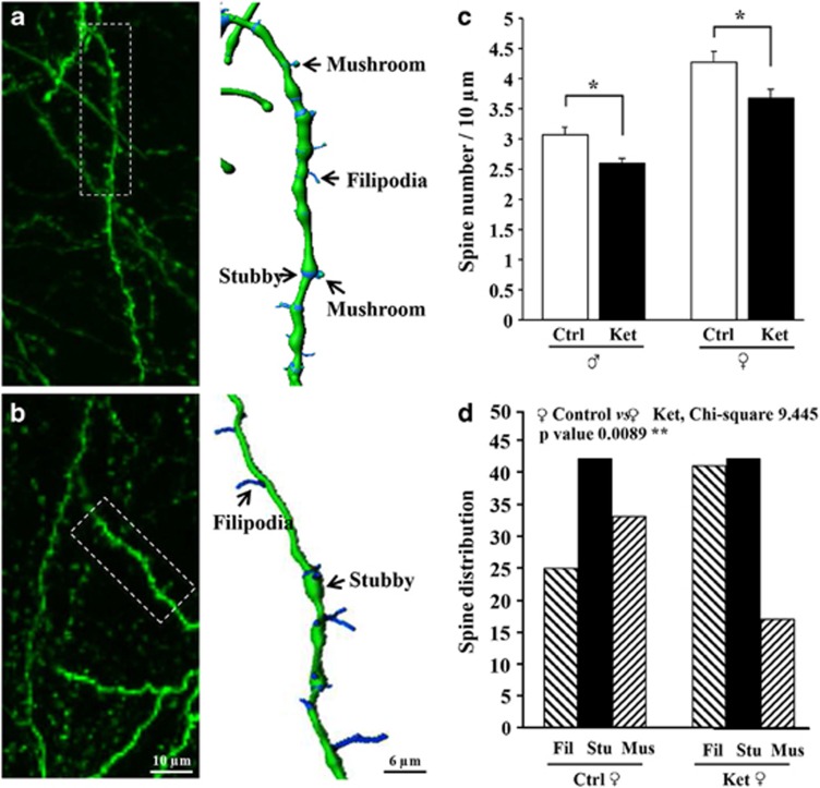
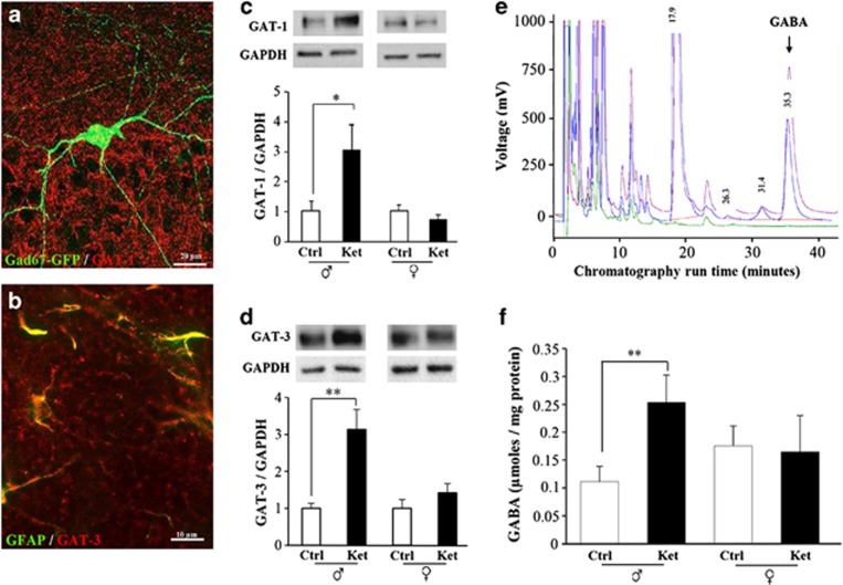
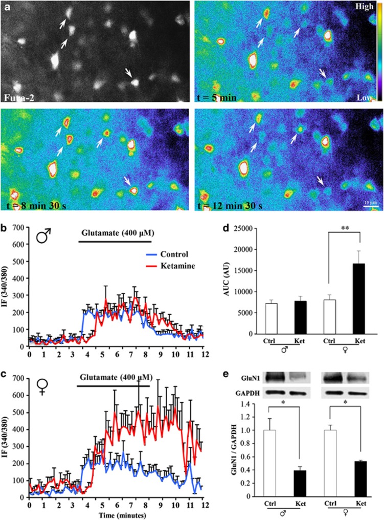
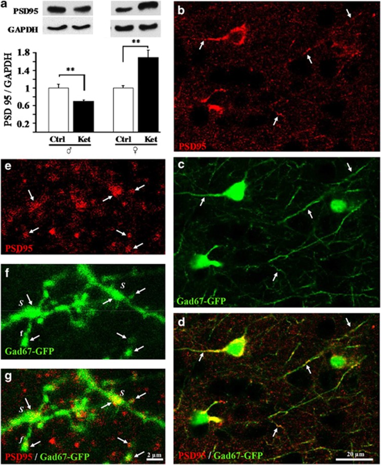
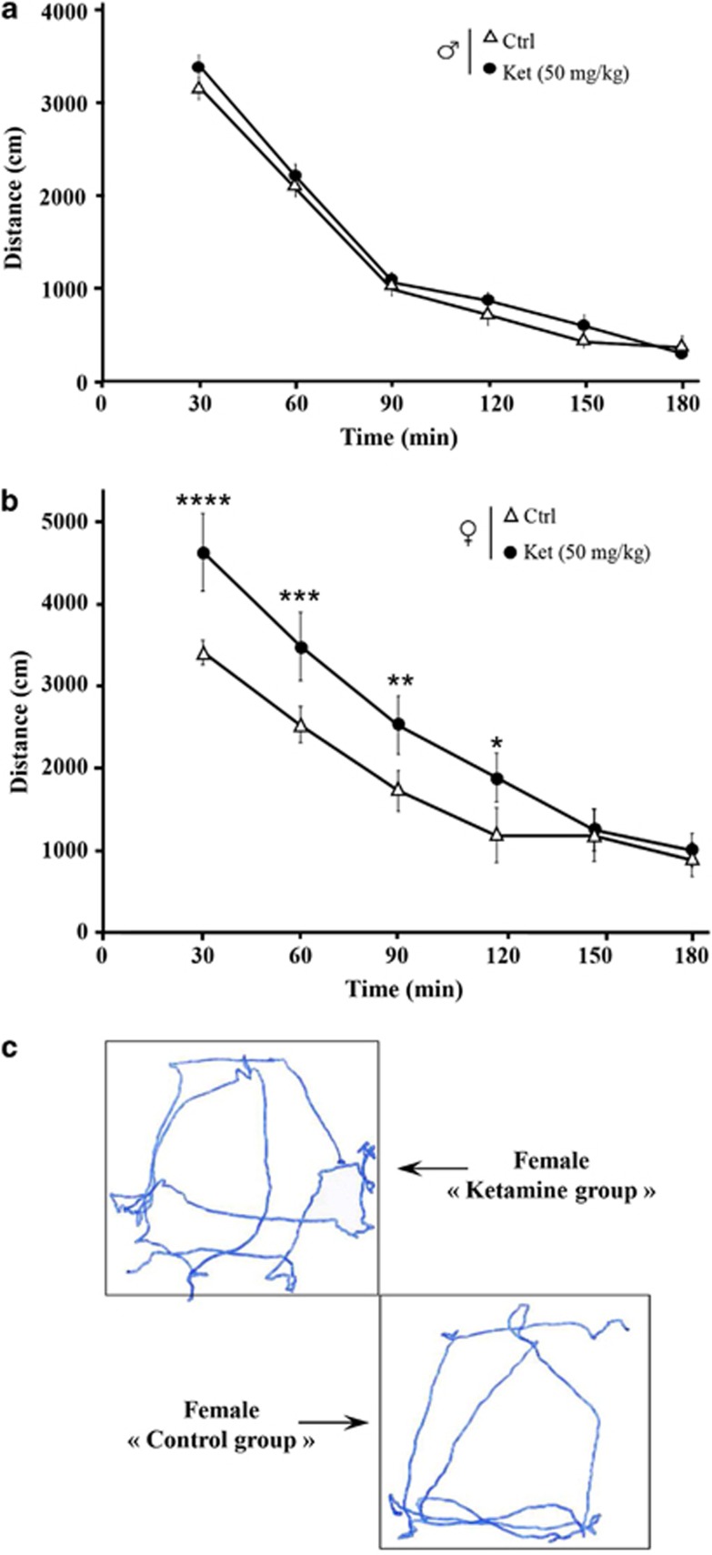
Ketamine, a non-competitive N-methyl-D-aspartate (NMDA) antagonist, widely used as an anesthetic in neonatal pediatrics, is also an illicit drug named Super K or KitKat consumed by teens and young adults. In the immature brain, despite several studies indicating that NMDA antagonists are neuroprotective against excitotoxic injuries, there is more and more evidence indicating that these molecules exert a deleterious effect by suppressing a trophic function of glutamate. In the present study, we show using Gad67-GFP mice that prenatal exposure to ketamine during a time-window in which GABAergic precursors are migrating results in (i) strong apoptotic death in the ganglionic eminences and along the migratory routes of GABAergic interneurons; (ii) long-term deficits in interneuron density, dendrite numbers and spine morphology; (iii) a sex-dependent deregulation of γ-aminobutyric acid (GABA) levels and GABA transporter expression; (iv) sex-dependent changes in the response to glutamate-induced calcium mobilization; and (v) the long-term sex-dependent behavioral impairment of locomotor activity. In conclusion, using a preclinical approach, the present study shows that ketamine exposure during cortical maturation durably affects the integration of GABAergic interneurons by reducing their survival and differentiation. The resulting molecular, morphological and functional modifications are associated with sex-specific behavioral deficits in adults. In light of the present data, it appears that in humans, ketamine could be deleterious for the development of the brain of preterm neonates and fetuses of addicted pregnant women.


Figures

Figure 1
Figure 1
Effect of ketamine on induction of apoptosis in E15 GE. (a) Quantification of caspase-3 activity in cultured E15 GEs after 6 and 24 h of exposure to ketamine (100 μM). (b) Quantification by western blotting of the effect of ketamine (100 μM) on caspase-3 cleavage after 6 and 24 h of exposure. Visualization at low (c and d) and high magnifications (e and f; insets) of cleaved caspase-3-positive cells in a control E15 GE (c and e) and after a 24-h exposure to ketamine (100 μM; d and f). GZ, germinative zone; PMZ, premigratory zone; Str, striatum; V, ventricle. (g) Quantification by western blotting of BAX levels in cultured E15 GEs after 6 and 2 h of ketamine treatment (100 μM). Visualization of BAX immunoreactivity in a control E15 GE (h) and after a 24-h exposure to ketamine (100 μM; i). Note that immunolabeling is preferentially localized in the PMZ and not the GZ of the GE. Inset: BAX labeling at high magnification. (j) Quantification of the fluorescent signals (590 nm/530 nm) emitted by the JC-1 probe after a 12-h treatment of the E15 GE with ketamine (100 μM). Visualization of the mitochondrial (orange) and cytosolic (green) forms of JC-1 in a control E15 GE (k) and after a 12-h exposure to ketamine (100 μM; l). Each value represents the means (±S.E.M.) of at least four independent experiments. *P<0.05; **P<0.01 for ketamine versus control groups, Mann–Whitney U-test and Wilcoxon signed-rank test
Figure 2
Figure 2
Effect of ketamine on protein levels of the GABAergic lineage markers Nkx2.1, DLX1 and LHX6. Quantification by western blotting of Nkx2.1 (a), DLX1 (b) and LHX6 (c) levels in cultured E15 GEs after 6 and 24 h of ketamine treatment (100 μM). Double labeling of cleaved caspase-3- (d) and Nkx2.1-positive cells (e) in the developing cortex of E15 brain slices after a 24-h treatment with ketamine (100 μM). Note that Nkx2.1 labeling partially overlaps with the cleaved caspase-3 signal (f), which is preferentially localized in the previously characterized migratory routes of GABAergic precursors (dashed arrow). Visualization of cleaved caspase-3 and Nkx2.1 double-labeled cells in the developing cortex from control (g) and ketamine-treated (h) E15 slices. Note the strong co-localization of the two signals after ketamine exposure. The total number of nuclei was visualized by Hoechst labeling. (i) High magnification confocal acquisition visualizing Nkx2.1 cells immunopositive (arrows) or immunonegative (arrow heads) for the cleaved caspase-3. (j) Quantification of the effect of a 24 h (100 μM) ketamine exposure on Nkx2.1/cleaved caspase-3 immunoreactive cells. For western blot studies, each value represents the mean (±S.E.M.) of at least six independent experiments. For cell quantification, each value represents the mean (±S.E.M.) of 18 ROI from 6 different slices. *P<0.05; **P<0.01 for ketamine versus control groups, Mann–Whitney U-test
Figure 3
Figure 3
Long-term effect of in vivo prenatal exposure to ketamine on the density of Gad67-GFP cells in the superficial cortical layers II–IV. Cresyl violet staining revealing the different cortical layers in P45 mice (a) and the preferential localization of Gad67-GFP cells in layers II–IV (b). (c) Quantification of the effect of prenatal ketamine exposure on the density of Gad67-GFP neurons in layers II–IV of female and male P45 mice. (d) 3D reconstruction of a Gad67-GFP cell from the neocortex of a P45 mouse. Arrow indicates a primary neurite. (e) Quantification of the effect of prenatal exposure to ketamine on the number of primary neurites in Gad67-GFP interneurons in layers II–IV of female and male P45 mice. (f) Quantification by western blotting of the effect of prenatal exposure to ketamine on GFP levels in female and male P45 mice. (g) Quantification by western blotting of the effect of prenatal exposure to ketamine on Gad levels in female and male P45 mice. For western blotting experiments, each value represents the mean (±S.E.M.) of five independent experiments. For morphometric analyses, each value represents the mean (±S.E.M.) of at least five animals per group. *P<0.05; **P<0.01; ***P<0.001; ****P<0.0001 for ketamine versus control groups, Mann–Whitney U-test
Figure 4
Figure 4
Long-term effect of in vivo prenatal exposure to ketamine on dendritic spines of Gad67-GFP neurons in cortical layers II–IV. Confocal microscopic photographs (left panels) showing dendritic spines in Gad67-GFP interneurons from control (a) and ketamine-treated (b) mice. Dashed rectangles illustrate dendrites used for 3D reconstruction and spine characterization (right panels). (c) Quantification of spine density on dendrites from Gad67-GFP interneurons in cortical layers II–IV of female and male P45 mice. Each value represents the mean (±S.E.M.) of 40 dendrites (10 dendrites analyzed per animal). *P<0.05 for ketamine versus control groups, Mann–Whitney U-test. (d) Distribution of the different spine morphologies on dendrites from Gad67-GFP interneurons in layers II–IV of female P45 mice; χ2-analysis of spine-type distribution obtained from quantification of three slices per animals and three animals per group
Figure 5
Figure 5
Long-term effect of in vivo prenatal exposure to ketamine on the expression levels of GABA transporters and GABA in the cortex of Gad67-GFP mice. (a) Photomicrographs showing GAT-1 immunolabeling in the cortex of P45 mice. GAT-1 puncta are widely distributed in the cortex, including on Gad67-GFP neurons. (b) Photomicrographs showing GAT-3 and GFAP immunolabeling in the cortex of P45 mice. (c) Quantification by western blotting of the effect of prenatal exposure to ketamine on GAT-1 levels in the cortex of male and female P45 mice. (d) Quantification by western blotting of the effect of prenatal exposure to ketamine on GAT-3 levels in the cortex of male and female P45 mice. (e) Representative HPLC chromatograms obtained by the elution solvent alone (green trace), the GABA standard (pink trace) and a sample from a mouse cortex (blue trace). (f) Quantification of GABA content in the cortex of male and female P45 mice. For western blotting experiments, each value represents the mean (±S.E.M.) of at least four independent experiments. For the quantification of GABA concentrations, each value represents the mean (±S.E.M.) of six animals per group. *P<0.05; **P<0.01 for ketamine versus control groups, Mann–Whitney U-test
Figure 6
Figure 6
Long-term effect of in vivo prenatal exposure to ketamine on glutamate-induced intracellular calcium mobilization and GluN1 expression levels. (a) Photomicrographs showing Fura-2 loaded cells in the superficial layers of P15 brain slices (arrows) and false-color photomicrographs visualizing intracellular calcium variations after exposure to glutamate (400 μM). Mean recordings of the 340/380 ratio of the Fura-2 probe after exposure to glutamate (400 μM) in cortical slices from P15 males (b) and females (c) previously exposed in utero to ketamine (red lines) or from the control group (blue lines). (d) Quantification of the areas under the curves obtained by the measurement of intracellular calcium levels. (e) Quantification by western blotting of the effects of prenatal exposure to ketamine on GluN1 levels in the frontal cortex of male and female P15 mice. For calcimetry experiments, each value represents the mean (±S.E.M.) of 16 slices. *P<0.05; **P<0.01 for ketamine versus control groups, Mann–Whitney U-test
Figure 7
Figure 7
Immunohistochemical visualization of PSD95 expression in Gad67-GFP mice and quantification of the long-term effect of in vivo prenatal exposure to ketamine on PSD95 expression levels. (a) Quantification by western blotting of the effect of prenatal exposure to ketamine on PSD95 levels in the cortex of male and female Gad67-GFP mice at P15. (bd) Low magnification photomicrographs visualizing PSD95 immunolabeling in the cortex of a Gad67-GFP mouse. Although PSD95 immunolabeling is not exclusively found in Gad67-GFP expressing neurons (b), numerous puncta co-localize with soma and dendrites (arrows) of the GABA interneurons (c and d). (eg) High-magnification photomicrographs visualizing PSD95 immunolabeling and eGFP fluorescent dendrites. Note that several PSD95-immunoreactive puncta (e) co-localize with eGFP-positive dendrites (arrows) and are associated with different spine subtypes such as stubby (S) or filipodia (f) (f and g). For western blotting experiments, each value represents the mean (±S.E.M.) of at least four independent experiments. **P<0.01 for ketamine versus control groups, Mann–Whitney U-test
Figure 8
Figure 8
Long-term effect of in vivo prenatal exposure to ketamine on spontaneous horizontal locomotor activity in male and female P45 mice. Quantification over 3 h of the total distance covered every 30 min by P45 Gad67-GFP males (a) and females (b). Animals were exposed (Δ) or not (•) to ketamine in utero. (c) Visualization of the distance covered by female Gad67-GFP mice from the control and ketamine groups. Each value represents the mean (±S.E.M.) of 15 animals per group. *P<0.05; **P<0.01; ***P<0.001; ****P<0.0001 for ketamine versus control groups, Mann–Whitney U-test

References

    1. Volpe JJ. Brain injury in premature infants: a complex amalgam of destructive and developmental disturbances. Lancet Neurol. 2009;8:110–124. - PMC - PubMed
    1. Jégou S, El Ghazi F, de Lendeu PK, Marret S, Laudenbach V, Uguen A, et al. Prenatal alcohol exposure affects vasculature development in the neonatal brain. Ann Neurol. 2012;72:952–960. - PubMed
    1. Degos V, Peineau S, Nijboer C, Kaindl AM, Sigaut S, Favrais G, et al. G protein-coupled receptor kinase 2 and group I metabotropic glutamate receptors mediate inflammation-induced sensitization to excitotoxic neurodegeneration. Ann Neurol. 2013;73:667–678. - PMC - PubMed
    1. Manning SM, Talos DM, Zhou C, Selip DB, Park HK, Park CJ, et al. NMDA receptor blockade with memantine attenuates white matter injury in a rat model of periventricular leukomalacia. J Neurosci. 2008;28:6670–6678. - PMC - PubMed
    1. Cho GS, Lee JC, Ju C, Kim C, Kim WK. N-Methyl-D-aspartate receptor antagonists memantine and MK-801 attenuate the cerebral infarct accelerated by intracorpus callosum injection of lipopolysaccharides. Neurosci Lett. 2013;538:9–14. - PubMed

Publication types

MeSH terms