

Natural mineral particles are cytotoxic to rainbow trout gill epithelial cells in vitro
- PMID: 24991818
- PMCID: PMC4081506
- DOI: 10.1371/journal.pone.0100856
Natural mineral particles are cytotoxic to rainbow trout gill epithelial cells in vitro
Abstract
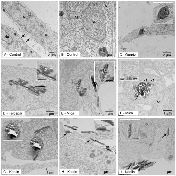
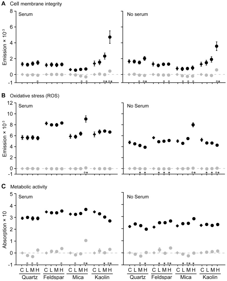
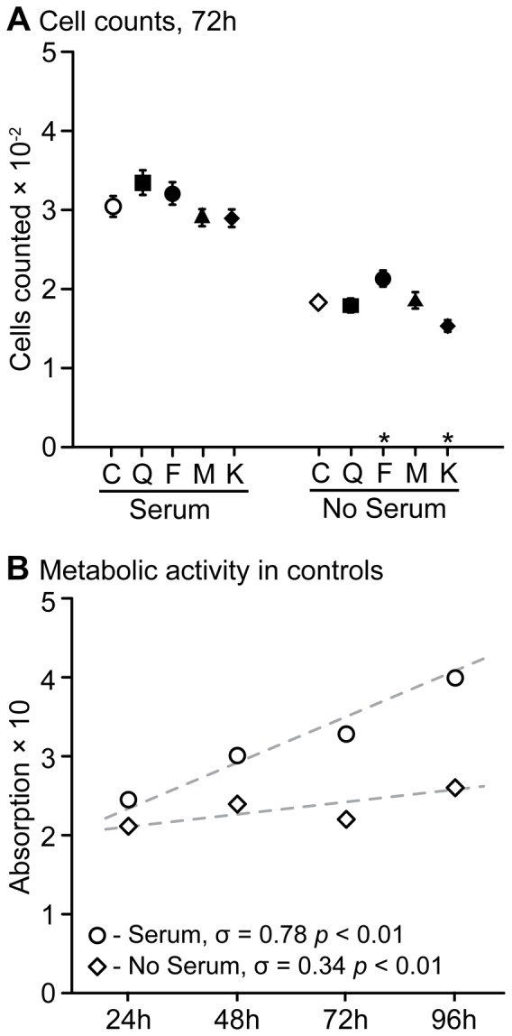
Worldwide increases in fluvial fine sediment are a threat to aquatic animal health. Fluvial fine sediment is always a mixture of particles whose mineralogical composition differs depending on the sediment source and catchment area geology. Nonetheless, whether particle impact in aquatic organisms differs between mineral species remains to be investigated. This study applied an in vitro approach to evaluate cytotoxicity and uptake of four common fluvial mineral particles (quartz, feldspar, mica, and kaolin; concentrations: 10, 50, 250 mg L(-1)) in the rainbow trout epithelial gill cell line RTgill-W1. Cells were exposed for 24, 48, 72, and 96 h. Cytotoxicity assays for cell membrane integrity (propidium iodide assay), oxidative stress (H2DCF-DA assay), and metabolic activity (MTT assay) were applied. These assays were complemented with cell counts and transmission electron microscopy. Regardless of mineral species, particles ≤ 2 µm in diameter were taken up by the cells, suggesting that particles of all mineral species came into contact and interacted with the cells. Not all particles, however, caused strong cytotoxicity: Among all assays the tectosilicates quartz and feldspar caused sporadic maximum changes of 0.8-1.2-fold compared to controls. In contrast, cytotoxicity of the clay particles was distinctly stronger and even differed between the two particle types: mica induced concentration-dependent increases in free radicals, with consistent 1.6-1.8-fold-changes at the 250 mg L(-1) concentration, and a dilated endoplasmic reticulum. Kaolin caused concentration-dependent increases in cell membrane damage, with consistent 1.3-1.6-fold increases at the 250 mg L(-1) concentration. All effects occurred in the presence or absence of 10% fetal bovine serum. Cell numbers per se were marginally affected. Results indicate that (i.) natural mineral particles can be cytotoxic to gill epithelial cells, (ii.) their cytotoxic potential differs between mineral species, with clay particles being more cytotoxic, and (iii.) some clays might induce effects comparable to engineered nanoparticles.
Conflict of interest statement
Figures
Similar articles
-
Cellular uptake and intracellular localization of poly (acrylic acid) nanoparticles in a rainbow trout (Oncorhynchus mykiss) gill epithelial cell line, RTgill-W1.Aquat Toxicol. 2017 Nov;192:58-68. doi: 10.1016/j.aquatox.2017.09.008. Epub 2017 Sep 7. Aquat Toxicol. 2017. PMID: 28917946
-
Ammonia-containing industrial effluents, lethal to rainbow trout, induce vacuolisation and Neutral Red uptake in the rainbow trout gill cell line, RTgill-W1.Altern Lab Anim. 2009 Feb;37(1):77-87. doi: 10.1177/026119290903700111. Altern Lab Anim. 2009. PMID: 19292578
-
Agglomeration of tungsten carbide nanoparticles in exposure medium does not prevent uptake and toxicity toward a rainbow trout gill cell line.Aquat Toxicol. 2009 Jun 28;93(2-3):91-9. doi: 10.1016/j.aquatox.2009.04.003. Epub 2009 Apr 17. Aquat Toxicol. 2009. PMID: 19439373
-
Flow cytometric analysis of BDE 47 mediated injury to rainbow trout gill epithelial cells.Aquat Toxicol. 2010 Apr 1;97(1):42-50. doi: 10.1016/j.aquatox.2009.11.013. Epub 2009 Dec 11. Aquat Toxicol. 2010. PMID: 20053465 Free PMC article.
-
Co-exposure to polystyrene plastic beads and polycyclic aromatic hydrocarbon contaminants in fish gill (RTgill-W1) and intestinal (RTgutGC) epithelial cells derived from rainbow trout (Oncorhynchus mykiss).Environ Pollut. 2019 May;248:706-714. doi: 10.1016/j.envpol.2019.02.066. Epub 2019 Feb 22. Environ Pollut. 2019. PMID: 30849588 Free PMC article.
Cited by
-
Evaluation of the Adsorption Efficacy of Bentonite on Aflatoxin M1 Levels in Contaminated Milk.Toxins (Basel). 2023 Jan 26;15(2):107. doi: 10.3390/toxins15020107. Toxins (Basel). 2023. PMID: 36828421 Free PMC article.
-
Silica Nanoparticle-Generated ROS as a Predictor of Cellular Toxicity: Mechanistic Insights and Safety by Design.Environ Sci Nano. 2016 Feb 1;3(1):56-66. doi: 10.1039/C5EN00179J. Epub 2015 Nov 10. Environ Sci Nano. 2016. PMID: 26998307 Free PMC article.
-
Potential adverse effects on animal health and performance caused by the addition of mineral adsorbents to feeds to reduce mycotoxin exposure.Mycotoxin Res. 2020 Feb;36(1):115-126. doi: 10.1007/s12550-019-00375-7. Epub 2019 Sep 13. Mycotoxin Res. 2020. PMID: 31515765 Free PMC article. Review.
-
Microplastics in fecal samples of whale sharks (Rhincodon typus) and from surface water in the Philippines.Microplast nanoplast. 2021;1(1):17. doi: 10.1186/s43591-021-00017-9. Epub 2021 Sep 26. Microplast nanoplast. 2021. PMID: 34939039 Free PMC article.
-
Microplastics affect assimilation efficiency in the freshwater amphipod Gammarus fossarum.Environ Sci Pollut Res Int. 2016 Dec;23(23):23522-23532. doi: 10.1007/s11356-016-7584-2. Epub 2016 Sep 10. Environ Sci Pollut Res Int. 2016. PMID: 27614640
References
-
- Giller PS, Malmqvist B (1998) The Biology of Streams and Rivers. New York: Oxford University Press.
-
- Kemp P, Sear D, Collins A, Naden P, Jones I (2011) The impacts of fine sediment on riverine fish. Hydrological Processes 25: 1800–1821.
-
- Owens PN, Batalla RJ, Collins AJ, Gomez B, Hicks DM, et al. (2005) Fine-grained sediment in river systems: Environmental significance and management issues. River Research and Applications 21: 693–717.
-
- Syvitski JPM, Vorosmarty CJ, Kettner AJ, Green P (2005) Impact of humans on the flux of terrestrial sediment to the global coastal ocean. Science 308: 376–380. - PubMed
-
- Waters TF (1995) Sediment in streams. Sources, biological effects, and control. Bethesda, MD: American Fisheries Society.
Publication types
MeSH terms
Substances
LinkOut - more resources
Full Text Sources
Other Literature Sources
Medical
