. 2014 Jul 2;83(1):189-201.
doi: 10.1016/j.neuron.2014.05.018.

Hippocampal memory traces are differentially modulated by experience, time, and adult neurogenesis

Affiliations

Hippocampal memory traces are differentially modulated by experience, time, and adult neurogenesis

Christine A Denny et al. Neuron. .

Abstract

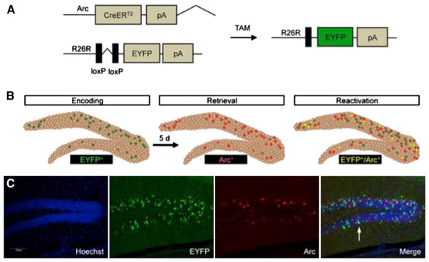
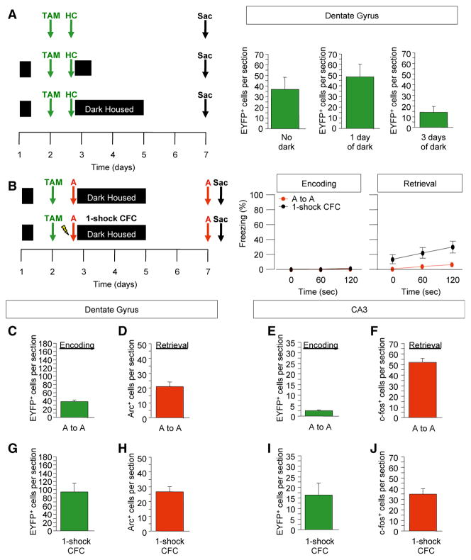
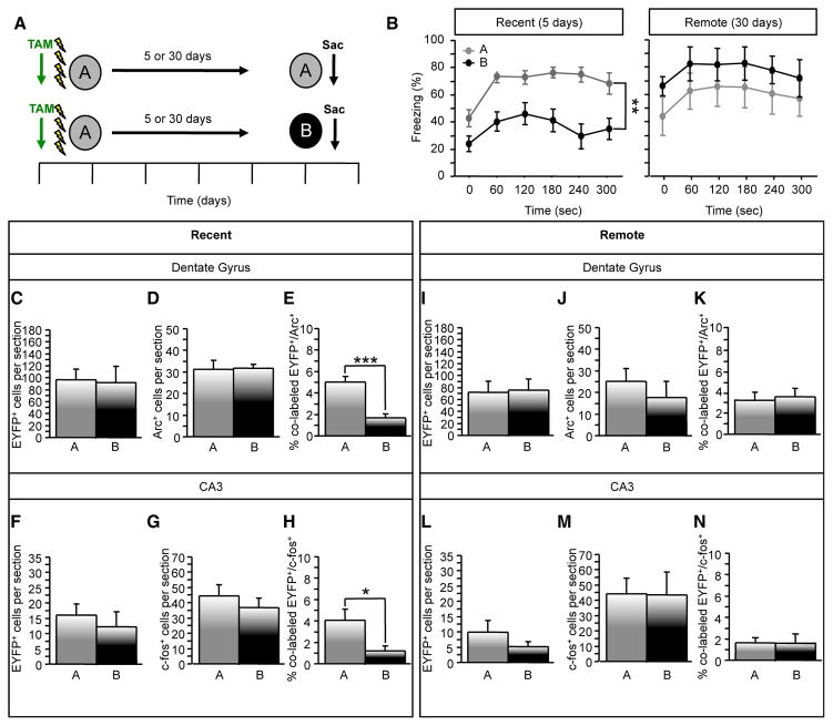
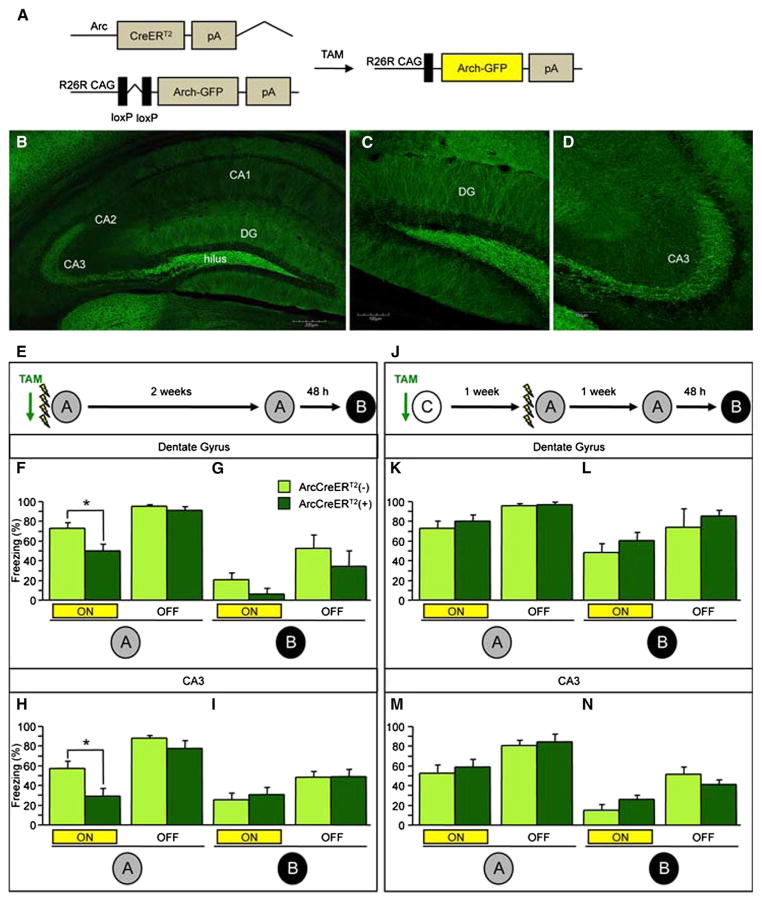
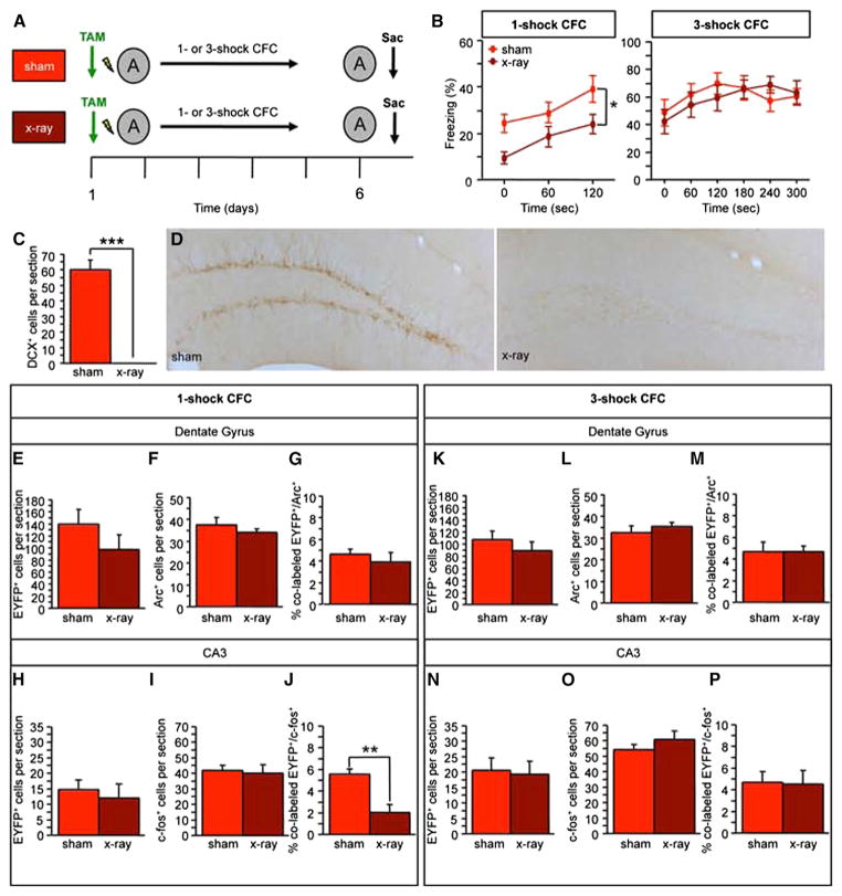
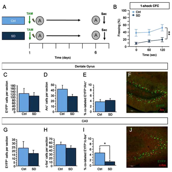
Memory traces are believed to be ensembles of cells used to store memories. To visualize memory traces, we created a transgenic line that allows for the comparison between cells activated during encoding and expression of a memory. Mice re-exposed to a fear-inducing context froze more and had a greater percentage of reactivated cells in the dentate gyrus (DG) and CA3 than mice exposed to a novel context. Over time, these differences disappeared, in keeping with the observation that memories become generalized. Optogenetically silencing DG or CA3 cells that were recruited during encoding of a fear-inducing context prevented expression of the corresponding memory. Mice with reduced neurogenesis displayed less contextual memory and less reactivation in CA3 but, surprisingly, normal reactivation in the DG. These studies suggest that distinct memory traces are located in the DG and in CA3 but that the strength of the memory is related to reactivation in CA3.


Figures

Figure 1
Figure 1. Indelible Labeling of Arc+ Cells following Learning
(A) Administration of TAM to ArcCreERT2 x R26R-STOP-floxed-EYFP mice results in an indelible EYFP label in the initially activated Arc+ cells. (B) The ArcCreERT2 line allows for a comparison between the indelibly labeled cells activated during memory encoding and memory expression. (C) Representative 10× images. The scale bar represents 100 μm. See also Figure S1.
Figure 2
Figure 2. Indelible, Specific Labeling of Arc+ Cells following Context Exposure and Contextual Fear Conditioning
(A) Experimental design. Mice that were dark housed for 3 days following the TAM injection had the least number of EYFP+ DG cells. (B) Experimental design to assess the impact of context exposure and one-shock CFC on behavior and cell labeling. Mice exposed to the context without a foot shock did not freeze, whereas mice administered a one-shock CFC paradigm froze approximately 20% of the time. (C–J) The numbers of EYFP+, Arc+, and c-fos+ cells were assessed in both conditions. The number of EYFP+ cells increased with context exposure and with one-shock CFC from the number of EYFP+ cells labeled in the home cage. n = 4–7 mice per group. Error bars represent ± SEM. See also Figure S2.
Figure 3
Figure 3. Recent and Remote Re-exposure to a Fearful Environment Differentially Reactivates DG and CA3 Cells
(A) Experimental design. (B) In the recent exposure, context-elicited freezing was significantly greater in mice exposed to context A compared to context B (F(1,17) = 22.4398, p < 0.001). In the remote exposure, mice did not differ in their levels of context-elicited freezing (F(1,9) = 1.217, p = 0.30). (C and D) For the recent exposure, the number of (C) EYFP+ and (D) Arc+ DG cells did not differ between groups. (E) Mice re-exposed to context A had a greater percent of colabeled EYFP+/Arc+ DG cells than did mice exposed to context B (p < 0.0001). (F and G) The number of (F) EYFP+ and (G) c-fos+ CA3 cells was similar between the groups. (H) The percent of colabeled EYFP+/c-fos+ was significantly greater in mice re-exposed to context A (p = 0.03). n = 9–10 mice per group. (I–K) For the remote exposure, the number of (I) EYFP+ and (J) Arc+ DG cells and (K) the percent of colabeled EYFP+/Arc+ DG cells did not differ between the groups. (L–N) The number of (L) EYFP+ and (M) c-fos+ CA3 cells and (N) the percent of colabeled EYFP+/c-fos+ was similar between the groups. n = 5–6 mice per group. Error bars represent ± SEM. See also Figure S3.
Figure 4
Figure 4. In Vitro Optogenetic Inhibition of the DG and CA3 in ArcCreERT2 x R26R-CAG-STOP-floxed-Arch-GFP Mice
(A) Genetic design. (B) Representative Arch-GFP+ DG cell. (C and D) In vitro photostimulation (500 ms, yellow bar) of DG cells resulted in 162.49 ± 38.26 pA steady-state current in Arch-GFP+ neurons (n = 18). This corresponds to −13.1 ± 2.5 mV hyper-polarization, when the cells were current clamped at their resting potentials (−73.47 ± 1 mV) (n = 18). (E) Photostimulation-evoked hyperpolarization was able to abolish current injection-induced action potentials (APs). Example traces represent 150 pA somatic current injection without or with light stimulation (n = 5). (F) Arch photostimulation was able to abolish trains of current injection-evoked APs. (G) The graph represents the increasing amount of steady-state current as a function of laser power (n = 3). (H) Representative Arch-GFP+ CA3 neuron. (I and J) In CA3, in vitro photostimulation also resulted in complete inhibition of APs. Voltage (upper trace) and current clamp (lower trace) recording of an Arch-GFP+ CA3 neuron. In vitro photostimulation resulted in 408.23 ± 54.79 pA steady-state current in all of the Arch-GFP+ CA3 neurons, which corresponds to −25.93 ± 6.66 mV hyperpolarization (n = 3). Error bars represent ± SEM.
Figure 5
Figure 5. In Vivo Optogenetic Inhibition of the DG and CA3 Impairs Expression of Initially Encoded Memory
(A) Genetic design. (B–D) Representative images of the (B) hippocampus (the scale bar represents 200 μm), (C) DG (the scale bar represents 100 μm), and (D) CA3 (the scale bar represents 100 μm). (E) The experimental design consisted of all mice being trained in four-shock CFC paradigm and then being re-exposed to context A and B Each context exposure consisted of 3 min of laser stimulation and 3 min without laser stimulation. (F and G) Optogenetic inhibition of Arch-GFP+ DG neurons impaired expression of the corresponding fear memory in context A (F) in ArcCreERT2(+) mice when compared with ArcCreERT2(−) mice (p = 0.02) but had no effect in (G) context B (n = 6–9 mice per group). (H and I) Optogenetic inhibition of Arch-GFP+ CA3 neurons impaired expression of the corresponding fear memory in context A (H) in ArcCreERT2(+) mice when compared with ArcCreERT2(−) mice (p < 0.02) but had no effect in (I) context B (n = 9–12 mice per group). (J–L) Experimental design. Optogenetic inhibition of Arch-GFP+ DG neurons labeled in context C (J) did not impair freezing in ArcCreERT2(+) mice when compared with ArcCreERT2(−) mice in (K) context A or in (L) context B (n = 4–5 mice per group). (M and N) Optogenetic inhibition of Arch-GFP+ CA3 neurons labeled in context C had no effect on memory expression in ArcCreERT2(+) mice when compared with ArcCreERT2(−) mice in (M) context A or in (N) context B (n = 5 mice per group). Error bars represent ± SEM. See also Figures S4 and S5.
Figure 6
Figure 6. The X-Irradiation Impairment in One-Shock CFC Is Paralleled by Decreased CA3 Reactivation, and Both Impairments Can be Rescued by Using a Three-Shock CFC Paradigm
(A) Experimental design (B) In one-shock CFC, context-elicited freezing was significantly reduced in x-irradiated mice (F(1,22) = 4.509, p < 0.05) (n = 11–13 mice per group). In three-shock CFC, context-elicited freezing was comparable in both groups (F(1,25) = 1.063, p = 0.31) (n = 13–14 mice per group). (C and D) The number of DCX+ cells was significantly reduced in x-irradiated mice. (E and F) In the one-shock CFC cohort, (E) the number of EYFP+ and (F) Arc+ DG cells was similar. (G) Both groups had a comparable percent of colabeled EYFP+/Arc+ cells. (H and I) The number of (H) EYFP+ and (I) c-fos+ CA3 cells was similar. (J) X-irradiated mice had a significantly smaller percent of colabeled EYFP+/c-fos+ CA3 cells (p < 0.01). (K–M) In the three-shock CFC cohort, (K) the number of EYFP+ and (L) Arc+ cells and (M) the percent of colabeled EYFP+/Arc+ DG cells did not differ between the groups. (N–P) In CA3, the number of (N) EYFP+ and (O) c-fos+ cells and (P) the percent of colabeled EYFP+/c-fos+ cells did not differ between the groups. Error bars represent ± SEM.
Figure 7
Figure 7. Social Defeat Causes Impairments in One-Shock CFC and CA3 Reactivation
(A) Mice were socially defeated (SD) for 2 weeks. Ten days following the end of SD, mice were administered a one-shock CFC paradigm, re-exposed to context A 5 days later, and sacrificed 1 hr later. (B) Context-elicited freezing was significantly reduced in SD mice when compared with Ctrl mice (F(1,18) = 11.717, p < 0.01). (C and D) In the DG, (C) the number of EYFP+ (p = 0.69) and (D) Arc+ cells (p = 0.10) did not differ between Ctrl and SD mice. (E) Ctrl and SD mice had a comparable percent of colabeled EYFP+/Arc+ DG cells (p = 0.63). (F) Representative image of the immunohistochemistry in the DG. The scale bar represents 100 μm. (G and H) In CA3, the number of (G) EYFP+ (p = 0.45) and (H) c-fos+ (p = 0.33) cells did not differ between the groups. (I) SD mice exhibited a significantly smaller percent of colabeled EYFP+/c-fos+ CA3 cells than did Ctrl mice (p < 0.05). (J) Representative image of immunohistochemistry in CA3. The scale bar represents 100 μm. n = 4–10 mice per group. *p < 0.05, **p < 0.01. Error bars represent ± SEM. See also Figure S6.

Comment in

Similar articles

Cited by

References

    1. Bakker A, Kirwan CB, Miller M, Stark CEL. Pattern separation in the human hippocampal CA3 and dentate gyrus. Science. 2008;319:1640–1642. - PMC - PubMed
    1. Bartlett FC. Remembering: A Study in Experimental and Social Psychology. Cambridge: Cambridge University Press; 1932.
    1. Deng W, Aimone JB, Gage FH. New neurons and new memories: how does adult hippocampal neurogenesis affect learning and memory? Nat Rev Neurosci. 2010;11:339–350. - PMC - PubMed
    1. Denny CA, Burghardt NS, Schachter DM, Hen R, Drew MR. 4- to 6-week-old adult-born hippocampal neurons influence novelty-evoked exploration and contextual fear conditioning. Hippocampus. 2012;22:1188–1201. - PMC - PubMed
    1. Drew MR, Denny CA, Hen R. Arrest of adult hippocampal neurogenesis in mice impairs single- but not multiple-trial contextual fear conditioning. Behav Neurosci. 2010;124:446–454. - PMC - PubMed

Publication types