. 2014 Aug;9(8):1792-802.
doi: 10.1038/nprot.2014.119. Epub 2014 Jul 3.

Activation and measurement of free whisking in the lightly anesthetized rodent

Affiliations

Activation and measurement of free whisking in the lightly anesthetized rodent

Jeffrey D Moore et al. Nat Protoc. 2014 Aug.

Abstract

The rodent vibrissa system is a widely used experimental model of active sensation and motor control. Vibrissa-based touch in rodents involves stereotypic, rhythmic sweeping of the vibrissae as the animal explores its environment. Although pharmacologically induced rhythmic movements have long been used to understand the neural circuitry that underlies a variety of rhythmic behaviors, including locomotion, digestion and ingestion, these techniques have not been available for active sensory movements such as whisking. However, recent work that delineated the location of the central pattern generator for whisking has enabled pharmacological control over this behavior. Here we specify a protocol for the pharmacological induction of rhythmic vibrissa movements that mimic exploratory whisking. The rhythmic vibrissa movements are induced by local injection of a glutamatergic agonist, kainic acid. This protocol produces coordinated rhythmic vibrissa movements that are sustained for several hours in the anesthetized mouse or rat and thus provides unprecedented experimental control in studies related to vibrissa-based neuronal circuitry.


Conflict of interest statement

COMPETING FINANCIAL INTERESTS

The authors declare no competing financial interests.

Figures

Figure 1
Figure 1. Target site for local injection of kainic acid to produce rhythmic vibrissa movements
The pons and medulla contain pools of motoneurons (background) that control the jaw (orange), tongue (green), face (red), and airway (yellow). The intermediate reticular formation (IRt) contains neuronal oscillators for licking (hIRt, green), whisking (vIRt, red), and breathing (black). The target injection site is shown in white in the sagittal (a) and (b) frontal planes.
Figure 2
Figure 2. Diagram of experimental procedures to induce and measure kainic-acid induced vibrissa movements
(a) A rat is placed in a stereotaxic holding frame and a craniotomy is made in the bone dorsal to the intermediate reticular formation of the brainstem. Kainic acid is injected through a micropipette which is lowered into the brainstem via a micromanipulator. (b) Following the injection, the rat is implanted with a head restraining device and transferred to a jig which holds the body and head in place. A camera captures the resulting vibrissa movements. Other physiological measures such as EMG recordings from the mystacial pad and breathing measurements through a thermocouple can be monitored simultaneously. The apparatus shown in this panel is for measurements in rats. Adapted from reference . (c) A similar apparatus for mice. All animal procedures were approved by the IACUC at UC San Diego.
Figure 3
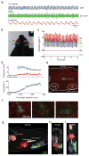
Figure 3. Kainic-acid injection produces rhythmic vibrissa movements in rat
(a) Vibrissa position (blue), EMG activity as measured from the mystacial pad (green), and breathing as measured with a thermocouple placed in the nose (red), following kainic-acid injection. Similar results for vibrissa position were obtained in a total of 21 out of 27 rats (77%). EMG activity was monitored in 4 rats, all of which produced similar results. All rats represented in the present and subsequent figures were Long Evans females, 250 to 350 grams, purchased from Charles River Laboratories. (b) Six C-row vibrissae in the head-restrained, lightly anesthetized rat were tracked using high speed videography.(c) Angular position of each of the tracked vibrissae relative to the x-axis in panel b, 5 hours post-injection. Videography of multiple vibrissae was recorded in 8 rats, all of which produced similar results. (d) Time-course of vibrissa movement frequency (top, blue) and amplitude (bottom, blue) after kainic-acid injection. The frequency is defined as (1/2π)·dΦ(t)/dt averaged over 30 s intervals, where Φ(t) represents the instantaneous phase from the Hilbert transform of the vibrissa angle in time 31. The amplitude is defined as 2·A(t) averaged over the same interval, where A(t) represents the amplitude of the Hilbert transform. The breathing frequency (top, red) is similarly defined. Frequency was defined only for movements that had an amplitude of greater than 5°/s. Vibrissa movements with amplitudes less than this are shown in black. Continuous vibrissa monitoring was performed in 23 rats, 20 of which produced similar results. (e) Sagittal section containing the injection site. The injection site was identified as described in Protocol Steps 16 to 20, and counterstained with anti-NeuN. (f) Magnified view of the injection site in panel e (white box) is shown in the left panel. Similar injection sites in two other rats in which continuous vibrissa movements were also observed are shown in the middle and right panels. The image in the middle panel was published previously as Supplementary Data . NeuN histology was performed on a total of 5 rats after labeling the injection site with BDA. Labeling was successful in 4 of the 5 attempts, and all 4 cases showed similar NeuN staining around the labeled site. (g) Sagittal view of a three-dimensional reconstruction of the injection site in panel e relative to anatomical landmarks: trigeminal (orange), facial (red), and ambiguus (yellow) motor nuclei, the inferior olive (IO, light blue), and the lateral reticular nucleus (LRt, dark blue). (h) Frontal and (i) horizontal views of the reconstruction in panel g. Reconstructions were made by scanning all sections on a Nanozoomer Slide Scanner (Hamamatsu) and tracing the anatomical boundaries using Neurolucida software (Microbrightfield). All animal procedures were approved by the IACUC at UC San Diego.
Figure 4
Figure 4. Kainic-acid injection produces rhythmic vibrissa movements in mouse
(a) Vibrissa position (blue) following kainic-acid injection. Similar results for vibrissa position were obtained in a total of 7 out of 12 mice (58%). All mice were C57Bl6 females, 20 to 30 grams, purchased from Jackson Laboratories. (b) Time-course of vibrissa movement frequency (top) and amplitude (bottom) after kainic-acid injection. Conventions are as in Figure 3d. (c) Sagittal section containing the injection site, prepared as in Figure 3e. NeuN histology was performed on a total of 5 mice after labeling the injection site with BDA. Labeling was successful in 5 of the 5 attempts, and all 5 cases showed similar NeuN staining around the labeled site. (d–f) Three-dimensional reconstruction of the injection site in panel c relative to anatomical landmarks, conventions are as in Figure 3g–i, respectively. All animal procedures were approved by the IACUC at UC San Diego.
Figure 5
Figure 5. Juxtacellular recordings in somatosensory brain regions during kainic-acid induced vibrissa movements
(a) Spiking activity of a single unit in VPM thalamus (black) and simultaneous vibrissa movement (blue). (b) Spike rate versus phase in the whisk cycle for the unit in panel a. Instantaneous phase is defined using the Hilbert transform, as in Figure 3d. A total of 15 single units in or near VPM were recorded in 4 rats, 10 of which were significantly modulated by phase in the whisk cycle (Kuiper test, , p<0.01) (c) Anatomical location of the recording in panels a,b. The location was marked with Chicago sky blue dye, and the section was counterstained for cytochrome oxidase activity. Labeling of the recording location was obtained in one rat. (d–e) Spiking activity of a single unit in vS1 cortex and simultaneous vibrissa movement. Conventions are as in panels a,b, respectively. A total of 10 single units were recorded in vS1 in 2 rats, 6 of which were significantly modulated by phase in the whisk cycle (Kuiper test, p<0.01). All animal procedures were approved by the IACUC at UC San Diego.
Figure 6
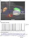
Figure 6. Intracellular recording in a facial motoneuron during kainic-acid induced vibrissa movements
(a) Schematic of the recording set-up. Conventions are as in Figure 1a. (b) Example record with membrane potential shown in black and vibrissa motion shown in blue. Similar recordings were obtained in a total of 9 cells in 4 rats. All animal procedures were approved by the IACUC at Laval University.
Figure 7
Figure 7. Bilateral kainic-acid injection produces independent vibrissa movements on the left and right sides of the face
(a) Movement of the left (dark blue) and right (light blue) C2 vibrissae following kainic acid injections. Similar results were obtained in a total of 2 rats. (b) Power spectra (top; dark and light blue) and spectral coherence (bottom; black) between the movements of each of the vibrissae in panel a. The two signals are show low coherence in the band of whisking frequencies relative to control data for bilateral active whisking in alert animals (bottom; gray). Control data are from Fee et al.. All animal procedures were approved by the IACUC at UC San Diego.

Similar articles

Cited by

References

    1. Cullen KE. Sensory signals during active versus passive movement. Current Opinion in Neurobiology. 2004;14:698–706. - PubMed
    1. von Holst E. Relations between the central nervous system and the peripheral organ. British Journal of Animal Behavior. 1954;2:89–94.
    1. Vincent SB. The tactile hair of the white rat. Journal of Comparative Neurology. 1913;23:1–23.
    1. Rice FL, Fundin BT, Arvidsson J, Aldskogius H, Johansson O. Comprehensive immunofluoresce and lectin binding study of the innervation of vibrissae follicle sinus complexes on the mystacial pad of the rat. Journal of Comparative Neurology. 1997;385:149–184. - PubMed
    1. Prescott TJ, Diamond ME, Wing AM. Active touch sensing. Philosophical Transactions of the ROyal Society of London B: Biology. 2011;366:2989–2995. - PMC - PubMed

Publication types

LinkOut - more resources