Review
. 2014 Jul 2;6(7):2571-601.
doi: 10.3390/v6072571.

New aspects of the pathogenesis of canine distemper leukoencephalitis

Affiliations
Review

New aspects of the pathogenesis of canine distemper leukoencephalitis

Charlotte Lempp et al. Viruses. .

Abstract

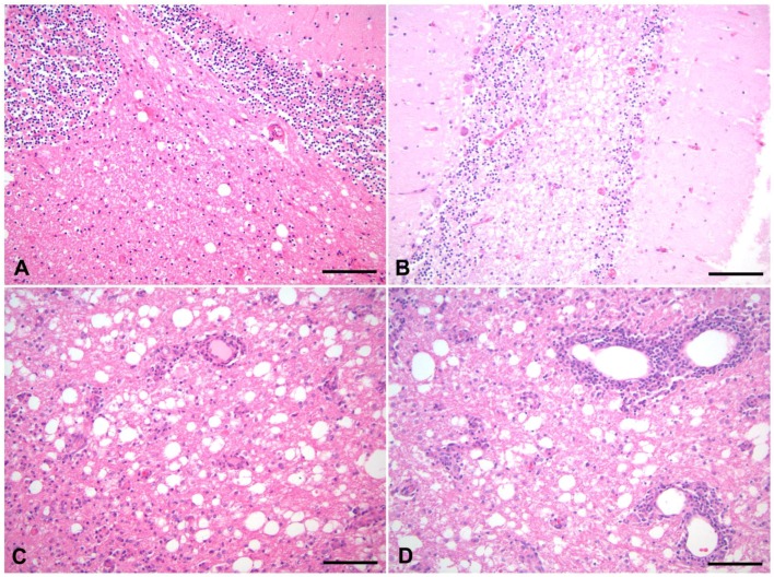
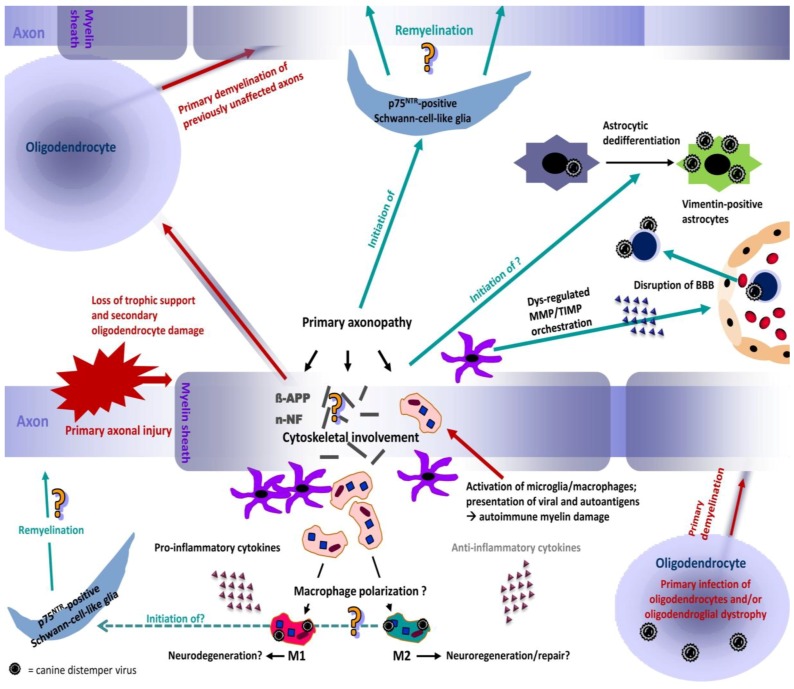
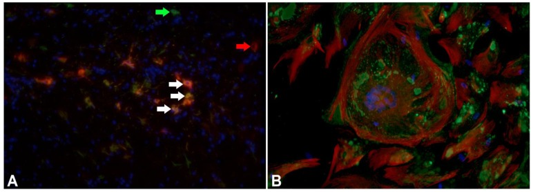
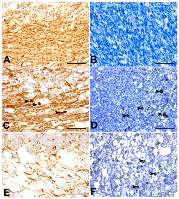
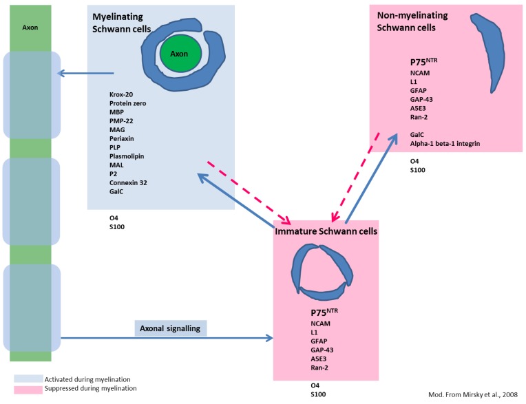
Canine distemper virus (CDV) is a member of the genus morbillivirus, which is known to cause a variety of disorders in dogs including demyelinating leukoencephalitis (CDV-DL). In recent years, substantial progress in understanding the pathogenetic mechanisms of CDV-DL has been made. In vivo and in vitro investigations provided new insights into its pathogenesis with special emphasis on axon-myelin-glia interaction, potential endogenous mechanisms of regeneration, and astroglial plasticity. CDV-DL is characterized by lesions with a variable degree of demyelination and mononuclear inflammation accompanied by a dysregulated orchestration of cytokines as well as matrix metalloproteinases and their inhibitors. Despite decades of research, several new aspects of the neuropathogenesis of CDV-DL have been described only recently. Early axonal damage seems to represent an initial and progressive lesion in CDV-DL, which interestingly precedes demyelination. Axonopathy may, thus, function as a potential trigger for subsequent disturbed axon-myelin-glia interactions. In particular, the detection of early axonal damage suggests that demyelination is at least in part a secondary event in CDV-DL, thus challenging the dogma of CDV as a purely primary demyelinating disease. Another unexpected finding refers to the appearance of p75 neurotrophin (NTR)-positive bipolar cells during CDV-DL. As p75NTR is a prototype marker for immature Schwann cells, this finding suggests that Schwann cell remyelination might represent a so far underestimated endogenous mechanism of regeneration, though this hypothesis still remains to be proven. Although it is well known that astrocytes represent the major target of CDV infection in CDV-DL, the detection of infected vimentin-positive astrocytes in chronic lesions indicates a crucial role of this cell population in nervous distemper. While glial fibrillary acidic protein represents the characteristic intermediate filament of mature astrocytes, expression of vimentin is generally restricted to immature or reactive astrocytes. Thus, vimentin-positive astrocytes might constitute an important cell population for CDV persistence and spread, as well as lesion progression. In vitro models, such as dissociated glial cell cultures, as well as organotypic brain slice cultures have contributed to a better insight into mechanisms of infection and certain morphological and molecular aspects of CDV-DL. Summarized, recent in vivo and in vitro studies revealed remarkable new aspects of nervous distemper. These new perceptions substantially improved our understanding of the pathogenesis of CDV-DL and might represent new starting points to develop novel treatment strategies.


Figures

Figure 1
Figure 1
Histological lesions of CDV-DL in the time course of disease. (A) Acute lesions are characterized by vacuolization of the white matter due to myelin sheath edema. Inflammation is nearly lacking and restricted to a sparse intraparenchymal infiltration of T cells; (B,C) Progressive myelin loss (pallor and lack of eosinophilia) is present in subacute lesions. Subacute lesions are further divided into lesions without (B) and with (C) inflammatory cell infiltration; (D) Chronic lesions invariably display more than three layers of perivascular inflammatory cells, accompanying ongoing demyelination and axonal loss. Hematoxylin and eosin staining. Bars = 50 µm.
Figure 2
Figure 2
Proposed neuropathogenesis of CDV-DL. The model is based on the concept of primary axonal injury (inside-out model), leading to disturbances in the axonal cytoskeleton and axonal transport mechanisms. The axonal damage induces secondary myelin loss and may induce oligodendrocyte death due to loss of trophic support. In consequence, previously non-affected axons undergo primary demyelination as one oligodendrocyte myelinates multiple axons. Another possible but less frequent mechanism of primary demyelination is the primary viral infection of oligodendrocytes. CDV-DL infection does not necessarily lead to oligodendroglial death but may induce an oligodendrocytic dystrophy with reduced translation of myelin proteins leading to demyelination. Axonal and myelin injury leads to activation of microglia/macrophages, phagocytosis of myelin fragments and presentation of viral antigens and autoantigens, potentially triggering another cascade of autoimmune mediated bystander demyelination presumably by “epitope spreading”. Resident activated astrocytes and microglia as well as attracted leukocytes from the peripheral blood can produce pro- and anti-inflammatory cytokines that can favour either neurodegeneration or neuroregeneration, respectively. The role of potential macrophage polarization towards a neurodegenerative (M1) or -regenerative (M2) phenotype remains undetermined so far and could represent an important pathogenetic mechanism. In parallel, the evident dysregulation of matrix metalloproteinases (MMPs) and their inhibitors (TIMPs) induces a disruption of the blood-brain-barrier (BBB), which favors the invasion of leukocytes and virus particles via cell associated viremia, which represents one possible mode of neuroinvasion. Dedifferentiation of astrocytes into immature vimentin expressing cells enhances their viral infection, leading to further virus spread. Pre-myelinating p75NTR-positive Schwann-cell-like glia arise during CDV-DL, potentially triggered by early axonal damage and activated microglia/macrophages. These cells may represent an important therapeutic target. However, whether manifest Schwann cell mediated remyelination occurs during CDV-DL remains elusive so far.
Figure 3
Figure 3
Immature astrocytes are characterized by expression of vimentin in CDV-DL. (A) Detection of glial fibrillary acidic protein (GFAP; green; Cy2-labeled secondary antibody; green arrows) and vimentin (red; Cy3-labeled secondary antibody; red arrow) expression in a chronic distemper lesion. Co-localization of GFAP and vimentin is displayed by a yellowish color in the cytoplasm of astrocytes (white arrows). Counterstaining of nuclei with bisbenzimide (blue). 200 fold magnification; (B) Immunostaining for vimentin and CDV antigen in adult canine mixed primary cell cultures infected with CDV strain R252 at 30 days post infection. Vimentin expression in mono- and multinucleated cells characterized by a fine fibrillary network (red: vimentin, Cy3-labeled secondary antibody). CDV antigen is mainly found in the cytoplasm (green: CDV nucleoprotein, Cy2-labeled secondary antibody). 400 fold magnification.
Figure 4
Figure 4
Axonal pathology during the time course of CDV-DL. (A), (B) cerebellum of control dogs; (C),(D): acute lesions of canine distemper virus induced demyelinating leukoencephalitis (CDV-DL); (E),(F) subacute lesions of CDV-DL; (A),(C),(E) Immunohistochemical labelling of phosphorylated neurofilament (p-NF) in the cerebellar white matter; (B),(D),(F) Luxol Fast Blue staining for myelin in the cerebellar white matter; (D),(F) Double-labelling of myelin with LFB and immunohistochemical detection of beta amyloid precursor protein (β-APP). (A) Expression of p-NF in the cerebellar white matter of a control dog. Note the normal density of axons, which physiologically express p-NF; (B) LFB staining reveals normal myelin content in the cerebellar white matter of a control dog; (C) Expression of p-NF in the cerebellar white matter in an acute lesion of CDV-DL. The morphologically unremarkable axons stain positive for p-NF. Additionally, a slight decrease of the axonal density, represented by loss of p-NF-immunoreactivity is visible. Moreover, single swollen axonal structures also stain positive for p-NF (arrowheads), which might be attributed to accumulation of neurofilaments due to impaired axonal transport; (D) In an acute lesion of CDV-DL, staining with LFB demonstrates normal myelin density, while axonally transported β-APP is expressed by several swollen axons (arrowheads) in the cerebellar white matter. This observation is consistent with the inside-out theory based on the assumption of primary axonopathy; (E) Expression of p-NF in a subacute lesion of CDV-DL. A marked loss of p-NF-positive axonal structures (decreased axonal density) is evident; (F) Expression of β-APP in a subacute lesion of CDV-DL. As an expression of ongoing axonal damage, β-APP-immunoreactive axons (arrowheads) are present within demyelinated areas as demonstrated by reduced staining intensity for LFB. Bar = 50 µm. (A),(C),(D),(E),(F): Avidin-Biotin-Peroxidase Complex Method, 3.3’diaminobenzidine-tetrahydrochloride.
Figure 5
Figure 5
Differentiation stages of Schwann cells and commonly expressed molecules. Unlike oligodendrocytes, immature Schwann cells may differentiate into two distinct phenotypes, myelinating (left) and non-myelinating (right) Schwann cells. However, differentiation is reversible as indicated by the dotted arrows. Blue boxes represent markers of myelinating Schwann cells and molecules promoting their differentiation. All of these markers display strong up-regulation in the stage of myelination. Red boxes include markers of non-myelinating and immature Schwann cells, which are down-regulated during myelination. Note that p75NTR is expressed in all differentiation stages except in myelinating Schwann cells. Whether the detected p75NTR expressing bipolar cells in CDV-DL differentiate into myelinating Schwann cells is currently not known. Markers and scheme modified from Mirsky et al., 2008 [141].
Figure 6
Figure 6
Organotypic slice cultures of the canine cerebellum, infected with the R252 strain of CDV and cultivated for nine days. (A) Hematoxylin and eosin staining reveals a remarkable response of activated, large phagocytic Gitter cells, presumably originating from resident microglia; (B) Immunohistochemistry for CDV shows that CDV-antigen is predominantly expressed by large, phagocytic cells reminiscent of microglia/macrophages. Whether these cells are infected by replicating virus or if they have phagocytozed viral antigen, remains to be determined in future studies; (C) Highly similar to axonopathy in spontaneous CDV-DL, axonal damage is characterized by enhanced expression of non-phosphorylated neurofilament. Similar axonal expression of n-NF is however also observed in non-infected slices, presumably due to mechanical transection of axons as a result of slice preparation; (D) p75NTR-positive bi- to multipolar cells emerge in response to prolonged culturing, potentially indicating occurrence of pre-myelinating Schwann cells. (BD) Avidin-Biotin-Peroxidase Complex Method, 3.3’diaminobenzidine tetrahydrochloride. Bars = 50 µm.

References

    1. Deem S.L., Spelman L.H., Yates R.A., Montali R.J. Canine Distemper in Terrestrial Carnivores: A Review. J. Zoo Wildl. Med. 2000;31:441–451. - PubMed
    1. Davies M. Risk of Re-Emergence of Canine Distemper. Vet. Rec. 2014;174:178. doi: 10.1136/vr.g1493. - DOI - PubMed
    1. Beineke A., Puff C., Seehusen F., Baumgärtner W. Pathogenesis and Immunopathology of Systemic and Nervous Canine Distemper. Vet. Immunol. Immunopathol. 2009;127:1–18. doi: 10.1016/j.vetimm.2008.09.023. - DOI - PubMed
    1. Greene C.E. Infectious Diseases of the Dog and Cat. 4th ed. Elsevier/Saunders; St. Louis, MO, USA: 2012. p. 1354.
    1. Maeda H., Ozaki K., Takagi Y., Sawashima K., Narama I. Distemper Skin Lesions in a Dog. Zentralbl. Veterinarmed. A. 1994;41:247–250. doi: 10.1111/j.1439-0442.1994.tb00091.x. - DOI - PubMed

Publication types

MeSH terms

Substances

LinkOut - more resources