

Comparison of epidermal/dermal damage between the long-pulsed 1064 nm Nd:YAG and 755 nm alexandrite lasers under relatively high fluence conditions: quantitative and histological assessments
- PMID: 24992273
- PMCID: PMC4082359
- DOI: 10.1089/pho.2013.3665
Comparison of epidermal/dermal damage between the long-pulsed 1064 nm Nd:YAG and 755 nm alexandrite lasers under relatively high fluence conditions: quantitative and histological assessments
Abstract
Objective: The purpose of this study was to compare degrees of epidermal/dermal tissue damage quantitatively and histologically after laser irradiation, to find ideal treatment conditions with relatively high fluence for skin rejuvenation.
Background data: A number of recent studies have evaluated the clinical efficacy and safety of therapeutic lasers under relatively low fluence conditions.
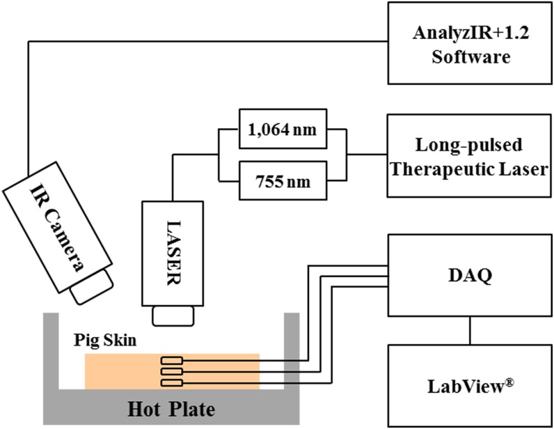
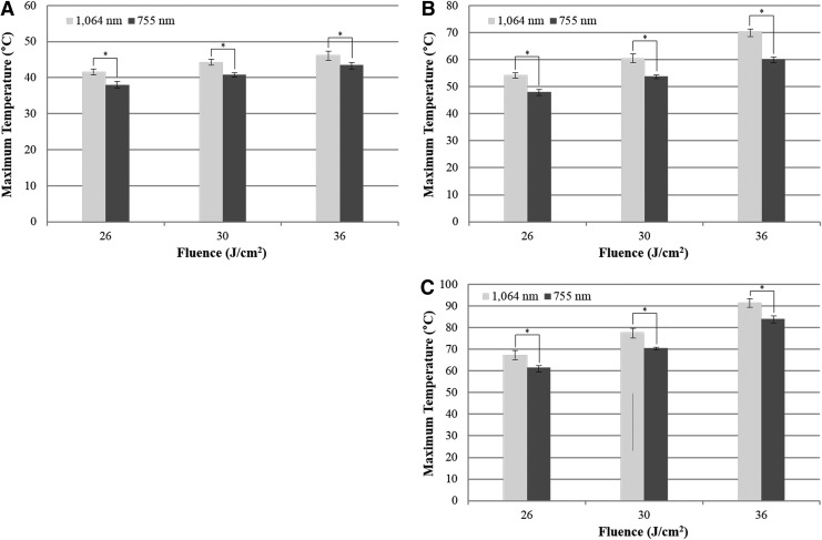
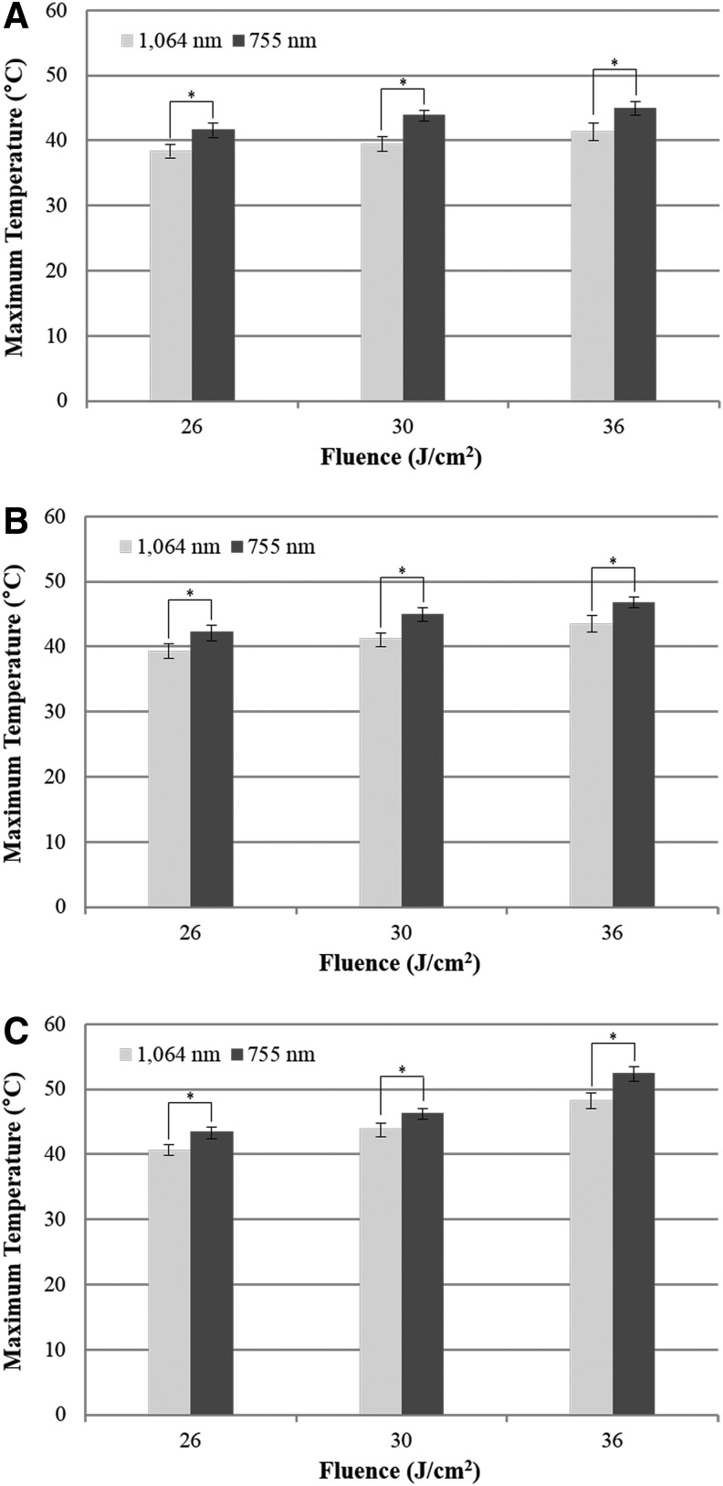
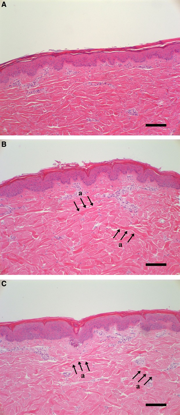
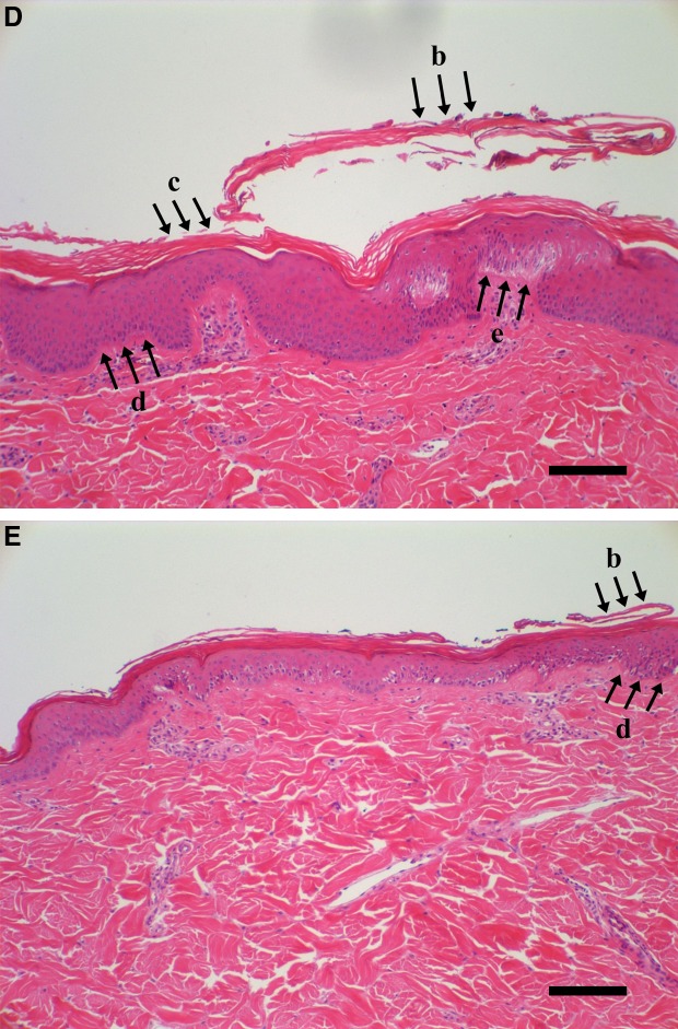
Methods: We transmitted the long-pulsed 1064 nm Nd:YAG and 755 nm Alexandrite lasers into pig skin according to different fluences and spot diameters, and estimated epidermal/dermal temperatures. Pig skin specimens were stained with hematoxylin and eosin for histological assessments. The fluence conditions comprised 26, 30, and 36 J/cm2, and the spot diameter conditions were 5, 8, and 10 mm. Pulse duration was 30 ms for all experiments.
Results: Both lasers produced reliable thermal damage on the dermis without any serious epidermal injuries, under relatively high fluence conditions. The 1064 nm laser provided more active fibrous formations than the 755 nm laser, while higher risks for tissue damages simultaneously occurred.
Conclusions: The ideal treatment conditions for skin rejuvenation were 8 mm diameter with 30 J/cm2 and 10 mm diameter with 26 J/cm2 for the 1064 nm laser, and 8 mm diameter with 36 J/cm2 and 10 mm diameter with 26 J/cm2 for the 755 nm laser.
Figures
References
-
- Munavalli G.S., Weiss R.A., and Halder R.M. (2005). Photoaging and nonablative photorejuvenation in ethnic skin. Dermatol. Surg. 31, 1250–1260 - PubMed
-
- Goldberg D.J., and Samady J.A. (2001). Intense pulsed light and Nd:YAG laser non-ablative treatment of facial rhytids. Lasers Surg. Med. 28, 141–144 - PubMed
-
- Newman J. (2001). Nonablative laser skin tightening. Facial Plast. Surg. Clin. North Am. 9, 343–349 - PubMed
-
- Ross E.V., Sajben F.P., Hsia J., Barnette D., Miller C.H., and McKinlay J.R. (2000). Nonablative skin remodeling: selective dermal heating with a mid-infrared laser and contact cooling combination. Lasers Surg. Med. 26, 186–195 - PubMed
-
- Doshi S.N., and Alster T.S. (2005). Combination radiofrequency and diode laser for treatment of facial rhytides and skin laxity. J. Cosmet. Laser Ther. 7, 11–15 - PubMed
Publication types
MeSH terms
LinkOut - more resources
Full Text Sources
Other Literature Sources
