. 2014 Jul 3;10(7):e1004216.
doi: 10.1371/journal.ppat.1004216. eCollection 2014 Jul.

Nsp9 and Nsp10 contribute to the fatal virulence of highly pathogenic porcine reproductive and respiratory syndrome virus emerging in China

Affiliations

Nsp9 and Nsp10 contribute to the fatal virulence of highly pathogenic porcine reproductive and respiratory syndrome virus emerging in China

Yan Li et al. PLoS Pathog. .

Erratum in

  • PLoS Pathog. 2014 Aug;10(8):e1004344

Abstract

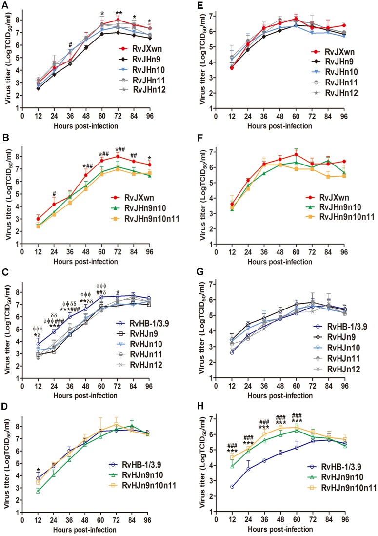
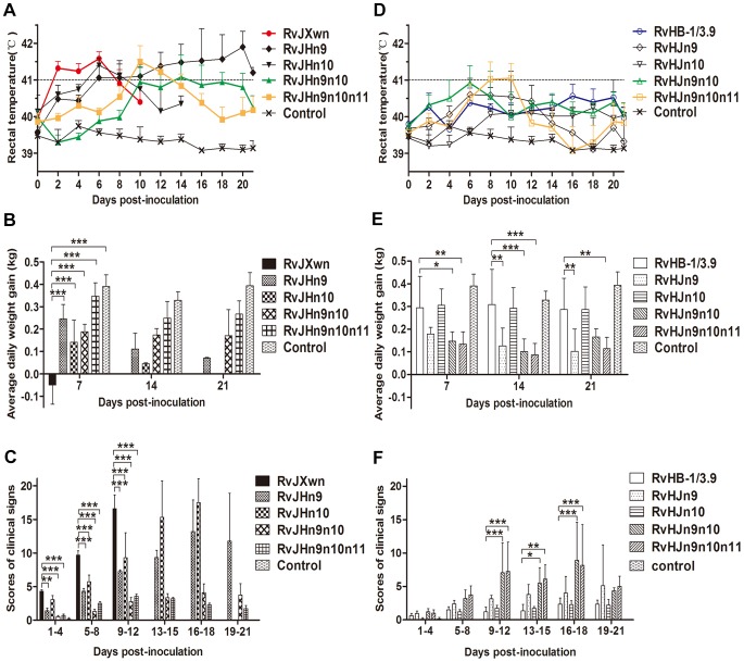
Atypical porcine reproductive and respiratory syndrome (PRRS), which is caused by the Chinese highly pathogenic PRRS virus (HP-PRRSV), has resulted in large economic loss to the swine industry since its outbreak in 2006. However, to date, the region(s) within the viral genome that are related to the fatal virulence of HP-PRRSV remain unknown. In the present study, we generated a series of full-length infectious cDNA clones with swapped coding regions between the highly pathogenic RvJXwn and low pathogenic RvHB-1/3.9. Next, the in vitro and in vivo replication and pathogenicity for piglets of the rescued chimeric viruses were systematically analyzed and compared with their backbone viruses. First, we swapped the regions including the 5'UTR+ORF1a, ORF1b, and structural proteins (SPs)-coding region between the two viruses and demonstrated that the nonstructural protein-coding region, ORF1b, is directly related to the fatal virulence and increased replication efficiency of HP-PRRSV both in vitro and in vivo. Furthermore, we substituted the nonstructural protein (Nsp) 9-, Nsp10-, Nsp11- and Nsp12-coding regions separately; or Nsp9- and Nsp10-coding regions together; or Nsp9-, Nsp10- and Nsp11-coding regions simultaneously between the two viruses. Our results indicated that the HP-PRRSV Nsp9- and Nsp10-coding regions together are closely related to the replication efficiency in vitro and in vivo and are related to the increased pathogenicity and fatal virulence for piglets. Our findings suggest that Nsp9 and Nsp10 together contribute to the fatal virulence of HP-PRRSV emerging in China, helping to elucidate the pathogenesis of this virus.


Conflict of interest statement

The authors have declared that no competing interests exist.

Figures

Figure 1
Figure 1. Construction strategy of the full-length cDNA clones.
(A) Full-length infectious clones with exchanged 5′UTR+ORF1a, ORF1b, and structural proteins (SPs)-coding regions. (B) Full-length infectious clones with exchanged nonstructural protein (Nsp)-coding region within ORF1b. The boxes represent the genomic fragments of parental backbone viruses RvJXwn (black) or RvHB-1/3.9 (white). Unique restriction enzyme sites used for cloning are shown above the bars. The designations of each full-length plasmid are shown on the left side and each rescued virus on the right side.
Figure 2
Figure 2. The growth kinetics of the rescued viruses with the exchanged 5′UTR+ORF1a, ORF1b and SP-coding regions.
(A) and (C) in MARC-145 cells; (B) and (D) in primary PAMs. Asterisk indicates a significant difference in the virus titers between RvJXwn (red) and RvJH1a (black) or between RvHB-1/3.9 (blue) and RvHJ1a (black) (*P<0.05; **P<0.01; ***P<0.001). Pound (#) indicates a significant difference between RvJXwn and RvJH1b (green) or between RvHB-1/3.9 and RvHJ1b (green) (#P<0.05; ##P<0.01; ###P<0.001). RvJHSP and RvHJSP (yellow).
Figure 3
Figure 3. The rectal temperatures and the mortality of piglets inoculated with the rescued virus with the exchanged coding regions.
Body temperatures of piglets inoculated with rescued viruses with the RvJXwn backbone (A) and with the RvHB-1/3.9 backbone (B). Body temperatures are shown as the means ± standard deviations (error bars), except the number of survival piglets in each group was less than two. The survival curves (C) and mortalities (D) of infected piglets in each group (n = 5) are shown.
Figure 4
Figure 4. Viral loads in sera of piglets inoculated with the rescued viruses with the exchanged coding regions.
Virus titers were determined by a microtitration infectivity assay. The data are shown as the means ± standard deviations (error bars), except only one piglet in RvJXwn-infected group survived on day 10 pi. Asterisk indicates significant differences in viral loads between the chimeric virus and RvJXwn or RvHB-1/3.9 (*P<0.05; **P<0.01; ***P<0.001).
Figure 5
Figure 5. Growth kinetics of the rescued viruses with the exchanged Nsp-coding region within ORF1b.
The growth curves of each chimeric virus, RvJXwn and RvHB-1/3.9 in MARC-145 cells (A, B, C and D) and in primary PAMs (E, F, G and H) are shown. Virus titers from 12 h to 96 h pi were determined by microtitration infectivity assays. The data are shown as the means ± standard deviations (error bars) from three independent trials. Asterisk (*) indicates a significant difference in virus titers between RvJXwn (red) and RvJHn9 (black), or RvJHn9n10 (green), or between RvHB-1/3.9 (blue) and RvHJn9 (black) or RvHJn9n10 (green) (*P<0.05; **P<0.01; ***P<0.001). Pound (#) indicates a significant difference between RvJXwn and RvJHn10 (light blue) or RvJHn9n10n11 (yellow), or between RvHB-1/3.9 and RvHJn10 (light blue) or RvHJn9n10n11 (yellow) (##P<0.01; ###P<0.001). Phi (Φ) indicates a significant difference between RvHB-1/3.9 and RvHJn11 (gray) (ΦΦP<0.01; ΦΦΦP<0.001). Delta (δ) indicates a significant difference between RvHB-1/3.9 and RvHJn12 (gray) (δP<0.05; δδP<0.01).
Figure 6
Figure 6. The rectal temperatures, clinical scores and average daily gains of piglets inoculated with the rescued viruses.
The body temperatures, average clinical scores and average daily gains (ADG) of piglets inoculated with the rescued viruses with the RvJXwn backbone (A, C and E) or with the RvHB-1/3.9 backbone (B, D and F) are shown. The data are shown as the means ± standard deviations (error bars). The clinical scoring included the gross clinical score (GCS), respiratory clinical score (RCS) and nervous signs score (NSS). Total scores for each piglet represented the sum of the GCS, RCS and NSS. An additional five score was calculated in the total scores when the piglet died. Each piglet was scored from 0–20, and the mean values of day 1 to 4 pi, 5 to 8 pi, 9 to 12 pi, 13 to 15 pi, 16 to 18 pi and 19 to 21 pi were calculated. Asterisk indicates a significant difference between the chimeric virus and its parental backbone virus, RvJXwn or RvHB-1/3.9 (*P<0.05; **P<0.01; ***P<0.001).
Figure 7
Figure 7. The mortality and survival curve of piglets inoculated with the rescued viruses.
The mortalities and survival curves of piglets infected with rescued viruses in each group are shown (n = 5).
Figure 8
Figure 8. Scores of gross lung lesions and microscopic lung lesions of piglets inoculated with the rescued viruses.
The mean scores of gross lung lesions, (A) from dead piglets during the experiment and (B) from euthanized piglets at the end of experiment in each group are shown. The gross lesions were graded based on the percentage of lung area affected. Lung sections were stained with hematoxylin and eosin (H&E). Representative pictures of the histopathological lung lesions and the average scores of dead piglets during the experiment (C) and euthanized piglets at the end of experiment (D) in each group. The microscopic lesions were scored based on the severity of interstitial pneumonia. Solid arrow indicates thickening of the interlobular septal or infiltration of inflammatory cells around the bronchiole. Solid triangle indicates inflammatory cells, necrotic debris and exfoliated epithelial cells infiltrate in the bronchiole. Triangle indicates hemorrhage or infiltration of inflammatory cells within alveolar septa, and alveolar spaces. Asterisk indicates significant differences in gross lesion scores and microscopic lesion scores between RvHB-1/3.9 and RvHJn9n10n11 (*P<0.05).
Figure 9
Figure 9. Immunohistochemical examination of inoculated piglets for PRRSV antigen.
Lung sections were examined by immunohistochemistry (IHC) using monoclonal antibodies (SDOW17) specific for the N protein of PRRSV, and numbers of positive cells in lungs were scored. Representative pictures of immunohistochemistry examinations and mean scores of lungs of the dead piglets during the experiment (A) and of euthanized piglets by the end of experiment (B) in each group are shown. The macrophages stain intensely dark brown for the PRRSV antigen. Hollow arrow indicates positive signals in macrophages within or around alveolus and bronchus. Asterisk indicates a significant difference in IHC scores between RvHB-1/3.9 and RvHJn9n10n11 (*P<0.05).
Figure 10
Figure 10. Viral loads and antibody kinetics in the sera of piglets inoculated with the rescued viruses.
Virus titers in the sera of piglets inoculated with chimeric viruses with the RvJXwn backbone (A) and with the RvHB-1/3.9 backbone (B) are shown. Virus titers were determined by microtitration infectivity assay. The data are shown as the means ± standard deviations (error bars). Asterisk indicates significant differences in viral loads between chimeric virus and its parental backbone virus RvJXwn or RvHB-1/3.9 (*P<0.05; **P<0.01; ***P<0.001). The antibody kinetics of piglets inoculated with chimeric viruses with the RvJXwn backbone (C) and with the RvHB-1/3.9 backbone (D) are shown. The antibodies specific for PRRSV were detected using an IDEXX Herdchek PRRS 2XR ELISA kit, and the antibody level was expressed as a sample value/positive value (S/P) ratio. A ratio of ≥0.4 was regarded as seroconversion. Asterisk indicates significant differences in the antibody level between RvJXwn and RvJHn9 or between RvHB-1/3.9 and RvHJn9 (*P<0.05; ***P<0.001). Pound (#) indicates a significant difference between RvJXwn and RvJHn10 or between RvHB-1/3.9 and RvHJn10 (##P<0.01; ###P<0.001). Phi (Φ) indicates significant difference between RvJXwn and RvJHn9n10 or between RvHB-1/3.9 and RvHJn9n10 (ΦΦΦP<0.001). Delta (δ) indicates significant difference between RvJXwn and RvJHn9n10n11 or between RvHB-1/3.9 and RvHJn9n10n11 (δδP<0.01; δδδP<0.001).

References

    1. Albina E (1997) Epidemiology of porcine reproductive and respiratory syndrome (PRRS): an overview. Vet Microbiol 55: 309–316. - PubMed
    1. Pejsak Z, Stadejek T, Markowska-Daniel I (1997) Clinical signs and economic losses caused by porcine reproductive and respiratory syndrome virus in a large breeding farm. Vet Microbiol 55: 317–322. - PubMed
    1. Keffaber KK (1989) Reproductive failure of unknown etiology. Am Assoc Swine Pract Newsl 1.2: 9.
    1. Bilodeau R, Dea S, Sauvageau RA, Martineau GP (1991) ‘Porcine reproductive and respiratory syndrome’ in Quebec. Vet Rec 129: 102–103. - PubMed
    1. Albina E, Baron T, Leforban Y (1992) Blue-eared pig disease in Brittany. Vet Rec 130: 58–59. - PubMed

Publication types

MeSH terms