. 2014 Jul 3;9(7):e100236.
doi: 10.1371/journal.pone.0100236. eCollection 2014.

Amniotic membrane grafts for the prevention of esophageal stricture after circumferential endoscopic submucosal dissection

Affiliations

Amniotic membrane grafts for the prevention of esophageal stricture after circumferential endoscopic submucosal dissection

Maximilien Barret et al. PLoS One. .

Abstract

Background and aims: The prevention of esophageal strictures following circumferential mucosal resection remains a major clinical challenge. Human amniotic membrane (AM) is an easily available material, which is widely used in ophthalmology due to its wound healing, anti-inflammatory and anti-fibrotic properties. We studied the effect of AM grafts in the prevention of esophageal stricture after endoscopic submucosal dissection (ESD) in a swine model.

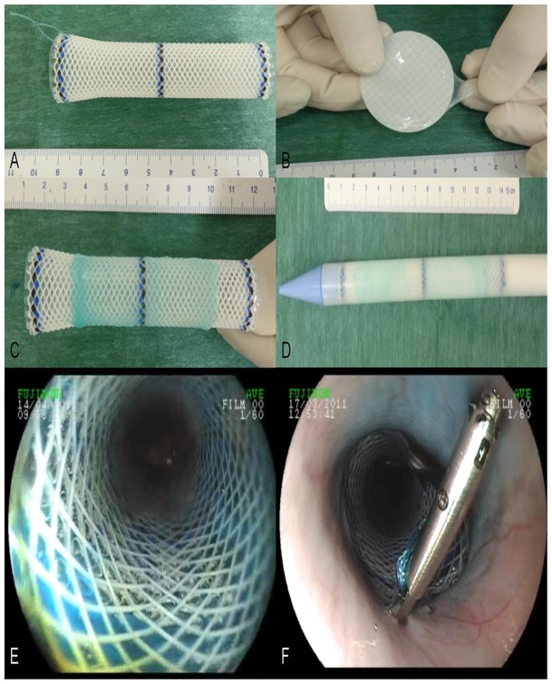
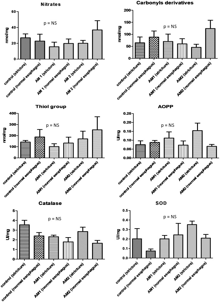
Animals and methods: In this prospective, randomized controlled trial, 20 swine underwent a 5 cm-long circumferential ESD of the lower esophagus. In the AM Group (n = 10), amniotic membrane grafts were placed on esophageal stents; a subgroup of 5 swine (AM 1 group) was sacrificed on day 14, whereas the other 5 animals (AM 2 group) were kept alive. The esophageal stent (ES) group (n = 5) had ES placement alone after ESD. Another 5 animals served as a control group with only ESD.

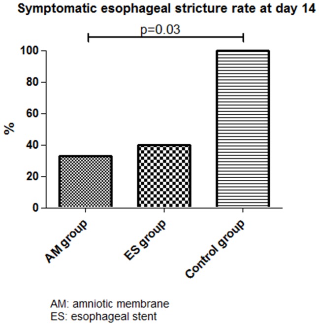
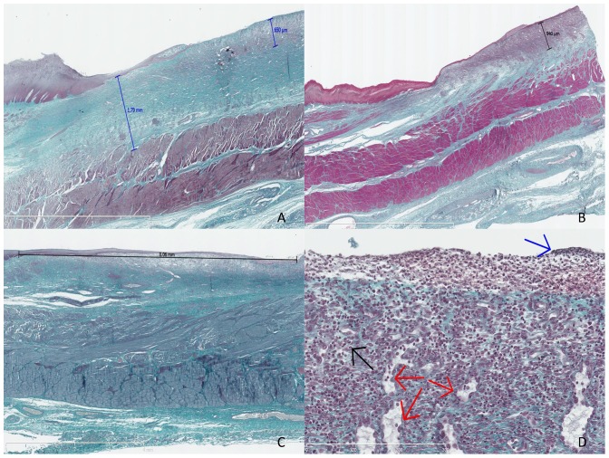
Results: The prevalence of symptomatic strictures at day 14 was significantly reduced in the AM group and ES groups vs. the control group (33%, 40% and 100%, respectively, p = 0.03); mean esophageal diameter was 5.8±3.6 mm, 6.8±3.3 mm, and 2.6±1.7 mm for AM, ES, and control groups, respectively. Median (range) esophageal fibrosis thickness was 0.87 mm (0.78-1.72), 1.19 mm (0.28-1.95), and 1.65 mm (0.7-1.79) for AM 1, ES, and control groups, respectively. All animals had developed esophageal strictures by day 35.

Conclusions: The anti-fibrotic effect of AM on esophageal wound healing after ESD delayed the development of esophageal stricture in our model. However, this benefit was of limited duration in the conditions of our study.


Conflict of interest statement

Competing Interests: The authors have declared that no competing interests exist.

Figures

Figure 1
Figure 1. Application of amniotic membrane grafts on esophageal wounds.
A: esophageal stent with attached non-absorbable suture; B: blue-stained amniotic graft on a nitrocellulose sheet after defrosting; C: amniotic membrane apposition on the external side of a Polyflex stent; D: esophageal stent coated with amniotic membrane graft loaded in the stent catheter; E: endoscopic view of the coated esophageal stent in the esophagus; F: esophageal stent clipped to the esophageal wall using the suture.
Figure 2
Figure 2. Symptomatic esophageal strictures rates at day 14.
Figure 3
Figure 3. Histological analysis of the swine esophagus after Masson's trichrome staining.
A: swine from the control group, sacrificed at day 14, with major fibrosis measured at 1.79 mm and thick granulation tissue measured at 0.65 mm, original magnification 12.5x; B: swine from the AM 1 group (amniotic membrane graft and early sacrifice scheduled at day 14), without esophageal stricture, with minimal fibrosis and mostly granulation tissue, measured at 0.94 mm, original magnification 10x; C: swine from the AM 2 group sacrificed at day 21, exhibiting major re-epithelialization measured at 8.06 mm, original magnification 15x; D: granulation tissue with features of acute inflammation, as observed in the early phase of esophageal wounds: high cell density, predominance of polynuclear cells (black arrow), fibrino-leucocytary network (blue arrow), and typical palissadic vascular growth (red arrows), original magnification 400x.
Figure 4
Figure 4. Immunohistochemistry staining with anti-αSMA antibody, original magnification ×200.
Strong signal (brown spots) attesting high myofibroblastic activity (panel A, black arrow) and high vascular density (panel A, white arrow) in control (A); nearly absent signal in MA-treated swine (B).
Figure 5
Figure 5. Oxidative stress markers measurements in tissue samples of strictured or normal esophagus; performed in control, AM1 and AM 2 groups.

References

    1. Bennett C, Vakil N, Bergman J, Harrison R, Odze R, et al. (2012) Consensus statements for management of Barrett's dysplasia and early-stage esophageal adenocarcinoma, based on a Delphi process. Gastroenterology 143: 336–346. - PMC - PubMed
    1. Shaheen NJ, Richter JE (2009) Barrett's oesophagus. Lancet 373: 850–861. - PubMed
    1. Shaheen NJ, Overholt BF, Sampliner RE, Wolfsen HC, Wang KK, et al. (2011) Durability of radiofrequency ablation in Barrett's esophagus with dysplasia. Gastroenterology 141: 460–468. - PMC - PubMed
    1. Titi M, Overhiser A, Ulusarac O, Falk GW, Chak A, et al. (2012) Development of subsquamous high-grade dysplasia and adenocarcinoma after successful radiofrequency ablation of Barrett's esophagus. Gastroenterology 143: : 564–566 e561. - PMC - PubMed
    1. Chennat J, Konda VJ, Ross AS, de Tejada AH, Noffsinger A, et al. (2009) Complete Barrett's eradication endoscopic mucosal resection: an effective treatment modality for high-grade dysplasia and intramucosal carcinoma–an American single-center experience. Am J Gastroenterol 104: 2684–2692. - PubMed

Publication types