. 2014 Jul 3;10(7):e1004413.
doi: 10.1371/journal.pgen.1004413. eCollection 2014 Jul.

Meiosis-specific cohesin component, Stag3 is essential for maintaining centromere chromatid cohesion, and required for DNA repair and synapsis between homologous chromosomes

Affiliations

Meiosis-specific cohesin component, Stag3 is essential for maintaining centromere chromatid cohesion, and required for DNA repair and synapsis between homologous chromosomes

Jessica Hopkins et al. PLoS Genet. .

Abstract

Cohesins are important for chromosome structure and chromosome segregation during mitosis and meiosis. Cohesins are composed of two structural maintenance of chromosomes (SMC1-SMC3) proteins that form a V-shaped heterodimer structure, which is bridged by a α-kleisin protein and a stromal antigen (STAG) protein. Previous studies in mouse have shown that there is one SMC1 protein (SMC1β), two α-kleisins (RAD21L and REC8) and one STAG protein (STAG3) that are meiosis-specific. During meiosis, homologous chromosomes must recombine with one another in the context of a tripartite structure known as the synaptonemal complex (SC). From interaction studies, it has been shown that there are at least four meiosis-specific forms of cohesin, which together with the mitotic cohesin complex, are lateral components of the SC. STAG3 is the only meiosis-specific subunit that is represented within all four meiosis-specific cohesin complexes. In Stag3 mutant germ cells, the protein level of other meiosis-specific cohesin subunits (SMC1β, RAD21L and REC8) is reduced, and their localization to chromosome axes is disrupted. In contrast, the mitotic cohesin complex remains intact and localizes robustly to the meiotic chromosome axes. The instability of meiosis-specific cohesins observed in Stag3 mutants results in aberrant DNA repair processes, and disruption of synapsis between homologous chromosomes. Furthermore, mutation of Stag3 results in perturbation of pericentromeric heterochromatin clustering, and disruption of centromere cohesion between sister chromatids during meiotic prophase. These defects result in early prophase I arrest and apoptosis in both male and female germ cells. The meiotic defects observed in Stag3 mutants are more severe when compared to single mutants for Smc1β, Rec8 and Rad21l, however they are not as severe as the Rec8, Rad21l double mutants. Taken together, our study demonstrates that STAG3 is required for the stability of all meiosis-specific cohesin complexes. Furthermore, our data suggests that STAG3 is required for structural changes of chromosomes that mediate chromosome pairing and synapsis, DNA repair and progression of meiosis.


Conflict of interest statement

The authors have declared that no competing interests exist.

Figures

Figure 1
Figure 1. Stag3 mutation results in gonadal failure.
(A) Image of Stag3+/− and Stag3−/− testes at 8 weeks of age. The average testis to body weight ratio of six Stag3+/− and Stag3−/− 8 week old mice was 0.72% (+/− 0.05%) and 0.18% (+/− 0.02%) respectively. (B) Haemoxylin and eosin staining of 5 micron thick testis sections of 8 week old Stag3+/− and Stag3−/− mice, scale bar  = 100 µm. (C) TUNEL staining of paraffin embedded 5 micron thick testis sections of 8 week old Stag3+/− and Stag3−/− mice; scale bar  = 100 µm. (D) Haemoxylin and eosin staining of 5 micron thick testis sections of 18 days postpartum (dpp) Stag3+/− and Stag3−/− mice. The star represents a tubule that contains germ cells undergoing apoptosis, scale bar  = 100 µm. (E) Image of Stag3+/− and Stag3−/− ovaries at 8 weeks of age. The average ovary to body weight ratio of six Stag3+/− and Stag3−/− 8 week old mice was 0.044% (+/−0.0064%) and 0.0048% (+/−0.001%) respectively. (F) Haemoxylin and eosin staining of 5 micron thick ovary sections of 8 week old Stag3+/− and Stag3−/− mice; scale bar  = 50 µm. (G) Haemoxylin and eosin staining of 5 micron thick ovary sections of 6 dpp Stag3+/− and Stag3−/− mice, scale bar  = 100 µm. All images in this figure are from mice with the Stag3OV mutant allele.
Figure 2
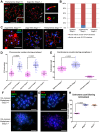
Figure 2. Stag3 mutation results in abnormal meiosis progression, atypical synapsis between sister chromatids, and absence of pachytene stage germ cells.
Chromatin spreads from (A) purified testicular germ cells of Stag3+/− and Stag3−/− mice aged 16 dpp and (B) embryonic ovarian germ cells of Stag3+/− and Stag3−/− mice aged 16.5 days post coitum were stained with DAPI (blue, DNA) and immunolabeled using antibodies against the SC lateral element protein SYCP3 (red) and the transverse filament of the central region of the SC SYCP1 (green). Meiotic prophase stages are indicated across the top; Stag3−/− spermatocytes and oocytes were deemed to be at a leptotene-like (lepto-like) stage when SYCP1 was not evident and at a zygotene-like stage (zygo-like) when SYCP1 colocalized with the SYCP3 signal. XY label represents the sex chromosome pair. Images in (A) and (B) are of spermatocytes carrying the Stag3OV mutant allele, but similar phenotypes were observed for spermatocytes with the Stag3JAX mutant allele and mice carrying the Stag3OV and Stag3JAX alleles combined (Fig. S2). (C) Scatter dot-plot graph of the number of SYCP3 linear stretches per spermatocyte chromatin spread during leptotene (lepto; average  = 154, N = 40), early zygotene (early zygo; average  = 43, N = 50), late zygotene (late zygo; average  = 25, N = 50) and pachytene (average  = 20, N = 40) stages for the Stag3+/− control and lepto-like (average  = 41, N = 50) and zygo-like (average  = 42, N = 51) stages for the Stag3−/− mice. Similar results were obtained when assessing oocyte chromatin spreads, summarized in Fig. S3. (D) Scatter dot-plot graph of the average SYCP3 length per spermatocyte chromatin spread during early zygo (7.1 µm), late zygo (6.7 µm) and pachytene (7.4 µm) stages for the Stag3+/− control and zygo-like (2.4 µm) stage for the Stag3−/− mice. Similar results were obtained when assessing oocyte chromatin spreads, summarized in Fig. S3. (E) Chromatin spreads from purified testicular germ cells of Stag3+/− and Stag3−/− mice aged 16 dpp were immunolabeled using an antibody against the SC lateral element protein SYCP3 (blue) and then hybridized to two pre-labelled FISH probes, one that detects the entire X chromosome (green) and the other detects 200 kilobases of mouse chromosome 11 (TK [11qE1]) distal to the centromere (red, white arrows). Mean and standard deviation of the columns of each graph are represented by the black bars and P values are given for indicated comparisons (Mann-Whitney, one-tailed). Experiments were performed using 4 separate littermate pairs of mutant and control mice. Scale bars  = 10 µm
Figure 3
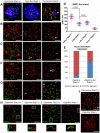
Figure 3. Stag3 mutation results in circular SYCP3 stretches, disrupted heterochromatin pericentromeric clustering (chromocenters), and premature loss of centromere cohesion between sister chromatids.
(A-E) Chromatin spreads were prepared from purified testicular germ cells of Stag3+/− and Stag3−/− mice aged 16 dpp. (A) Chromatin spreads were immunolabeled with antibodies against the SC lateral element protein SYCP3 (red), the centromere-kinetochore (blue, CEN) and the telomeric protein TRF1 (green). The left most panel is a Stag3+/− chromatin spread at pachytene stage. XY label represents the sex chromosome pair. Inset image on the bottom right corner is a 2× zoom of a synapsed autosome pair with a telomere signal at each end and a centromere signal at one end. The middle panel is a Stag3−/− chromatin spread at a zygo-like stage. The diamond and triangle arrow heads point to the SYCP3 stretches that are magnified in the right most panels. The top right most panel is a 5× zoom of a Stag3−/− SYCP3 stretch with a telomere signal at each end and a centromere signal at one end. The bottom right most panel is a 5× zoom of a Stag3−/− circular SYCP3 stretch. (B) Quantification of nuclei with circular SYCP3 stretches. No circular SYCP3 stretches were observed during zygotene or pachytene stages for the Stag3+/− control (N = 179 and 224 respectively), whereas 10.9% of zygo-like chromatin spreads from the Stag3−/− mice were recorded to have circular SYCP3 stretches (N = 212). This experiment was performed in triplicate and positive and negative error bars represent the highest and lowest percentage of circular SYCP3 stretches obtained. (C) Chromatin spreads were immunolabeled with antibodies against the SC lateral element protein SYCP3 (red), the centromere-kinetochore (green, CEN) and SMC6 protein which localizes to the pericentromeric heterochromatin clusters also known as “chromocenters” (blue). Meiotic prophase stages are indicated across the top. (D) Scatter dot-plot graph of the number of chromocenters per spermatocyte chromatin spread during leptotene (average  = 8.4, N = 56), zygotene (average  = 6.9, N = 89) and pachytene (average  = 8.2, N = 55) stages for the Stag3+/− control and lepto-like (average  = 16.6, N = 74) and zygo-like (17.7, N = 102) stages for the Stag3−/− mice. Similar results were obtained when assessing oocyte chromatin spreads, summarized in Fig. S4A and B. (E) Scatter dot-plot graph of the number of centromere-kinetochore signals per spermatocyte chromatin spread during zygotene (average  = 36.1, N = 89) and pachytene (average  = 21.2, N = 55) stages for the Stag3−/− mice and zygo-like stage (average  = 43.8, N = 102) for the Stag3−/− mice. Experiments were performed using 3 separate littermate pairs of mutant and control mice. Images are from germ cells carrying the Stag3OV allele, comparable phenotypes were observed for germ cells carrying the Stag3JAX mutant allele (Fig. S2). Similar results for centromere counts were obtained when assessing oocyte chromatin spreads summarized in Fig. S4A and C. (F and G) Chromatin spreads from purified and short-term cultured testicular germ cells of Stag3+/− and Stag3−/− mice aged 20 dpp following treatment with 5 µM of okadaic acid. (F) Chromatin spreads stained with DAPI (blue, DNA) and immunolabeled with antibodies against the SC lateral element protein SYCP3 (red) and the pan-cohesin component SMC3 (green). (G) Chromatin spreads stained with DAPI (blue, DNA) and immunolabeled with antibodies against the SC lateral element protein SYCP3 (red) and the meiosis-specific α-kleisin cohesin component REC8 (green). (H) Scatter dot-plot graph of the number of centromere-kinetochore signals per spermatocyte chromatin spread following 5 hours of OA treatment for Stag3+/− (average  = 39.5, N = 40), Stag3−/− (average  = 78.5, N = 60) and Rec8−/− (average  = 77.6, N = 18) mice. Mean and standard deviation of the columns of each graph are represented by the black bars and P values are given for indicated comparisons (Mann-Whitney, one-tailed). Scale bar  = 10 µm
Figure 4
Figure 4. Mutation of Stag3 does not affect the localization of components of the mitotic cohesin complex, but is required for the localization and stability of meiosis-specific cohesin subunits.
Chromatin spreads were prepared from purified testicular germ cells of Stag3+/− and Stag3−/− mice aged 16 dpp. Chromatin spreads were immunolabeled with antibodies against the SC lateral element protein SYCP3 (red) and pan-cohesin component SMC3 (A), mitotic cohesin components SMC1α (B) and RAD21 (C) and meiosis-specific cohesin components RAD21L (D), REC8 (E) and SMC1β (F) in green. Meiotic prophase stages are indicated across the top. Experiments were performed using 3 separate littermate pairs of mutant and control mice. Images are from germ cells carrying the Stag3Ov allele. Similar results were obtained when assessing oocyte chromatin spreads, summarized in Fig. S5 and for the Stag3JAX allele mutants (Fig. S6). (G) Protein extracts from purified testicular germ cells of WT (Stag3+/+), HET (Stag3+/−) and KO (Stag3−/−) mice aged 16 dpp were prepared and western blot analyses performed for STAG3, RAD21, REC8, RAD21L, SMC3, SMC1α, SMC1β, STAG1 and STAG2. Tubulin was used as a loading control. (H) Quantification of protein levels of each cohesin component analyzed in (G). Tubulin was used to normalize the loading of each lane. Each western blot was repeated at least twice. Tubulin loading controls corresponding to each western blot analyzed is present in Fig. S7. Data shown for germ cell extracts from the Stag3OV homozygous mutants and littermate controls. Scale bar  = 10 µm
Figure 5
Figure 5. Stag3 mutants fail to repair meiotic DSBs and have an abnormal DNA damage response.
Chromatin spreads from purified testicular germ cells of Stag3+/− and Stag3−/− mice aged 16 dpp were prepared and immunolabeled. (A) Chromatin spreads were immunolabeled with antibodies against the SC lateral element protein SYCP3 (red), phosphorylated histone H2AFX (blue, γH2AX) and the transverse filament of the central region of the SC SYCP1 (green). (B) Chromatin spreads were immunolabeled with antibodies against the SC lateral element protein SYCP3 (red) and meiosis-specific single-end invasion protein DMC1 (green). (C) Chromatin spreads were immunolabeled with antibodies against the SC lateral element protein SYCP3 (red) and single-end invasion protein RAD51 (green). Arrows represent RAD51 aggregates not associated with SYCP3 stretches. (D) Scatter dot-plot graph of the number of DMC1 foci per spermatocyte chromatin spread during early zygotene (Early Z, average  = 220, N = 50), late zygotene (Late Z, average  = 129, N = 50) and early pachytene (Early P, average  = 39.5, N = 20) stages for the Stag3+/− control and zygo-like stage (Z-like average  = 112, N = 50) for the Stag3−/− mice. Mean and standard deviation of each column of the graph are represented by the black bars and P values are given for indicated comparisons (Mann-Whitney, one-tailed). (E) Bar graph of the percentage of chromatin spreads that contain RAD51 aggregates at the zygotene stage (average  = 11.2%, N = 179) for the Stag3+/− control and zygotene-like stage (average  = 61.8%, N = 212) for the Stag3−/− mice. The error bars represent the variation between three independent experiments. (F) Chromatin spreads were immunolabeled with antibodies against the SC lateral element protein SYCP3 (red) and DNA damage response protein ATR (green). Arrows represent ATR aggregates not associated with SYCP3 stretches. (G) Chromatin spreads were immunolabeled with antibodies against the SC lateral element protein SYCP3 (red) and DNA damage response protein ATRIP (green). Arrows represent ATRIP aggregates. (H and I) Chromatin spreads were immunolabeled using antibodies against the HORMA domain containing protein HORMAD1 (H, red) or HORMAD2 (I, red) and the SC central element protein TEX12 (green). The boxed regions are magnified 3× below the whole chromatin spread images. Images are from the Stag3Ov mutant allele, comparable phenotype was observed for the Stag3JAX mutant allele (Fig. S2). (J) Chromatin spreads were immunolabeled with antibodies against the SC lateral element protein SYCP3 (red) and crossover protein MLH1 (green). Each experiment was performed at least twice. Images are from cells with the Stag3Ov mutant allele. XY label represents the sex chromosome pair. Scale bars  = 10 µm
Figure 6
Figure 6. STAG3 is required for stability and loading of meiosis-specific cohesins to chromosome axes during meiosis.
(A) Diagram depicting the ring-like structure of the cohesin complex. The mitotic cohesin and meiosis-specific components are written in black and red text respectively. (B) Summary table of the phenotypes recorded for mutants of the four meiosis-specific cohesin components. 1 The axis length was not defined in these studies, but from our analysis it is ∼50% shorter. 2 Although the female phenotype was not reported in this study, it can be implied due to the phenotype of the Rec8−/− mutant. (C) Cartoon of mid-prophase of a wild type (pachytene stage) and Stag3 mutant (zygotene-like stage). All features are described within the key. At pachytene stage, homologous chromosomes are fully synapsed and an obligate crossover has formed. In zygotene-like staged Stag3 mutant germ cells, localization and stability of meiosis-specific cohesin complexes is aberrant and leads to synapsis between sister chromatids, DNA double strand breaks (DSBs) are not repaired and centromere cohesion between sister chromatids is perturbed. Chromatin loops are depicted to be larger in the Stag3 mutant as their chromosome axes are shorter compared to wild type, and is supported by analysis of the Smc1β mutant mouse .

Similar articles

Cited by

References

    1. Jordan P (2006) Initiation of homologous chromosome pairing during meiosis. Biochem Soc Trans 34: 545–549. - PubMed
    1. Nasmyth K (2011) Cohesin: a catenase with separate entry and exit gates? Nat Cell Biol 13: 1170–1177. - PubMed
    1. Ishiguro K-i, Kim J, Fujiyama-Nakamura S, Kato S, Watanabe Y (2011) A new meiosis-specific cohesin complex implicated in the cohesin code for homologous pairing. EMBO Rep 12: 267–275. - PMC - PubMed
    1. Gómez R, Valdeolmillos A, Parra M, Viera A, Carreiro C, et al. (2007) Mammalian SGO2 appears at the inner centromere domain and redistributes depending on tension across centromeres during meiosis II and mitosis. EMBO Rep 8: 173–180. - PMC - PubMed
    1. Prieto I, Suja JA, Pezzi N, Kremer L, Martinez-A C, et al. (2001) Mammalian STAG3 is a cohesin specific to sister chromatid arms in meiosis I. Nat Cell Biol 3: 761–766. - PubMed

Publication types

LinkOut - more resources