. 2014 Jul 3;9(7):e99881.
doi: 10.1371/journal.pone.0099881. eCollection 2014.

Transplanting supersites of HIV-1 vulnerability

Affiliations

Transplanting supersites of HIV-1 vulnerability

Tongqing Zhou et al. PLoS One. .

Erratum in

  • PLoS One. 2014;9(8); e105659. doi: 10.1371/journal.pone.0105659. Kwon, Peter D [corrected to Kwong, Peter D]

Abstract

One strategy for isolating or eliciting antibodies against a specific target region on the envelope glycoprotein trimer (Env) of the human immunodeficiency virus type 1 (HIV-1) involves the creation of site transplants, which present the target region on a heterologous protein scaffold with preserved antibody-binding properties. If the target region is a supersite of HIV-1 vulnerability, recognized by a collection of broadly neutralizing antibodies, this strategy affords the creation of "supersite transplants", capable of binding (and potentially eliciting) antibodies similar to the template collection of effective antibodies. Here we transplant three supersites of HIV-1 vulnerability, each targeted by effective neutralizing antibodies from multiple donors. To implement our strategy, we chose a single representative antibody against each of the target supersites: antibody 10E8, which recognizes the membrane-proximal external region (MPER) on the HIV-1 gp41 glycoprotein; antibody PG9, which recognizes variable regions one and two (V1V2) on the HIV-1 gp120 glycoprotein; and antibody PGT128 which recognizes a glycopeptide supersite in variable region 3 (glycan V3) on gp120. We used a structural alignment algorithm to identify suitable acceptor proteins, and then designed, expressed, and tested antigenically over 100-supersite transplants in a 96-well microtiter-plate format. The majority of the supersite transplants failed to maintain the antigenic properties of their respective template supersite. However, seven of the glycan V3-supersite transplants exhibited nanomolar affinity to effective neutralizing antibodies from at least three donors and recapitulated the mannose9-N-linked glycan requirement of the template supersite. The binding of these transplants could be further enhanced by placement into self-assembling nanoparticles. Essential elements of the glycan V3 supersite, embodied by as few as 3 N-linked glycans and ∼ 25 Env residues, can be segregated into acceptor scaffolds away from the immune-evading capabilities of the rest of HIV-1 Env, thereby providing a means to focus the immune response on the scaffolded supersite.


Conflict of interest statement

Competing Interests: The authors have declared that no competing interests exist.

Figures

Figure 1
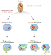
Figure 1. Supersite transplants.
Select locations of an antigen may comprise supersites of vulnerability, each of which is comprised of the overlapping epitopes of effective neutralizing antibodies that arise commonly in multiple donors as a result of the immune response to natural infection. One such site, surrounded by a dotted outline with epitopes shown as colored circles, is depicted at top. Supersite transplantation involves the placement of the supersite into a protein scaffold capable of preserving recognition to broadly neutralizing antibodies, elicited in multiple donors (right). By contrast, epitope scaffolds involve the transplantation of an epitope targetted by a single rare or subdominant antibody (left).
Figure 2
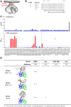
Figure 2. MPER-supersite transplants.
Supersite transplants were designed to maintain the 10E8 epitope in its antibody bound conformation. (A) Location of MPER supersite on HIV-1 viral spike. (B) Sequence alignment of MPER sequences from HIV-1 clades A, B, and C, covering residues 656–683. Boxes shown represent subregions of the MPER supersite that were transplanted. (C) 96-well expression and 10E8 antibody binding of 42 site transplants. 2F5-ES2 was used as expression control. (D) Superposition of 3 best site transplants (grey) with the 10E8 MPER epitope (red), and the variable domains of 10E8 (heavy chain: blue; light chain: green). The full epitope (residues 659–683) is shown for MPER-ST02 and MPER-ST05, whereas predominantly the turn and C-terminal helix (residues 664–683) for MPER-ST07, for which the full N-terminal helix is not matched. Reactivity of the site transplants with antibodies from different donors is shown.
Figure 3
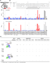
Figure 3. V1V2-supersite transplants.
Supersite transplants were designed to maintain the PG9-interacting strands B and C in the antibody bound conformation. Four design ideas were explored for transplanting the B and C strands: matching (i) strands B and C (residues 154–177) to small structurally characterized peptides; (ii) strands B and C to surface-exposed β-hairpin regions; (iii) the entire Greek key motif (residues 126–196) comprising four strands to surface-exposed patches; and (iv) extending the base of the B and C strands (A) V1V2-supersite location on viral spike. (B) Sequence alignment of the V1V2 domain from three clades and the transplanted strain ZM109. Regions of V1V2 used for transplant design are marked below. (right) The location of the supersite on the viral spike is displayed in green. (C) (top) 96-well expression of 67 V1V2 transplants and Ni-Sensor Octet quantitation of expression levels. Four full V1/V2 domain transplants: 1JO8_CAP45, 1JO8_ZM109, 1FD6_CAP45 and 1FD6_ZM109 (grey) and a monomeric gp120 (black) were used as expression controls. (bottom) PG9 ELISA of 67 V1V2 transplants. 10 of the PG9-epitope scaffolds showed substantial interaction with PG9 (red). (D) V1V2-supersite transplants which showed significant PG9 binding were tested for binding to additional V1V2-directed antibodies with two displaying weak binding to antibodies from different donors.
Figure 4
Figure 4. Glycan V3-supersite transplants.
Supersite transplants were designed to maintain the mini-V3 in its PG128-bound conformation. (A) Location of glycan V3 supersite on HIV-1 viral spike. (B) Sequence alignment of the V3 loop from clades A, B and C with potential glycosylations positions marked above and V3 region-structural elements marked below. The transplanted glycan V3-supersite sequence was shown with the mini-V3 highlighted in red. (C) (top) 96-well expression of 20 glycan V3-supersite transplants and Ni-Sensor Octet quantitation of expression levels. 2F5-ES2 was used as expression control. (bottom) ELISA of PGT128 antibody binding of the expressed glycan V3 transplants. 11 of the 20 glycan V3 transplants showed substantial interaction with PGT128 (red). (D) Glycan V3-supersite transplants which showed significant PGT128 binding were tested for binding to additional glycan V3-directed antibodies from different donors.
Figure 5
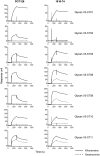
Figure 5. Glycosylation of glycan V3-supersite transplants and antibody reactivity.
SPR sensorgrams showed binding of PGT128 (donor 39) and 10–1074 (donor 39) to the 7 glycan V3 transplants expressed in 293F cells in the presence of glycosylation inhibitors kifunensine (solid line) or swainsonine (dashed line).
Figure 6
Figure 6. Contribution of the mini-V3 region to the epitopes of antibodies targeting the glycan V3 supersite.
(A) (left) HIV-1 viral spike with mini-V3 region shown in red. (middle and right) Different views of binding modes for glycan V3-targeting antibodies PGT122, PGT128 and PGT135. (B) Surface representation of HIV-1 gp120 with glycans involved in antibody binding shown in sticks. The antibody epitope from the mini-V3 region were colored red and epitope from non-glycan V3 regions were colored cyan. (C) Sequence alignment of the glycan V3 region with antibody interacting residues highlighted in red. The mini-V3 was boxed and the residue numbers above marked the potential glycosylation sites. (D) Comparison of epitope surface area contributed by the glycan V3 region and other regions for antibodies PGT122, PGT128 and PGT135. (E) Percentage of epitope contribution by transplanted glycan V3 site correlated with the reactivity of the glycan V3-supersite transplants to somatic variants of template antibodies.
Figure 7
Figure 7. Nanoparticle presentation enhances affinity but not diversity of antigenic recognition of glycan V3-supersite transplants.
(A) SPR sensorgrams showing the binding to template antibodies PGT128 and 10–1074 for glycan V3 ST11 in its native and ferritin nanoparticle forms. (B) Antigenicity profile of glycan V3 ST08-11 in their ferritin nanoparticle forms.

References

    1. Klein F, Mouquet H, Dosenovic P, Scheid JF, Scharf L, et al. (2013) Antibodies in HIV-1 vaccine development and therapy. Science 341: 1199–1204. - PMC - PubMed
    1. Burton DR, Desrosiers RC, Doms RW, Koff WC, Kwong PD, et al. (2004) HIV vaccine design and the neutralizing antibody problem. Nat Immunol 5: 233–236. - PubMed
    1. Starcich BR, Hahn BH, Shaw GM, McNeely PD, Modrow S, et al. (1986) Identification and characterization of conserved and variable regions in the envelope gene of HTLV-III/LAV, the retrovirus of AIDS. Cell 45: 637–648. - PubMed
    1. Wyatt R, Kwong PD, Desjardins E, Sweet RW, Robinson J, et al. (1998) The antigenic structure of the HIV gp120 envelope glycoprotein. Nature 393: 705–711. - PubMed
    1. Kwong PD, Doyle ML, Casper DJ, Cicala C, Leavitt SA, et al. (2002) HIV-1 evades antibody-mediated neutralization through conformational masking of receptor-binding sites. Nature 420: 678–682. - PubMed

Publication types

MeSH terms

Substances