. 2014 Aug 6;32(36):4672-80.
doi: 10.1016/j.vaccine.2014.06.002. Epub 2014 Jun 30.

Evaluation of a multisubunit recombinant polymorphic membrane protein and major outer membrane protein T cell vaccine against Chlamydia muridarum genital infection in three strains of mice

Affiliations

Evaluation of a multisubunit recombinant polymorphic membrane protein and major outer membrane protein T cell vaccine against Chlamydia muridarum genital infection in three strains of mice

Hong Yu et al. Vaccine. .

Abstract

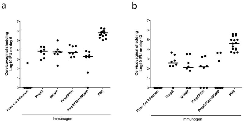
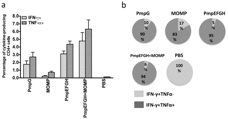
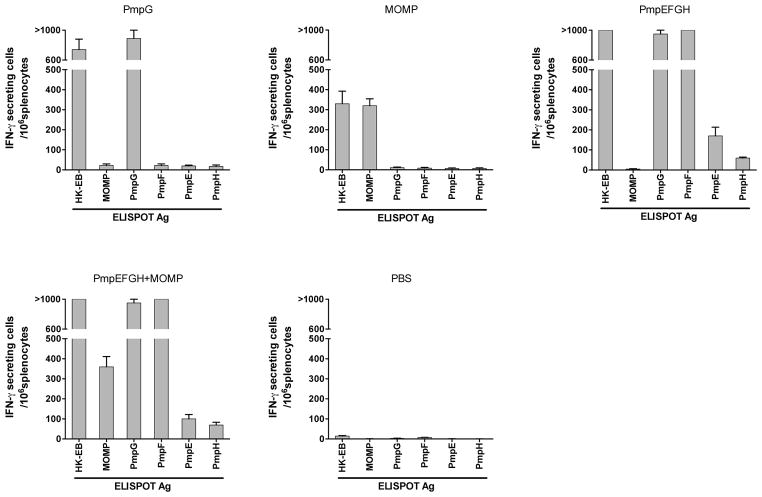
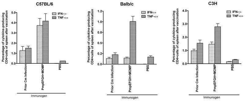
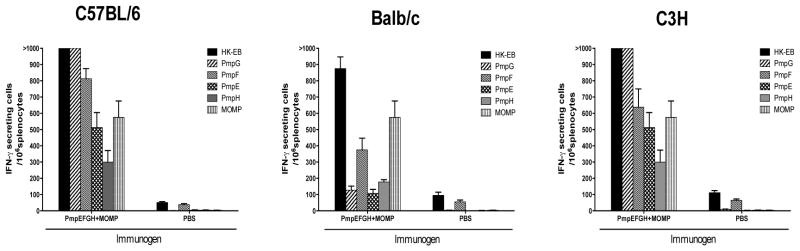
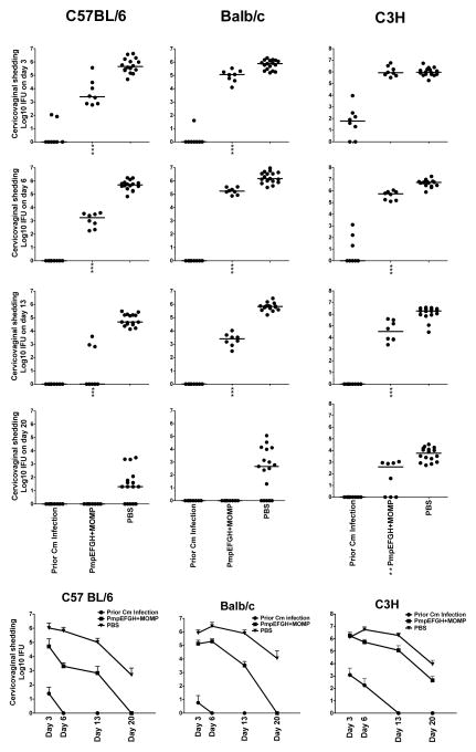
An efficacious vaccine is needed to control Chlamydia trachomatis infection. In the murine model of Chlamydia muridarum genital infection, multifunctional mucosal CD4 T cells are the foundation for protective immunity, with antibody playing a secondary role. We previously identified four Chlamydia outer membrane proteins (PmpE, PmpF, PmpG and PmpH) as CD4 T cell vaccine candidates using a dendritic cell-based immunoproteomic approach. We also demonstrated that these four polymorphic membrane proteins (Pmps) individually conferred protection as measured by accelerated clearance of Chlamydia infection in the C57BL/6 murine genital tract model. The major outer membrane protein, MOMP is also a well-studied protective vaccine antigen in this system. In the current study, we tested immunogenicity and protection of a multisubunit recombinant protein vaccine consisting of the four Pmps (PmpEFGH) with or without the major outer membrane protein (MOMP) formulated with a Th1 polarizing adjuvant in C57BL/6, Balb/c and C3H mice. We found that C57BL/6 mice vaccinated with PmpEFGH+MOMP elicited more robust cellular immune responses than mice immunized with individual protein antigens. Pmps elicited more variable cellular immune responses than MOMP among the three strains of mice. The combination vaccine accelerated clearance in the three strains of mice although at different rates. We conclude that the recombinant outer membrane protein combination constitutes a promising first generation Chlamydia vaccine construct that should provide broad immunogenicity in an outbred population.

Keywords: Chlamydia; Immunoproteomics; Recombinant protein; T cell antigens; Vaccine.


Figures

Figure 1
Figure 1
Vaccine elicited protection against C. muridarum genital tract infection in C57BL/6 mice after immunization with different vaccine formulations in DDA/MPL adjuvant. Four weeks after the final immunization, mice were challenged intravaginally with 1,500 IFU of C. muridarum. Cervicovaginal washes were taken at day 6 (a), day 13 (b) post infection, and bacterial shedding was measured by inclusion counts on HeLa 229 cells. Mice immunized with PBS were used as a negative control, and mice previously infected with 1,500 IFU of live C. muridarum intravaginally were used as a positive control. Horizontal bar represents median value for each group. All vaccine groups significantly reduced Chlamydia shedding compared to the PBS group P value <0.001.
Figure 2
Figure 2
C. muridarum -specific cytokine responses after immunization with different vaccine formulations in C57BL/6 mice. Two weeks after the final immunization, splenocytes from different vaccine groups were harvested and stimulated with HK-EB (5×10⁵ IFU/ml). IFN-γ- or TNF-α-producing CD4 T cells were analyzed by multiparameter flow cytometry as described in Materials and Methods. (a) Percentage of IFN-γ- or TNF-α-producing CD4 T cells from each vaccine group. The results are expressed as means ± SEM for groups of six mice. (b) Fraction of IFN-γ single- and IFN-γ/TNF-α double-positive cells in total IFN-γproducing CD4 T cells. All vaccine formulations most commonly elicited double positive cytokine producing CD4 T cells.
Figure 3
Figure 3
C. muridarum antigen-specific IFN-γ responses after immunization with different vaccine formulations determined by ELISPOT assay in C57BL/6 mice. The vaccine immunogen is shown alone each figure panel. Two weeks after the final immunization, mouse splenocytes from different vaccine groups were harvested and stimulated in vitro for 20 h with 5×10⁵ IFU/ml of HK-EB or 1 μg/ml of recombinant protein as indicated as the ELISPOT Ag along the x-axis. The results are expressed as means ± SEM for groups of six mice. MOMP, PmpG and PmpF are immunodominant among the antigens tested in C57BL/6 mice.
Figure 4
Figure 4
C. muridarum -specific cytokine responses after immunization with different vaccine formulations in three different inbred strains of mice (C57BL/6, Balb/c, C3H). Two weeks after the final immunization or six weeks after live EB infection, mouse splenocytes were harvested and stimulated with HK-EB (5×10⁵ IFU/ml). IFN-γ- or TNF-α-producing CD4 T cells were analyzed by multiparameter flow cytometry. The results are expressed as means ± SEM for groups of four mice. The vaccine formulations were variably immunogenic among the three tested strains of mice.
Figure 5
Figure 5
C. muridarum individual antigen-specific IFN-γ responses determined by ELISPOT assay in C57BL/6, Balb/c and C3H mice after immunization with PmpEFGH+MOMP. Two weeks after the final immunization, mouse splenocytes were harvested and stimulated in vitro for 20 h with 5×10⁵ IFU/ml of HK-EB, or 1 μg/ml of indicated recombinant proteins. The results are expressed as means ± SEM for groups of four mice. All three mouse strains immunized with the PmpEFGH+MOMP vaccine elicited IFN-γ responses to five individual outer membrane proteins with Pmps displaying different patterns based on the genetic background and MOMP similar immunogenic in the three strains of mice.
Figure 6
Figure 6
Vaccine elicited protection against C. muridarum genital tract infection in C57BL/6, Balb/c or C3H mice immunized with different vaccine formulations. Four weeks after the final immunization or eight weeks after live EB infection, mice were challenged intravaginally with 1,500 IFU of C. muridarum. Cervicovaginal washes were taken at day 3, 6, 13 and 20 post infection and Chlamydia shedding was measured by inclusion forming units (IFU) on HeLa 229 cells. Mice immunized with PBS were used as a negative control, and mice infected with 1,500 IFU of live C. muridarum intravaginally were used as a positive control. (a) Individual Chlamydia sheddings for each group. The bar represents the median value. **, P<0.01; ***, P<0.001 vs the PBS group. (b) Infection curves for each group. The bar represents the mean Chlamydia IFU ± SD. The effect of vaccine on the rate of clearance differed among the strains of mice.

Similar articles

Cited by

References

    1. Starnbach MN, Roan NR. Conquering sexually transmitted diseases. Nat Rev Immunol. 2008;8:313–7. - PubMed
    1. Brunham RC, Pourbohloul B, Mak S, White R, Rekart ML. The unexpected impact of a Chlamydia trachomatis infection control program on susceptibility to reinfection. J Infect Dis. 2005;192:1836–44. - PubMed
    1. Grayston JT, Wang SP. The potential for vaccine against infection of the genital tract with Chlamydia trachomatis. Sex Transm Dis. 1978;5:73–7. - PubMed
    1. Wang SP, Grayston JT, Alexander ER. Trachoma vaccine studies in monkeys. Am J Ophthalmol. 1967;63(Suppl):1615–30. - PubMed
    1. Brunham RC, Rey-Ladino J. Immunology of Chlamydia infection: implications for a Chlamydia trachomatis vaccine. Nat Rev Immunol. 2005;5:149–61. - PubMed

Publication types