. 2014 Jul 3;18(4):R141.
doi: 10.1186/cc13969.

Modulation of chemokine gradients by apheresis redirects leukocyte trafficking to different compartments during sepsis, studies in a rat model

Modulation of chemokine gradients by apheresis redirects leukocyte trafficking to different compartments during sepsis, studies in a rat model

Zhi-Yong Peng et al. Crit Care. .

Abstract

Introduction: Prior work suggests that leukocyte trafficking is determined by local chemokine gradients between the nidus of infection and the plasma. We recently demonstrated that therapeutic apheresis can alter immune mediator concentrations in the plasma, protect against organ injury, and improve survival. Here we aimed to determine whether the removal of chemokines from the plasma by apheresis in experimental peritonitis changes chemokine gradients and subsequently enhances leukocyte localization into the infected compartment, and away from healthy tissues.

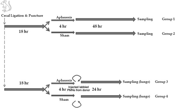
Methods: In total, 76 male adult Sprague-Dawley rats weighing 400 g to 600 g were included in this study. Eighteen hours after inducing sepsis by cecal ligation and puncture, we randomized these rats to apheresis or sham treatment for 4 hours. Cytokines, chemokines, and leukocyte counts from blood, peritoneal cavity, and lung were measured. In a separate experiment, we labeled neutrophils from septic donor animals and injected them into either apheresis or sham-treated animals. All numeric data with normal distributions were compared with one-way analysis of variance, and numeric data not normally distributed were compared with the Mann-Whitney U test.

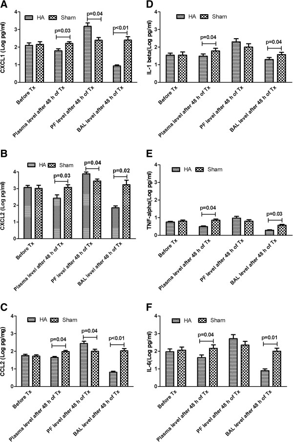
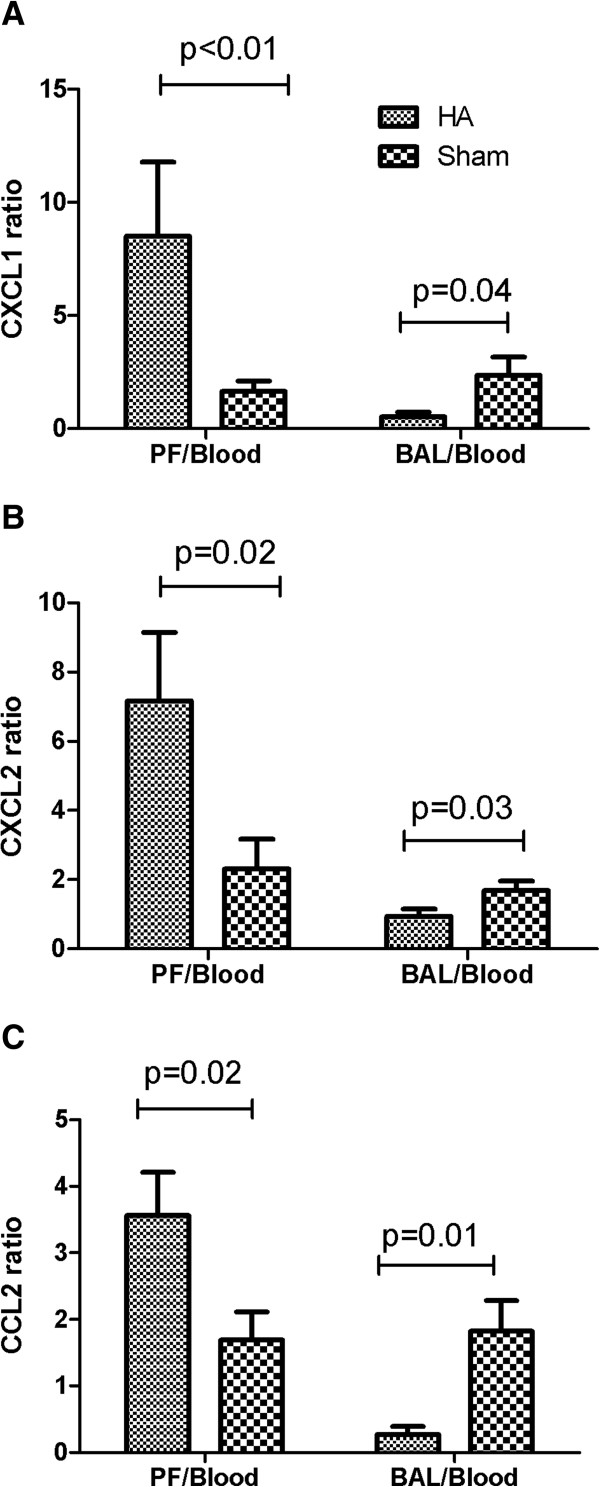
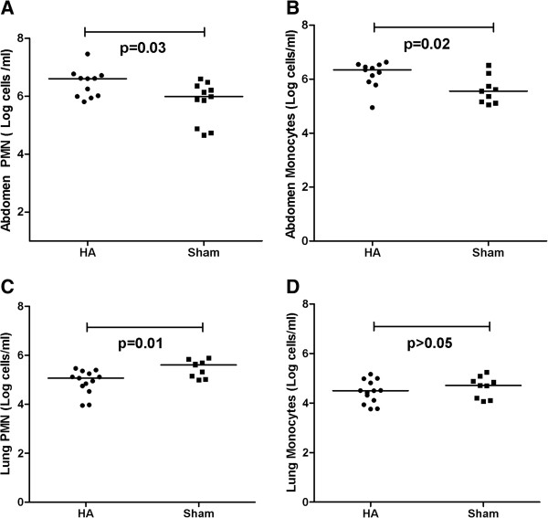
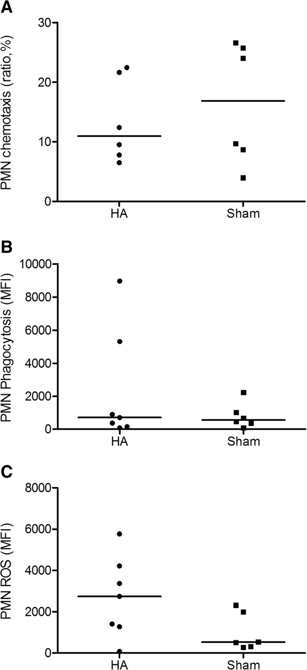
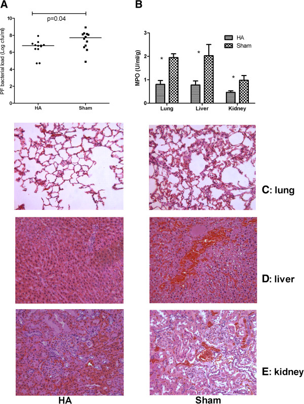
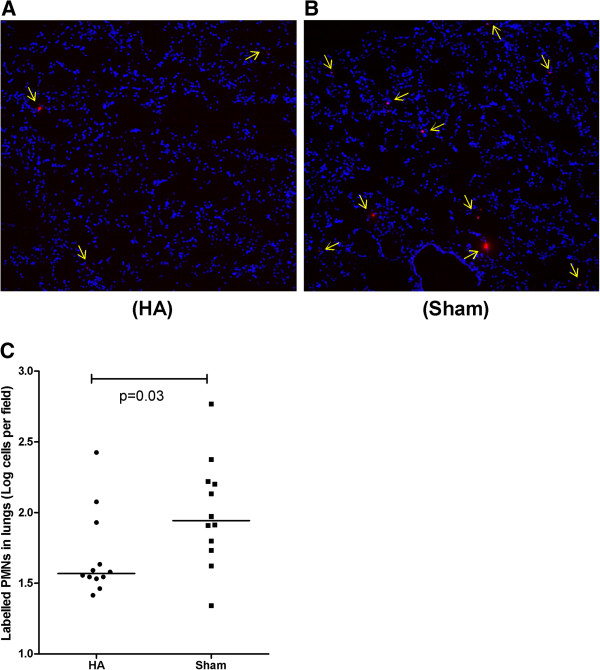
Results: Apheresis significantly removed plasma cytokines and chemokines, increased peritoneal fluid-to-blood chemokine (C-X-C motif ligand 1, ligand 2, and C-C motif ligand 2) ratios, and decreased bronchoalveolar lavage fluid-to-blood chemokine ratios, resulting in enhanced leukocyte recruitment into the peritoneal cavity and improved bacterial clearance, but decreased recruitment into the lung. Apheresis also reduced myeloperoxidase activity and histologic injury in the lung, liver, and kidney. These Labeled donor neutrophils exhibited decreased localization in the lung when infused into apheresis-treated animals.

Conclusions: Our results support the concept of chemokine gradient control of leukocyte trafficking and demonstrate the efficacy of apheresis to target this mechanism and reduce leukocyte infiltration into the lung.


Figures

Figure 1
Figure 1
Individual experimental groups. This study is composed of two experimental phases. In the first experiment, 18 hours after CLP, animals (groups 1 and 2) were randomly assigned to receive either apheresis by hemoadsorption (HA) or sham treatment for 4 hours. Animals were then killed for sampling 48 hours after treatment. In the second experiment, 18 hours after CLP, animals were randomly assigned to receive either apheresis by HA or sham treatment for 4 hours. Labeled neutrophils from a separate group of septic donor animals were injected into all the treated animals. At 24 hours after treatment, animals were then killed for measurement of labeled neutrophils in lung tissue.
Figure 2
Figure 2
Effects of apheresis on cytokines and chemokines in different compartments (data are expressed as mean ± SE after being natural log transformed, n= 14 to 18 each). Eighteen hours after CLP, animals were randomly assigned to receive either apheresis by hemoadsorption (HA) or sham treatment for 4 hours. Animals were then killed for sampling 48 hours after treatment. Shown are the comparisons among blood, peritoneal fluid (PF), and bronchoalveolar lavage fluid (BAL) after treatments. (A) CXCL1; (B) CXCL2; (C) CCL2; (D) IL-1β; (E) TNF-α; and (F) IL-6. Tx, treatment. Before Tx” is the time point when samples were obtained at 18 hours after CLP but before initiation of apheresis.
Figure 3
Figure 3
Effects of apheresis on the ratios of local to systemic chemokine concentrations (mean ± SE, n= 14 to 18 each). Eighteen hours after CLP, animals were randomly assigned to receive either apheresis by hemoadsorption (HA) or sham treatment for 4 hours. Animals were then killed for sampling 48 hours after treatment. Shown are peritoneal fluid (PF)-to-blood and bronchoalveolar lavage fluid (BAL)-to-blood ratios for (A) CXCL1; (B) CXCL2; and (C) CCL2.
Figure 4
Figure 4
Effects of apheresis on leukocyte influx into the peritoneal cavity and lung. Eighteen hours after CLP, animals were randomly assigned to receive either apheresis by HA or sham treatment for 4 hours. Animals were then killed for sampling 48 hours after treatment. Cells were washed and centrifuged for three cycles and then stained with PE-Cy5 mouse anti-rat CD45 (OX-1), PE mouse anti-rat granulocyte (RP-1) and Biotin mouse anti-rat mononuclear followed by FITC streptavidin staining. Cells were gated on CD45. CD45 cells were analyzed for positive expression of either the granulocyte or mononuclear markers. Shown are (A) neutrophils; and (B) monocytes in the peritoneal cavity; (C) neutrophils and (D) monocytes in the lung. Comparisons are between apheresis (HA) and sham. Medians and ranges of cells per milliliter (natural log transformed, n = 14 to 18 each) are shown.
Figure 5
Figure 5
Effects of apheresis on PMN function (median and ranges, n= 7). Eighteen hours after CLP, animals were randomly assigned to receive either apheresis by HA or sham treatment for 4 hours. Animals were then killed for sampling 48 hours after treatment. (A) PMN chemotaxis expressed as ratio (percentage) of cells migrating toward chemoattractant to cells migrating away from chemoattractant. (B) PMN phagocytosis expressed as mean fluorescent intensity (MFI). (C) PMN oxidative burst (ROS) expressed as mean fluorescent intensity (MFI).
Figure 6
Figure 6
Effects of apheresis on the bacterial clearance, neutrophil infiltration, and histopathology. Eighteen hours after CLP, animals were randomly assigned to receive either apheresis (HA) or sham treatment for 4 hours. Animals were then killed for sampling 48 hours after treatment. (A) Bacterial load in the peritoneal fluid (PF) (medians and ranges for colony-forming units per milliliter (natural log transformed, n = 12 to 16 each). (B) Myeloperoxidase (MPO) activity (mean ± SE, U/ml/mg; n = 20 each). *P < 0.05, apheresis (HA) versus sham in lung, liver, and kidney. (C) Lung histology (n = 4 to 6 each). Sham showed neutrophil infiltration and hemorrhage not seen with apheresis (HA). (D) Liver histology (n = 4 to 6 each). Apheresis (HA) showed milder swelling of hepatocytes and necrosis compared with sham. (E) Kidney histology (n = 4 to 6 each). Apheresis (HA) resulted in less vacuolization in tubules compared with sham.
Figure 7
Figure 7
Effects of apheresis on neutrophil influx into the lungs. Eighteen hours after CLP, animals were randomly assigned to receive either apheresis (HA) or sham treatment for 4 hours. Labeled neutrophils from septic donor animals were injected after treatment into (A) HA and (B) sham-treated animals (n = 12 each). Neutrophils in the lungs (red color, and arrows) were observed after 24 hours with immunofluorescence microscopy (20× magnifications). (C) Comparison of labeled neutrophils infiltrated in the lung between HA and sham-treated septic animals (data expressed as medians and ranges after natural log transformation). Fewer neutrophils were seen in the lungs of HA-treated animals (A) compared with sham (B).

References

    1. Alves-Filho JC, Sônego F, Soto FO, Freitas A, Verri WA, Auxiliadora-Martins M, Basile-Filho A, McKenzie AN, Xu D, Cunha FQ, Liew FY. Interleukin-33 attenuates sepsis by enhancing neutrophil influx to the site of infection. Nat Med. 2010;16:708–712. - PubMed
    1. Bouchon A, Facchetti F, Weigand MA, Colonna M. TREM-1 amplifies inflammation and is a crucial mediator of septic shock. Nature. 2001;410:1103–1107. - PubMed
    1. Guo RF, Riedemann NC, Laudes IJ, Sarma VJ, Kunkel RG, Dilley KA, Paulauskis JD, Ward PA. Altered neutrophil trafficking during sepsis. J Immunol. 2002;169:307–314. - PubMed
    1. Nathan C. Points of control in inflammation. Nature. 2002;420:846–852. - PubMed
    1. Brown KA, Brain SD, Pearson JD, Edgeworth JD, Lewis SM, Treacher DF. Neutrophils in development of multiple organ failure in sepsis. Lancet. 2006;368:157–169. - PubMed

Publication types