

Optimizing ocular vestibular evoked myogenic potential testing for superior semicircular canal dehiscence syndrome: electrode placement
- PMID: 24993062
- PMCID: PMC4310219
- DOI: 10.1159/000360124
Optimizing ocular vestibular evoked myogenic potential testing for superior semicircular canal dehiscence syndrome: electrode placement
Abstract
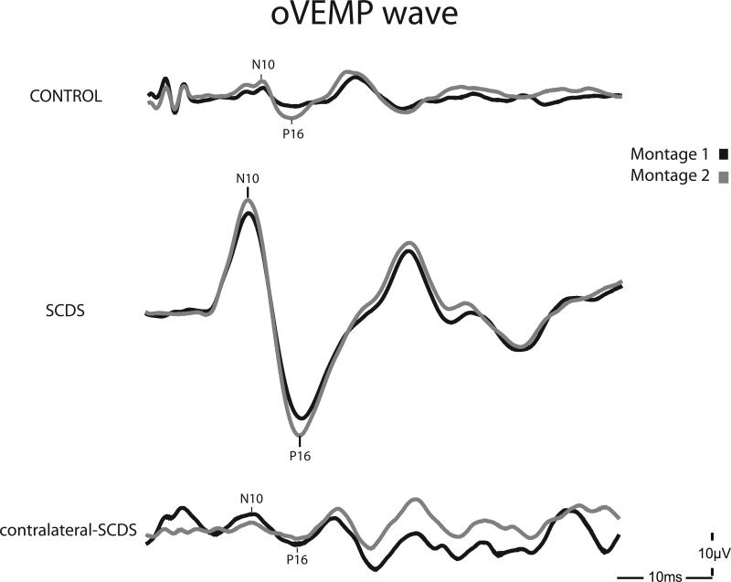
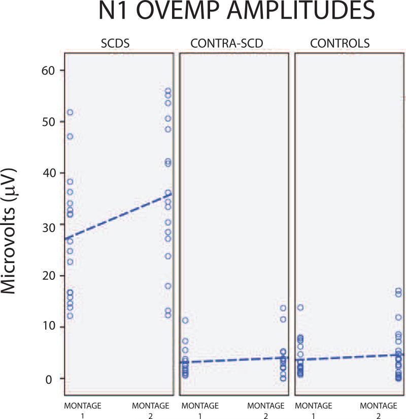
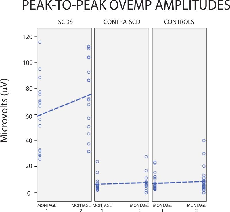
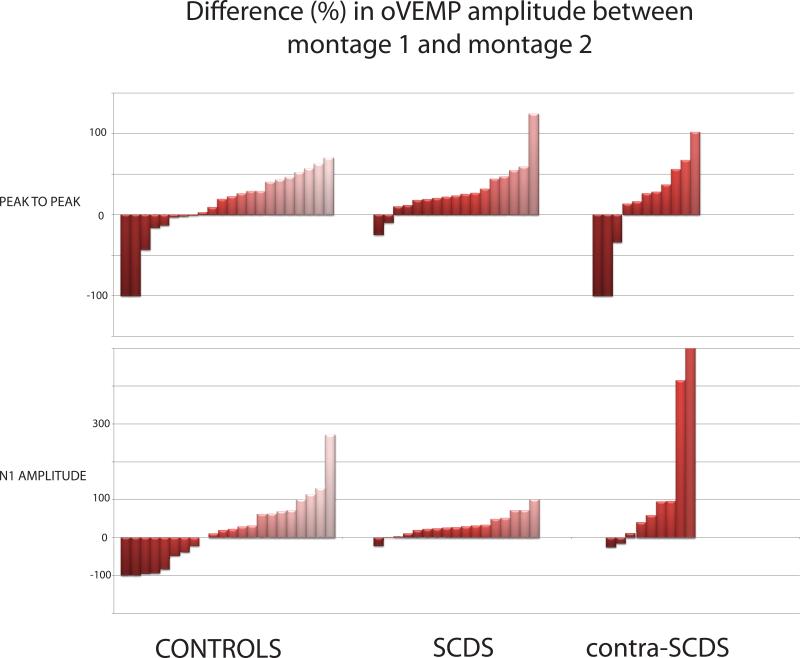
Objective: To compare the sensitivity and specificity of ocular vestibular evoked myogenic potentials (oVEMPs) using 2 electrode montages for the diagnosis of superior canal dehiscence syndrome (SCDS).
Subjects: 16 SCDS patients (17 affected-SCDS ears, 15 contralateral-SCDS ears) and 12 controls (24 ears).
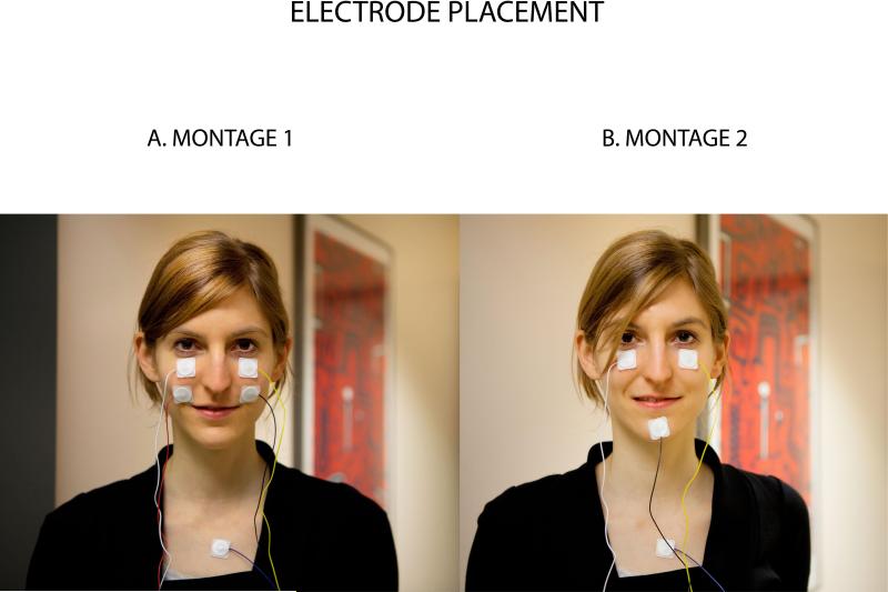
Methods: oVEMPs were recorded in response to 500-Hz tone bursts using 2 electrode montages. For both montages the active electrode was placed approximately 5 mm below each eye and a ground electrode on the sternum. For montage 1 (standard), the reference electrode was centered 2 cm below each active electrode. For montage 2, the reference electrode was placed on the chin.
Results: For either montage, the separation between oVEMP amplitudes in affected-SCDS ears and controls was significant (p < 0.001), with excellent sensitivity and specificity (>90%).
Conclusion: oVEMP recordings with the standard montage remain a reliable method for evaluation of SCDS.
Figures
Similar articles
-
A Comparison of Two Recording Montages for Ocular Vestibular Evoked Myogenic Potentials in Patients with Superior Canal Dehiscence Syndrome.J Am Acad Audiol. 2019 Apr;30(4):293-301. doi: 10.3766/jaaa.17093. Epub 2018 Feb 8. J Am Acad Audiol. 2019. PMID: 30461389
-
Development and Standardization of Modified Simultaneous Multifrequency Stimulus for Recording Ocular Vestibular-Evoked Myogenic Potential and Its Interaction with the Alternate Electrode Montages.J Am Acad Audiol. 2024 Oct;35(9-10):241-255. doi: 10.1055/a-2353-2797. Epub 2024 Jun 26. J Am Acad Audiol. 2024. PMID: 38925160
-
Reducing Sound Exposure During Ocular Vestibular Evoked Myogenic Potential Testing for Superior Semicircular Canal Dehiscence Syndrome.Otol Neurotol. 2021 Jul 1;42(6):e735-e743. doi: 10.1097/MAO.0000000000003084. Otol Neurotol. 2021. PMID: 33710145
-
Perilymphatic Fistulas and Superior Semi-Circular Canal Dehiscence Syndrome.Adv Otorhinolaryngol. 2019;82:93-100. doi: 10.1159/000490276. Epub 2019 Jan 15. Adv Otorhinolaryngol. 2019. PMID: 30947173 Review.
-
Characteristics and clinical applications of ocular vestibular evoked myogenic potentials.Hear Res. 2012 Dec;294(1-2):55-63. doi: 10.1016/j.heares.2012.10.008. Epub 2012 Oct 30. Hear Res. 2012. PMID: 23123220 Review.
Cited by
-
Optimizing Ocular Vestibular Evoked Myogenic Potentials With Narrow Band CE-Chirps.Ear Hear. 2021 Sep/Oct;42(5):1373-1380. doi: 10.1097/AUD.0000000000001031. Ear Hear. 2021. PMID: 33734171 Free PMC article.
-
Vestibular evoked myogenic potentials in practice: Methods, pitfalls and clinical applications.Clin Neurophysiol Pract. 2019 Feb 26;4:47-68. doi: 10.1016/j.cnp.2019.01.005. eCollection 2019. Clin Neurophysiol Pract. 2019. PMID: 30949613 Free PMC article. Review.
-
Air-Conducted Vestibular Evoked Myogenic Potential Testing in Children, Adolescents, and Young Adults: Thresholds, Frequency Tuning, and Effects of Sound Exposure.Ear Hear. 2019 Jan/Feb;40(1):192-203. doi: 10.1097/AUD.0000000000000607. Ear Hear. 2019. PMID: 29870520 Free PMC article.
-
On the impact of examiners on latencies and amplitudes in cervical and ocular vestibular-evoked myogenic potentials evaluated over a large sample (N = 1,038).Eur Arch Otorhinolaryngol. 2016 Feb;273(2):317-23. doi: 10.1007/s00405-015-3510-3. Epub 2015 Jan 28. Eur Arch Otorhinolaryngol. 2016. PMID: 25628238
References
-
- Belden CJ, Weg N, Minor LB, Zinreich SJ. CT evaluation of bone dehiscence of the superior semicircular canal as a cause f sound- and/or pressure-induced vertigo. Radiology. 2003;226(2):337–343. - PubMed
-
- Brantberg K, Bergenius J, Tribukait A. Vestibular-evoked myogenic potentials in patients with dehiscence of the superior semicircular canal. Acta Otolaryngol. 1999;119(6):633–640. - PubMed
-
- Curthoys IS. A critical review of the neurophysiological evidence underlying clinical vestibular testing using sound, vibration and galvanic stimuli. Clin. Neurophysiol. 2010;121(2):132–144. - PubMed
Publication types
MeSH terms
Grants and funding
LinkOut - more resources
Full Text Sources
Other Literature Sources
