. 2014;164(2):122-36.
doi: 10.1159/000363446. Epub 2014 Jun 28.

Epicutaneous immunization with TNP-Ig and Zymosan induces TCRαβ+ CD4+ contrasuppressor cells that reverse skin-induced suppression via IL-17A

Affiliations

Epicutaneous immunization with TNP-Ig and Zymosan induces TCRαβ+ CD4+ contrasuppressor cells that reverse skin-induced suppression via IL-17A

Monika Majewska-Szczepanik et al. Int Arch Allergy Immunol. 2014.

Abstract

Background: Our previous work showed that epicutaneous (EC) immunization with protein antigen e.g. TNP-conjugated mouse immunoglobulin (TNP-Ig) in the form of a patch prior to hapten sensitization inhibits Th1-mediated contact hypersensitivity (CHS) in mice. We also found that suppression of CHS was mediated by TCRαβ+ CD4+ CD8+ T suppressor cells producing TGF-β. The aim of this study was to investigate the role of innate immunity in the suppression of CHS.

Methods: Mice were immunized by applying gauze patches containing protein antigen alone or in the presence of zymosan, and were then tested for the CHS response. Adoptive cell transfer experiments were used to study the mechanisms involved in the reversal of skin-induced suppression. The influence of EC immunization on cytokine production by lymph node cells was measured by ELISA.

Results: We found that EC immunization with TNP-Ig and zymosan before trinitrophenyl chloride sensitization reverses skin-induced suppression, demonstrated in vivo and in vitro. The reversal of skin-induced suppression was transferable by antigen-specific TCRαβ+ CD4+ T contrasuppressor cells. Furthermore, we showed that the contrasuppression was IL-17A-dependent and TLR2- and MyD88-independent.

Conclusions: Our work strongly suggests that EC immunization with protein antigen and zymosan reverses skin-induced suppression and that this approach may be a potential tool to increase the immunogenicity of weakly immunogenic antigens.


Figures

Fig. 1
Fig. 1
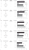
CBA/J mice were patched with PBS or 100 μg TNP-Ig alone or TNP-Ig plus 100μg of zymosan or zymosan alone on day 0 and 4. On day 7 patches were removed and mice were actively sensitized with 5% TNP-Cl. Four days later, animals were challenged on the ears with 0.4% TNP-Cl and tested for CHS 24 h later as described in Materials and Methods.
Fig. 2
Fig. 2
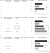
a Reversal of skin-induced suppression in vivo. CBA/J mice were EC exposed to PBS (Group A) or TNP-Ig (Group B) or TNP-Ig plus zymosan (Group C) or zymosan alone (Group D) prior to TNP-Cl sensitization. Four days later CHS response was elicited and tested 24 h later. Results shown as mean ± SE. n = 18. ***, p ≤ 0.001. b EC immunization with TNP-Ig and zymosan reverses decreased vascular permeability. CBA/J mice were patched with PBS (Group A) or TNP-Ig (Group B) or TNP-Ig plus zymosan (Group C) or zymosan alone (Group D) prior to TNP-Cl sensitization, challenge and injection of 1% Evans blue dye. One hour later mice were killed and ear punches were collected. Evans blue dye was extracted and the optical density was read at 565 nm. For details see Materials and Methods. Results shown as mean ± SE. n = 12. **, p ≤ 0.01; ***, p ≤ 0.001. c Patching with TNP-Ig and zymosan reverses decreased ear weight when compared to EC tolerized mice. Ears were removed and punch of the ear was made. The weight of ear punches was evaluated with the use of analytical scale as described in Materials and Methods. Results shown as mean ± SE. n = 9. *, p ≤ 0.05; **, p ≤ 0.01. d EC immunization with TNP-Ig and zymosan reverses inhibited MPO activity. CBA/J mice were EC treated with PBS (Group A) or TNP-Ig (Group B) TNP-Ig plus zymosan (Group C) or zymosan alone (Group D) prior to TNP-Cl sensitization followed by ear challenge. Ears were removed 24 h post challenge and punches were collected. Biopsies were homogenized and MPO activity was measured as described in Materials and Methods. MPO activity was expressed in units per protein concentration (U/mg of protein). Results shown as mean ± SE. n = 6. **, p ≤ 0.01; ***, p ≤ 0.001. e Patching with TNP-Ig and zymosan reverses IL-17A production in ear extracts. CBA/J mice were EC treated with PBS (Group A) or TNP-Ig (Group B) or TNP-Ig and zymosan (Group C) or zymosan alone (Group D) prior to TNP-Cl sensitization and challenge. 24 h post challenge ear punches were collected. Concentration of IL-17A was measured in tissue homogenates by ELISA. Results shown as mean ± SE. n = 15. *, p ≤ 0.05.
Fig. 3
Fig. 3
CBA/J mice were patched with PBS (Group A) or TNP-Ig (Group B) TNP-Ig plus zymosan (Group C) or zymosan alone (Group D) prior to TNP-Cl sensitization. Four days later inguinal and auxiliary LN were isolated and 3×10⁵ ALNC were cultured in triplicates in the presence of three fold dilutions of TNP-Ig for 48 h prior to the addition of [3H]-thymidine for an additional 18 h. [3H]-thymidine incorporation was determined by ß liquid scintillation counting. Results are presented as mean ± SE. n = 3. **, p ≤ 0.01; ***, p ≤ 0.001.
Fig. 4
Fig. 4
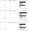
a Reversal of skin-induced suppression is transferable with ALNC – “transfer out” protocol. 7 × 10⁷ of the CHS-effector cells from TNP-Cl sensitized donors were incubated with 5×10⁷ ALNC used as a source of suppressor cells (Ts cells) isolated from mice (suppression control) (Group B) patched with TNP-Ig. After incubation the cell mixture was transferred i.v. into naïve recipients (suppression control) (Group B). Additionally, 7 × 10⁷ of the CHS-effector cells were incubated with 5×10⁷ ALNC (contrasuppressor T cells (Tcs)) from mice EC immunized with TNP-Ig plus zymosan prior to incubation with 5×10⁷ lymphoid cells from mice EC immunized with TNP-Ig (Ts) (suppression control) (Group C). Then resultant cells were transferred into naïve recipients. To determine whether ALNC from mice EC immunized with TNP-Ig and zymosan could transfer CHS, 5×10⁷ ALNC were transferred i.v. into naïve recipient mice (Group D). Recipients were subsequently ear challenged with TNP-Cl and tested for CHS at 24 h. Results shown as mean ± SE. n = 12. **, p ≤ 0.01; ***, p ≤ 0.001. b Transfer of skin-induced contrasuppression – “transfer in” protocol. CBA/J mice were EC exposed to PBS (Group A), TNP-Ig (Group B) or TNP-Ig plus zymosan (Group C). Additionally, one group of mice before patching with TNP-Ig plus zymosan received 5×10⁷ of EC induced Ts cells (Group D), whereas another group of animals was transferred with EC induced Tcs before EC immunization with TNP-Ig alone (Group E). After patch removal, mice in all experimental groups were sensitized with TNP-Cl and tested for CHS. Results shown as mean ± SE. n =10. ***, p ≤ 0.001. c EC immunization with zymosan alone does not reverse skin-induced suppression. CBA/J mice were i.v. transferred with 5× 10⁷ of ALNC isolated from donors patched with TNP-Ig plus zymosan (Group C) or zymosan alone (Group D) and recipient mice were subsequently patched with TNP-Ig. Control groups were EC exposed to PBS (Group A) or TNP-Ig (Group B) for one week. After patch removal all tested mice were sensitized with TNP-Cl and tested for CHS. Results shown as mean ± SE. n = 10. **, p ≤ 0.01; ***, p ≤ 0.001.
Fig. 5
Fig. 5
a TCRαβ+ lymphocytes are involved in skin-induced contrasuppression. ALNC from animals patched with TNP-Ig plus zymosan were treated with anti-TCRβ mAb and RC (Group D) or RC alone (Group C). Resultant cells were i.v. injected into recipients that were EC immunized with TNP-Ig prior to CHS induction. Animals in control groups were patched with PBS (Group A) or TNP-Ig (Group B) prior to TNP-Cl sensitization. Four days post sensitization mice were challenged and tested for CHS. Results shown as mean ± SE. n = 16. p ≤ 0.001. b CD4+ lymphocytes are required to reverse skin-induced suppression. ALNC from mice patched with TNP-Ig and zymosan were treated with anti-CD4 or anti-CD8 mAb and RC (Groups D and E respectively). In contrasuppression control, ALNC were treated with RC alone (Group C). Resultant cells were i.v. injected into recipients that were EC immunized with TNP-Ig prior CHS induction. Control mice were patched with PBS alone (Group A) or TNP-Ig (Group B) before TNP-Cl painting. Then mice were challenged and tested for CHS. Results shown as mean ± SE. n = 10. *, p ≤ 0.05, ***, p ≤ 0.001. c Contrasuppressor cells belong to the population of TCRαβ+ CD4+ lymphocytes. ALNC from donors patched with TNP-Ig and zymosan were used to obtain MACS purified TCRαβ+ CD4+ cells (Group D). Contarsuppression control contained whole ALNC (Group C). Then, 5×10⁷ ALNC or 2.1×10⁷ MACS purified TCRαβ+ CD4+ cells were i.v. injected into recipients that were EC immunized with TNP-Ig prior to CHS induction. Mice in control groups were patched with PBS (Group A) or TNP-Ig (Group B) prior to TNP-Cl sensitization. Four days post sensitization mice were challenged and tested for CHS. Results shown as mean ± SE. n = 14. ***, p ≤ 0.001.
Fig. 6
Fig. 6
CBA/J mice were EC immunized with TNP-Ig (Group B) or TNP-Ig+zymosan (Group C) or OX-Ig (Group D) OX-Ig+zymosan (Group E) prior to TNP-CL sensitization. Positive control group contained mice patched with PBS and then sensitized with TNP-Cl (Group A). Four days post TNP-Cl sensitization animals were tested for CHS. Results shown as mean ± SE. n = 16. ***, p ≤ 0.001.
Fig. 7
Fig. 7
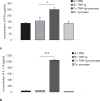
CBA/J mice were patched with PBS or TNP-Ig or TNP-Ig + zymosan or zymosan alone (Groups A-D respectively). One week later ALN were collected and then ALNC (3 × 10⁶) isolated from tested groups were cultured in the presence of 100 μg/ml TNP-Ig. After 48 h supernatants were collected and tested for cytokine concentration. a IFN-γ production. Results shown as mean ± SE. n = 4. *, p ≤ 0.05. b IL-17A production. Results shown as mean ± SE. n = 4. ***p ≤ 0.001.
Fig. 8
Fig. 8
a Mechanism of skin-induced contrasuppression (“transfer out” protocol). 5×10⁷ ALNC isolated from control C57Bl/6 (Group C) or IL-17A-/-B6 (Group D) or TLR2-/-B6 (Group E) or MyD88-/-B6 (Group F) mice EC treated with TNP-Ig and zymosan were incubated with 7×10⁷ TNP-specific CHS effector cells and then with 5×10⁷ EC induced Ts cells. Positive control recipients were transferred with immune cells alone (Group A) whereas suppression control group received immune cells co-transferred with EC induced Ts cells (Group B). The recipients were challenged and tested for CHS. Results shown as mean ± SE. n = 7. *, p ≤ 0.05; ***, p ≤ 0.001. b Reversal of skin-induced suppression is TLR2 independent (active immunization protocol). TLR2-/-B6 mice were patched with PBS or TNP-Ig or TNP-Ig plus zymosan (Groups A-C respectively) as described in Materials and Methods. Seven days later mice were TNP-Cl sensitized and then tested for CHS. Results shown as mean ± SE. n = 10. **, p ≤ 0.01. c Contrasuppression induced via EC immunization with TNP-Ig and zymosan is IL-17A dependent (active immunization protocol). IL-17A-/-B6 mice were patched with PBS or TNP-Ig or TNP-Ig plus zymosan or TNP-Ig and LPS (Groups A–D respectively) as described in Materials and Methods. Seven days later mice were sensitized with TNP-Cl and tested for CHS. Results shown as mean ± SE. n = 8. **, p≤ 0.01; ***, p ≤ 0.001. d, e C57Bl/6 or TLR2-/-B6 mice were patched with PBS or TNP-Ig or TNP-Ig plus zymosan (Groups A-C respectively). Seven days later ALN were isolated and ALNC (3 × 10⁶) were cultured in the presence of 100 μg/ml TNP-Ig. After 48 h supernatants were tested for IL-17A concentration. Results shown as mean ± SE. n = 4. ***, p ≤ 0.001.
Figure 9
Figure 9. EC immunization with TNP-Ig and curdlan reverses skin-induced suppression
a Reversal of skin-induced suppression in vivo. CBA/J mice were patched with PBS (Group A) or TNP-Ig (Group B) or TNP-Ig plus zymosan (Group C) or zymosan alone (Group D) or TNP-Ig plus curdlan (Group E) or curdlan alone (Group F) prior to TNP-Cl sensitization. Four days later CHS response was elicited and tested 24 h later. Results shown as mean ± SE. n = 18. **, p ≤ 0.01; ***, p ≤ 0.001. b EC immunization with TNP-Ig and curdlan reverses skin-induced suppression in vitro. CBA/J mice were EC treated with PBS (Group A) or TNP-Ig (Group B) or TNP-Ig plus curdlan (Group C) or curdlan alone (Group D) prior to TNP-Cl sensitization. Four days later ALNC were isolated and 3×10⁵ ALNC were cultured in triplicates in the presence of three fold dilutions of TNP-Ig for 48 h and then for additional 18 h with [3H]-thymidine. [3H]-thymidine incorporation was determined by ß liquid scintillation counting. Results are presented as mean ± SE. n = 3. *, p ≤ 0.05; **, p ≤ 0.01.

Similar articles

Cited by

References

    1. Askenase PW, Szczepanik M, Itakura A, Kiener C, Campos RA. Extravascular T-cell recruitment requires initiation begun by Vα14+ invariant NK T cells and B-1 cells. Trends Immunol. 2004;25:441–449. - PubMed
    1. Szczepanik M, Bryniarski K, Tutaj M, Ptak M, Skrzeczynska J, Askenase PW, Ptak W. Epicutaneous immunization induces αβ T-cell receptor CD4 CD8 double-positive non-specific suppressor T cells that inhibit contact sensitivity via transforming growth factor-β. Immunology. 2005;115:42–54. - PMC - PubMed
    1. Ptak W, Szczepanik M, Bryniarski K, Tutaj M, Ptak M. Epicutaneous application of protein antigens incorporated into cosmetic cream induces antigen-nonspecific unresponsiveness in mice and affects the cell-mediated immune response. Int Arch Allergy Immunol. 2002;128:8–14. - PubMed
    1. Majewska-Szczepanik M, Zemelka-Wiącek M, Ptak W, Wen L, Szczepanik M. Epicutaneous immunization with DNP-BSA induces CD4+ CD25+ Treg cells that inhibit Tc1-mediated CS. Immunol Cell Biol. 2012;90:784–795. - PubMed
    1. Zemelka-Wiącek M, Majewska-Szczepanik M, Ptak W, Szczepanik M. Epicutaneous immunization with protein antigen induces antigen-non-specific suppression of CD8 T cell mediated contact sensitivity. Pharmacol Rep. 2012;64:1485–1496. - PubMed

Publication types

MeSH terms