. 2014 Aug 6;34(4):e00129.
doi: 10.1042/BSR20140079.

The N-terminal portion of autoinhibitory element modulates human endothelial nitric-oxide synthase activity through coordinated controls of phosphorylation at Thr495 and Ser1177

Affiliations

The N-terminal portion of autoinhibitory element modulates human endothelial nitric-oxide synthase activity through coordinated controls of phosphorylation at Thr495 and Ser1177

Pei-Rung Wu et al. Biosci Rep. .

Abstract

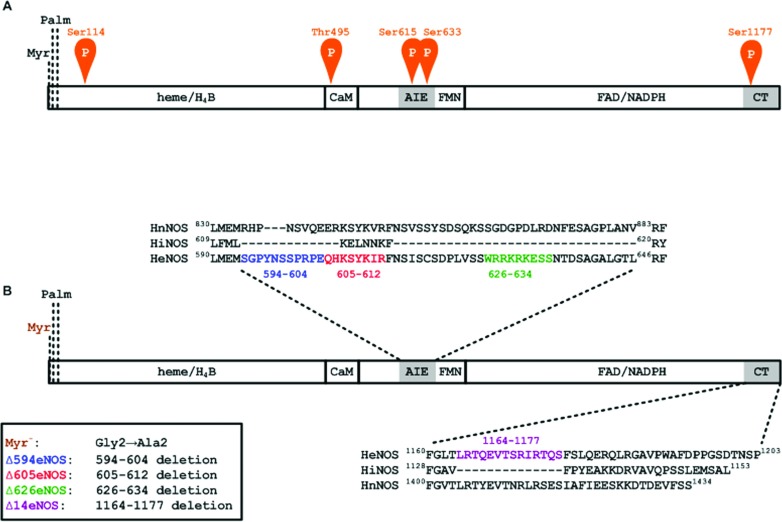
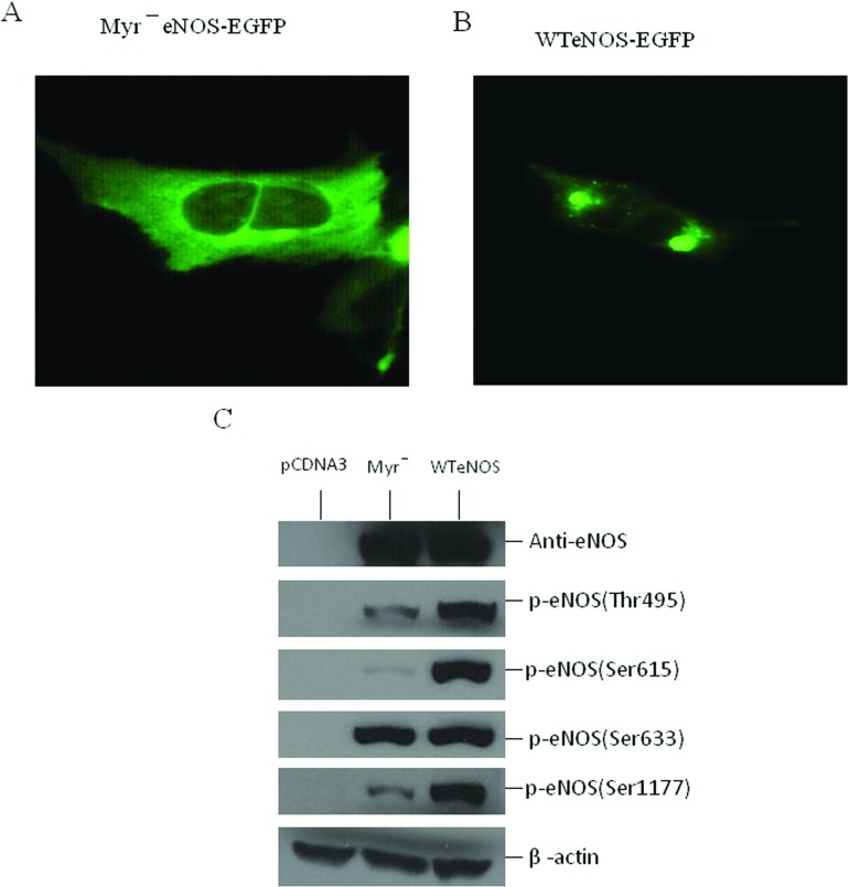
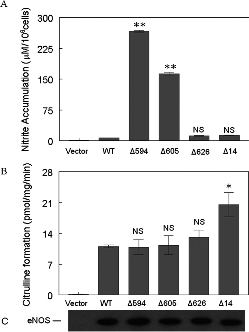
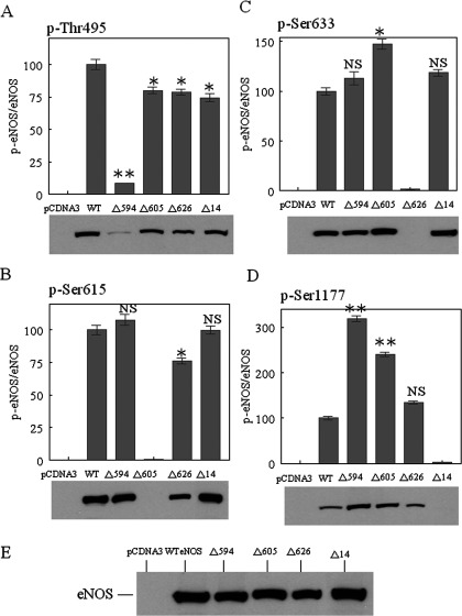
NO production catalysed by eNOS (endothelial nitric-oxide synthase) plays an important role in the cardiovascular system. A variety of agonists activate eNOS through the Ser1177 phosphorylation concomitant with Thr495 dephosphorylation, resulting in increased ·NO production with a basal level of calcium. To date, the underlying mechanism remains unclear. We have previously demonstrated that perturbation of the AIE (autoinhibitory element) in the FMN-binding subdomain can also lead to eNOS activation with a basal level of calcium, implying that the AIE might regulate eNOS activation through modulating phosphorylation at Thr495 and Ser1177. Here we generated stable clones in HEK-293 (human embryonic kidney 293) cells with a series of deletion mutants in both the AIE (Δ594-604, Δ605-612 and Δ626-634) and the C-terminal tail (Δ14; deletion of 1164-1177). The expression of Δ594-604 and Δ605-612 mutants in non-stimulated HEK-293 cells substantially increased nitrate/nitrite release into the culture medium; the other two mutants, Δ626-634 and Δ1164-1177, displayed no significant difference when compared with WTeNOS (wild-type eNOS). Intriguingly, mutant Δ594-604 showed close correlation between Ser1177 phosphorylation and Thr495 dephosphorylation, and NO production. Our results have indicated that N-terminal portion of AIE (residues 594-604) regulates eNOS activity through coordinated phosphorylation on Ser1177 and Thr495.


Figures

Figure 1
Figure 1. heNOS and the deletion constructs
(A) The relative positions of phosphorylation sites in heNOS. Blocks indicate the binding sites for haeme, H4B, CaM, FMN, FAD and NADPH in eNOS. AIE and CT refer to the AIE and the C-terminal tail, respectively. The myristoylation site (Myr.) and palmitoylation sites (Palm.) are noted. Potential phosphorylation sites Ser114, Thr495, Ser615, Ser633 and Ser1177 are indicated. (B) Constructs for the eNOS deletion mutants. The sequences of the AIE and the C-terminal tail are shown above and below, respectively. The Δ594 is a deletion of the AIE region encompassing residues Ser594–Glu604 (shown in blue); Δ605 is a deletion of the AIE region encompassing residues Gln605–Arg612 (shown in red); Δ626 is a deletion of the AIE region encompassing residues Trp626–Ser634 (shown in green); Δ14 is a deletion of 14 amino acid residues at the C-terminal tail encompassing residues Leu1164–Ser1177 (shown in violet).
Figure 2
Figure 2. Relationship between the subcellular localization and phosphorylation state of eNOS
Stable HEK-293 cell lines with (A) myreNOS-EGFP or (B) WTeNOS–EGFP were visualized in live cells by fluorescence microscopy. (C) cell lysates from HEK-293 cells transfected with empty vector (pCDNA3), MyreNOS or WTeNOS were immunoblotted for changes in phosphorylation state of Thr495, Ser615, Ser633 and Ser1177. The expression of eNOS and β-actin was used as internal loading control. Western blots shown are the representatives of at least three independent studies.
Figure 3
Figure 3. Enzymatic activities of various eNOS—HEK-293 stable clones
(A) Nitrate/nitrite accumulation in HEK-293 cell culture medium 48 h after plating. The nitrate/nitrite accumulation in culture medium was determined using a colorimetric assay kit obtained from Cayman Chemical Co. (B) L-citrulline formation activity was determined in the cell lysates of various eNOS–HEK-293 stable clones. Reactions were carried out as described in the ‘Materials and Methods’ section. (C) The eNOS–HEK-293 cell lysates were immunoblotted with an antibody specific to total eNOS to confirm the equal expression levels of each eNOS construct. Data shown are the mean±S.D. (*P<0.05; **P<0.01; NS, no significant difference versus WT eNOS). Each experiment was performed in triplicate and repeated three times.
Figure 4
Figure 4. The effects of deletion of the AIE and the C-terminal tail on eNOS phosphorylation
The cell lysates from various eNOS–HEK-293 stable clones, including empty pCDNA3.1(+), WT eNOS (WT), Δ594–604 (Δ594), Δ605–612 (Δ605) and Δ1164–1177 (Δ14) were immunoblotted with antibodies specific to phosphosites at (A) p-Thr495, (B) p-Ser615, (C) p-Ser633 and (D) p-Ser1177. (E) An antibody specific to total eNOS was used to confirm equal expression levels for the various eNOS constructs. The blots shown are representative of at least three experiments. Densitometry was used to quantify the phosphorylated eNOS relative to total eNOS of respective constructs. Data are presented as the percentage of WTeNOS and represent means±S.D. (*P<0.05; **P<0.01; NS, no significant difference versus WT eNOS).
Figure 5
Figure 5. Interaction of synthetic CBD peptides with calcium/CaM
(A) Peptide CBD sequences (residues 491–510, based on heNOS) with unmodified T495 (T495), phosphonull A substitution (A495, red colour), phosphomimetic substitution (D495, blue colour) and phosphorylated T495 (pT495, brown colour) are indicated. (B) The synthetic CBD variants were incubated with CaM (200 pmol) by increasing peptide:CaM molar ratios in the presence of 100 μM CaCl2 before electrophoresis. The samples were analysed on 18% non-denaturing gels and visualized with Coomassie Brilliant Blue R-250. Representative Coomassie Brilliant Blue-stained gels of samples containing CaM and increasing molar ratios of T495, A495, D495 and pT495 are shown. The first lane in each gel contains CaM only, i.e., CBD/CaM ratio is 0. The rest CBD/CaM ratios were 0.1:1 (2nd lane), 0.2:1 (3rd lane), 0.4:1 (4th lane), 0.8:1 (5th lane), 1.6:1 (6th lane), 3.2:1 (7th lane), 6.4:1 (8th lane) and 12.8:1 (9th lane). CBD–CaM complexes and free CaM are denoted. (C) The relative amount of CaM on the gel at each peptide concentration (I) was determined by densitometry and normalized to CaM in the absence of peptide (I0). The data are plotted as the mean I/I0±S.D. (n=4) versus peptide:CaM ratio. The graph shows CaM with increasing peptide concentration described by the following symbols: ■, T495; ▲, A495; ●, D495 and ▼, pT495.
Figure 6
Figure 6. Evaluation of inhibitory potency of CBD variants on eNOS activity
Modified or unmodified CBD peptides were assessed for their ability to inhibit (A) citrulline formation activity and (B) Cytochrome c reduction. CR denotes a complete reaction mixture; CR-CaM denotes omission of CaM from the complete reaction mixture; CR+T495 indicates the addition of 10 μM of unmodified CBD (T495) to the complete reaction mixture; CR+A495 denotes the addition of 10 μM of phosphonull CBD (A495) to the complete reaction mixture; CR+D495 indicates the addition of 10 μM of phosphomimetic CBD (D495) to the complete reaction mixture; CR+pT495 denotes the addition of 10 μM of phosphorylated CBD (pT495) to the complete reaction mixture. The complete reaction mixture for L-[3H]citrulline formation contains 25 mM Tris, pH 7.5, 100 mM NaCl, 0.5 μM CaM, 0.2 mM EDTA, 0.3 mM CaCl2, 100 μM β-NADPH, 10 μM H4B, 20 μM L-arginine, 1 μCi of L-[3H]arginine, 100 nM eNOS. The complete reaction mixture for cytochrome c reduction contains 25 mM Tris–HCl, pH 7.5, 100 mM NaCl, 10% glycerol, 50 μM cytochrome c, 0.5 μM CaM, 100 μM CaCl2 and 100 μM β-NADPH. The catalytic activity was normalized to give percentages relative to the reaction rate in the absence of each peptide. Each bar represents mean±S.D. of triplicate experiments. Under these conditions, the activities for eNOS bound to CaM were 12±2/min for citrulline formation, and 295±15/min for cytochrome c reduction. Data are presented as means±S.D.*denotes P<0.05, ** P<0.01, NS, not significant difference as compared with that in the absence of synthetic CBD peptides. Each experiment was performed in triplicate and repeated three times.
Figure 7
Figure 7. T495 phosphorylation and eNOS function
(A) Structure of CaM and its complex with eNOS CBD (PDB accession code 1NIW). The backbone ribbons of the CBD (sky blue) and of CaM (grey) are shown. The T495 and E498 in eNOS CBD are labelled in yellow, and the neighbouring glutamate residues located on CaM in the vicinity of Thr495 and Glu498 including E7, E11, E14 and E127 are labelled in green. (B) Close-up view showing residues surrounding the T495 phosphorylation site. The residues as ball-and-stick representation are coloured by atom type (nitrogen, blue; oxygen, red). The hydrogen bond between Thr495OG1 and Glu498 amide N is indicated by the violet coil. The selected distances between residues are measured from the structure (1NIW) and indicated in dash lines: Thr(495)Oγ-Glu(498)Cβ, 3.38 Å; Thr(495)Oγ-Glu(7)Oε, 6.53 Å; Thr(495)Oγ-Glu(127)Oε, 4.67 Å; Glu(498)Cβ-Glu(7)Oε,7.05Å; Glu(498)Cβ-Glu(11)Oε,4.94Å; Glu(498)Cβ-Glu(14) Oε, 6.59Å.

Similar articles

Cited by

References

    1. Dudzinski D. M., Michel T. Life history of eNOS: partners and pathways. Cardiovasc. Res. 2007;75:247–260. doi: 10.1016/j.cardiores.2007.03.023. - DOI - PMC - PubMed
    1. Pollock J. S., Förstermann U., Mitchell J. A., Warner T. D., Schmidt H. H., Nakane M., Murad F. Purification and characterization of particulate endothelium-derived relaxing factor synthase from cultured and native bovine aortic endothelial cells. Proc. Natl. Acad. Sci. U. S. A. 1991;88:10480–10484. doi: 10.1073/pnas.88.23.10480. - DOI - PMC - PubMed
    1. Bredt D. S., Hwang P. M., Glatt C. E., Lowenstein C., Reed R. R., Snyder S. H. Cloned and expressed nitric oxide synthase structurally resembles cytochrome P-450 reductase. Nature. 1991;351:714–718. doi: 10.1038/351714a0. - DOI - PubMed
    1. Xie Q. W., Cho H. J., Calaycay J., Mumford R. A., Swiderek K. M., Elliston K., Mumford R., Nathan C. Cloning and characterization of inducible nitric oxide synthase from mouse macrophages. Science. 1992;256:225–228. doi: 10.1126/science.1373522. - DOI - PubMed
    1. McMillan K., Bredt D. S., Hirsch D. J., Snyder S. H., Clark J. E., Masters B. S. Cloned, expressed rat cerebellar nitric oxide synthase contains stoichiometric amounts of heme, which binds carbon monoxide. Proc. Natl. Acad. Sci. U. S. A. 1992;89:11141–11145. doi: 10.1073/pnas.89.23.11141. - DOI - PMC - PubMed

Publication types