Review
. 2014 Jul 3;4(10):a013631.
doi: 10.1101/cshperspect.a013631.

Markers of epidermal stem cell subpopulations in adult mammalian skin

Affiliations
Review

Markers of epidermal stem cell subpopulations in adult mammalian skin

Kai Kretzschmar et al. Cold Spring Harb Perspect Med. .

Abstract

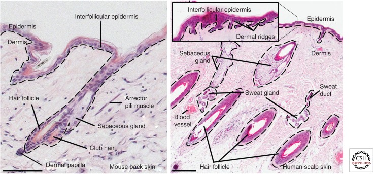
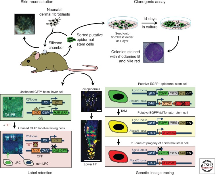
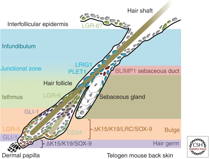
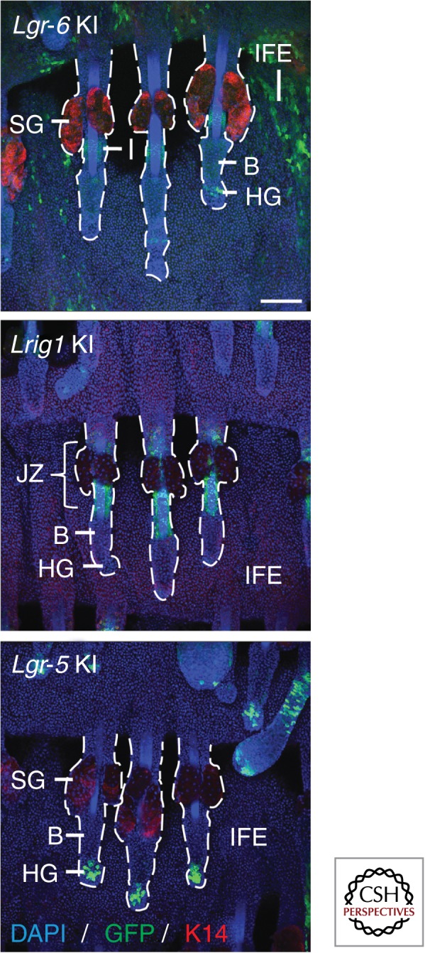
The epidermis is the outermost layer of mammalian skin and comprises a multilayered epithelium, the interfollicular epidermis, with associated hair follicles, sebaceous glands, and eccrine sweat glands. As in other epithelia, adult stem cells within the epidermis maintain tissue homeostasis and contribute to repair of tissue damage. The bulge of hair follicles, where DNA-label-retaining cells reside, was traditionally regarded as the sole epidermal stem cell compartment. However, in recent years multiple stem cell populations have been identified. In this review, we discuss the different stem cell compartments of adult murine and human epidermis, the markers that they express, and the assays that are used to characterize epidermal stem cell properties.


Figures

Figure 1.
Figure 1.
Histology of mammalian skin. Adult mouse (left) and human skin (right) stained with hematoxylin. Note the absence of eccrine sweat glands in mouse back skin. Dashed lines indicate position of the basement membrane. Scale bars, 100 µm (mouse skin) and 500 µm (human skin).
Figure 2.
Figure 2.
Strategies to study epidermal stem cells and their markers. Disaggregated epidermal cells can be either mixed with neonatal murine dermal fibroblasts and grafted onto immunocompromised mice to study their skin reconstitution potential in vivo or they can be seeded onto a feeder cell layer to study their clonogenic potential in culture. Slowly cycling cells in vivo can be identified through DNA label retention, either by injecting nucleotide labels such as 5-bromo-2-deoxyuridine (BrdU) or by using genetic approaches such as the tetracycline-regulated H2B-GFP system. (Images based on data from Mascre et al. 2012; reproduced, with permission, from C. Blanpain and Nature © 2012, Macmillan.) Genetic lineage tracing enables fate mapping of epidermal stem cells and their progeny during tissue homeostasis. CAG, chicken β-actin promoter with CMV enhancer; CMV, cytomegalovirus promoter; EGFP, enhanced GFP; ER, tamoxifen-inducible mutated estrogen receptor; GFP, green fluorescent protein; HF, hair follicle; H2B, histone H2B; IFE, interfollicular epidermis; IRES, internal ribosome entry site; K, keratin; LRC, label-retaining cell; TAM, tamoxifen; TET, tetracycline; tetO, tetracycline operator; tTA, tetracycline transactivator. Scale bars, 100 µm.
Figure 3.
Figure 3.
Markers of epidermal stem cell subpopulations in murine adult skin. Schematic of epidermal stem cell pools in murine telogen (hair follicle resting phase) back skin.
Figure 4.
Figure 4.
Genetically engineered mice carrying a bicistronic expression cassette containing a fluorescent reporter (such as green fluorescent protein) and a tamoxifen-inducible Cre recombinase under the control of an epidermal stem cell pool-specific promoter—such as the Lgr-6, Lrig1, and Lgr-5 knock-in mice—enable sorting of the stem cells and lineage tracing of their progeny. Whole mounts of murine tail epidermis stained for GFP (stem cell marker), K14 (basal layer marker) and DAPI (nuclei marker) are shown. B, bulge; DAPI, 4′,6-diamidino-2-phenylindole; GFP, green fluorescent protein; HG, hair germ; IFE, interfollicular epidermis; JZ, junctional zone; K14, keratin 14; KI, knock-in; SG, sebaceous gland. Scale bars, 100 µm.

References

    1. Alonso L, Fuchs E. 2006. The hair cycle. J Cell Sci 119: 391–393 - PubMed
    1. Arwert EN, Hoste E, Watt FM. 2012. Epithelial stem cells, wound healing and cancer. Nat Rev Cancer 12: 170–180 - PubMed
    1. Baker CM, Verstuyf A, Jensen KB, Watt FM. 2010. Differential sensitivity of epidermal cell subpopulations to β-catenin-induced ectopic hair follicle formation. Dev Biol 343: 40–50 - PMC - PubMed
    1. Barker N, van Es JH, Kuipers J, Kujala P, van den Born M, Cozijnsen M, Haegebarth A, Korving J, Begthel H, Peters PJ, et al. 2007. Identification of stem cells in small intestine and colon by marker gene Lgr5. Nature 449: 1003–1007 - PubMed
    1. Barker N, Huch M, Kujala P, van de Wetering M, Snippert HJ, van Es JH, Sato T, Stange DE, Begthel H, van den Born M, et al. 2010. Lgr5+ve stem cells drive self-renewal in the stomach and build long-lived gastric units in vitro. Cell Stem Cell 6: 25–36 - PubMed

Publication types