. 2014 Sep;32(3):1281-90.
doi: 10.3892/or.2014.3309. Epub 2014 Jul 3.

Long non-coding RNA urothelial carcinoma associated 1 induces cell replication by inhibiting BRG1 in 5637 cells

Affiliations

Long non-coding RNA urothelial carcinoma associated 1 induces cell replication by inhibiting BRG1 in 5637 cells

Xiujuan Wang et al. Oncol Rep. 2014 Sep.

Abstract

Long non-coding RNA urothelial carcinoma associated 1 (UCA1) was first identified in bladder cancer tissues. High expression of UCA1 in bladder cancer has suggested it may serve as a potential diagnostic molecular marker for bladder cancer. Subsequent research in bladder cancer cell lines showed that UCA1 can promote cell proliferation, but the underlying mechanism remains unknown. In the present study, we identified BRG1 as a UCA1 binding partner using an in vitro RNA pull-down assay, and RNA-binding protein immunoprecipitation assay confirmed UCA1-BRG binding in bladder cancer cells in vivo. BRG1 is a chromatin remodeling factor with reported tumor suppressor activities that directly upregulates levels of the p21 cell cycle inhibitor by binding sequences in the p21 promoter. Depletion of UCA1 by RNAi resulted in upregulated p21 levels and inhibition of cell replication, while overexpressed UCA1 reduced p21 protein and promoted cell growth. Notably, UCA1 downregulation of p21 and induction of cell proliferation antagonized the function of BRG1. UCA1 highly expressed tissue samples are often with BRG1 high expression. Furthermore, we found that UCA1 impairs both binding of BRG1 to the p21 promoter and chromatin remodeling activity of BRG1. Collectively, these results demonstrate that UCA1 promotes bladder cancer cell proliferation by inhibiting BRG1.


Figures
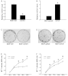
Figure 1
Figure 1
UCA1 promotes proliferation of 5637 cells. (A) Establishment of UCA1 knockdown 5637 cells. The 5637 cells were infected with pll3.7-NC (control) and pll3.7-iUCA1 viruses and selected with G418 for a week to generate 5637-NC and 5637-iUCA1 cells, respectively. Total RNA was isolated and the level of UCA1 was determined using real-time PCR. Data were normalized to GAPDH and expressed as the means ± SD of three independent experiments. *P<0.05 (t-test). (B) Establishment of UCA1 overexpressing 5637 cells. The 5637 cells were infected with pZsG or pZsG-UCA1 viruses and selected with puromycin for 7 days to generate 5637-pZsG and 5637-UCA1 cells, respectively. Total RNA was isolated and the level of UCA1 was determined using real-time PCR. Data are expressed as the means ± SD of three independent experiments. *P<0.05 (t-test). (C) Colony formation of 5637-NC and 5637-iUCA1 cells. The cells were cultured for 14 days. Colonies were stained using crystal violet. (D) Colony formation of 5637-pZsG and 5637-UCA1 cells. The cells were cultured for 14 days. Colonies were stained using crystal violet. (E) Growth curve of 5637-iUCA1 cells. Cellular proliferation was measured using MTT assays at 24, 48, 72, 96 and 120 h. The differences in data of 72, 96 and 120 h were significant, *P<0.05 (t-test). (F) Growth curves of 5637-UCA1 cells. Cellular proliferation was measured using MTT assays at 24, 48, 72, 96 and 120 h. The differences in data of 48, 72, 96 and 120 h were significant, *P<0.05 (t-test). UCA1, urothelial carcinoma associated 1.
Figure 2
Figure 2
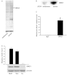
UCA1 binds to BRG1 in vitro and in vivo. (A) Electrophoresis of proteins bound to biotin-labeled sense and antisense UCA1 in NuPAGE 4–12% Bis-Tris gel. The biotin-labeled sense and antisense UCA1 were transcribed in vitro and incubated with HeLa cell lysate. The arrow shows the protein band for BRG1. (B) Western blot analysis of biotin-labeled sense and antisense UCA1-bound proteins using antibody against BRG1. (C) UCA1 binding BRG1 in 5637 cells was detected by RNA-binding protein immunoprecipitation assay. Antibody against BRG1 and mouse IgG (as a negative control) were used to pull-down RNAs in 5637 cells. The level of UCA1 was determined using real-time PCR. Data were normalized to input and are expressed as the means ± SD of three independent experiments *P<0.05 (t-test). (D) Expression profile analysis of three human bladder cancer cell lines. (Top) Total RNA was isolated and the level of UCA1 was determined using real-time PCR. GAPDH was used as an internal control. (Bottom) BRG1 was determined by western blotting. β-actin was used as an internal control. UCA1, urothelial carcinoma associated 1.
Figure 3
Figure 3
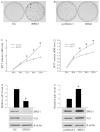
BRG1 plays a tumor suppressor role. (A) Colony formation of 5637-NC and 5637-iBRG1 cells. The cells were cultured for 14 days. Colonies were stained using crystal violet. (B) Colony formation of 5637-pcDNA3.1 and 5637-BRG1 cells. The cells were cultured for 14 days. Colonies were stained using crystal violet. (C) Growth curves of 5637 cells after transfection with BRG1 RNAi. Cellular proliferation was measured using MTT assay at 24, 48, 72, 96 and 120 h. The differences in data of 72, 96 and 120 h were significant, *P<0.05 (t-test). (D) Growth curves of 5637 cells overexpressing BRG1. Cellular proliferation was measured using MTT assay at 24, 48, 72, 96 and 120 h. The differences in data of 72, 96 and 120 h were significant, *P<0.05 (t-test). (E) The expression level of p21 in 5637-NC and 5637-iBRG1 cells was determined by real-time PCR. Data were normalized to GAPDH and are expressed as the means ± SD of three independent experiments, *P<0.05 (t-test). Western blot analysis of BRG1 and P21 in 5637-NC and 5637-iBRG1 cells. β-actin served as the internal control. (F) The expression level of p21 in 5637-pcDNA3.1 and 5637-BRG1 cells was determined by real-time PCR. Data were normalized to GAPDH and are expressed as the means ± SD of three independent experiments, *P<0.05 (t-test). Western blot analysis of BRG1 and p21 in 5637-pcDNA3.1 and 5637-BRG1 cells. β-actin served as the internal control.
Figure 4
Figure 4
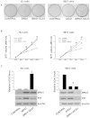
UCA1 antagonizes the tumor suppressing function of BRG1. (A) (Left) Colony formation of EJ control, EJ-BRG1 (overexpressed BRG1) and EJ-BRG1-UCA1 (overexpressed BRG1 and UCA1) cells. The cells were cultured with G418 for 10 days. Colonies were stained using crystal violet. (Right) Colony formation of 5637 control, 5637-iUCA1 (UCA1 knocked down) and 5637-iUCA1-iBRG1 (UCA1 and BRG1 both knocked down) cells. The cells were cultured with G418 and puromycin for 10 days. Colonies were stained using crystal violet. (B) (Left) Growth curves of EJ control, EJ-BRG1 and EJ-BRG1-UCA1 cells. Cellular proliferation was measured using MTT assays at 24, 48, 72, 96 and 120 h. The differences between EJ-BRG1 and EJ-BRG1-UCA1 were significant. *P<0.05 (t-test). (Right) Growth curves of 5637 control, 5637-iUCA1 and 5637-iUCA1-iBRG1 cells. Cellular proliferation was measured using MTT assays at 24, 48, 72, 96 and 120 h. The differences between 5637-iUCA1 and 5637-iUCA1-iBRG1, 5637-iUCA1 and control were significant *P<0.05 (t-test). (C) (Left) The expression levels of UCA1 in EJ cells control EJ-BRG1 and EJ-BRG1-UCA1 were determined by real-time PCR. Data were normalized to GAPDH and are expressed as the means ± SD of three independent experiments, the UCA1 level in EJ-BRG1-UCA1 was significantly high, *P<0.05 (t-test). The expression levels of BRG1 and p21 were determined by western blotting with β-actin as the internal control. (Right) The expression levels of UCA1 in 5637 cells control 5637-iUCA1 and 5637-iUCA1-iBRG1 were determined by real-time PCR. Data were normalized to GAPDH and are expressed as the means ± SD of three independent experiments. The effect of RNAi was significant *P<0.05 (t-test). The expression levels of BRG1 and p21 were determined by western blotting with β-actin as the internal control. UCA1, urothelial carcinoma associated 1.
Figure 5
Figure 5
UCA1 blocks recruitment of BRG1 to chromatin. (A) UCA1 does not affect the ATPase activity of BRG1. The kinetics of BRG1-induced ATP hydrolysis were analyzed in the presence or absence of UCA1. (B) ChIP analysis of BRG1 binding to the p21 promoter in 5637-iUCA1. 5637-NC cells were used as the control. Genomic DNA was fixed and immunoprecipitated using anti-BRG1 antibody, with IgG as a negative control. Real-time PCR was performed using a primer set specific to the BRG1-binding site of p21 promoter. Data were normalized to input and are expressed as the means ± SD of three independent experiments. *P<0.05 (t-test). (C) Micrococcal nuclease assay of 5637-NC, 5637-iUCA1. Same amounts of DNA were digested with micrococcal nuclease and electrophoresed. The image shows that nuclease digestion produced a laddering pattern. It is evident that DNA from 5637-iUCA1 is more sensitive to nuclease digestion. (D) Western blotting to detect histone proteins H3K4m3, H3K9m3 in 5637-NC, 5637-iUCA1. (E) ChIP analysis of BRG1 binding to the p21 promoter in EJ-BRG1, EJ-BRG1-UCA1. Genomic DNA was fixed and immunoprecipitated using anti-BRG1 antibody, with IgG as a negative control. Real-time PCR was performed using a primer set specific to the BRG1-binding site of p21 promoter. Data were normalized to input and are expressed as the means ± SD of three independent experiments. Overexpression of UCA1 in EJ-BRG1 led to decreased occupancy of p21 promoter by BRG1, *P<0.05 (t-test). UCA1, urothelial carcinoma associated 1.
Figure 6
Figure 6
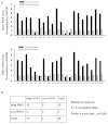
UCA1 expression correlates positively with BRG1 expression in bladder cancer tissue samples. (A) Real-time PCR was used to analyze UCA1 and BRG1 mRNA levels in the same samples. S, sample. (B) The status of UCA1 and BRG1 expression in bladder cancer specimens, >2-fold was identified as high expression. The 2×2 correlation table and Fisher’s exact test were used. The two-sided value P=0.009 was considered significant. UCA1, urothelial carcinoma associated 1.

Similar articles

Cited by

References

    1. ENCODE Project Consortium. Birney E, Stamatoyannopoulos JA, et al. Identification and analysis of functional elements in 1% of the human genome by the ENCODE pilot project. Nature. 2007;447:799–816. - PMC - PubMed
    1. Khachane AN, Harrison PM. Mining mammalian transcript data for functional long non-coding RNAs. PLoS One. 2010;5:e10316. - PMC - PubMed
    1. Tsang WP, Wong TW, Cheung AH, Co CN, Kwok TT. Induction of drug resistance and transformation in human cancer cells by the noncoding RNA CUDR. RNA. 2007;13:890–898. - PMC - PubMed
    1. Wilusz JE, Sunwoo H, Spector DL. Long noncoding RNAs: functional surprises from the RNA world. Genes Dev. 2009;23:1494–1504. - PMC - PubMed
    1. Wang KC, Chang HY. Molecular mechanisms of long noncoding RNAs. Mol Cell. 2011;43:904–914. - PMC - PubMed

Publication types

MeSH terms