. 2014 Aug 22;289(34):23520-33.
doi: 10.1074/jbc.M114.590638. Epub 2014 Jul 3.

Functional analysis of dishevelled-3 phosphorylation identifies distinct mechanisms driven by casein kinase 1ϵ and frizzled5

Affiliations

Functional analysis of dishevelled-3 phosphorylation identifies distinct mechanisms driven by casein kinase 1ϵ and frizzled5

Ondřej Bernatík et al. J Biol Chem. .

Abstract

Dishevelled-3 (Dvl3), a key component of the Wnt signaling pathways, acts downstream of Frizzled (Fzd) receptors and gets heavily phosphorylated in response to pathway activation by Wnt ligands. Casein kinase 1ϵ (CK1ϵ) was identified as the major kinase responsible for Wnt-induced Dvl3 phosphorylation. Currently it is not clear which Dvl residues are phosphorylated and what is the consequence of individual phosphorylation events. In the present study we employed mass spectrometry to analyze in a comprehensive way the phosphorylation of human Dvl3 induced by CK1ϵ. Our analysis revealed >50 phosphorylation sites on Dvl3; only a minority of these sites was found dynamically induced after co-expression of CK1ϵ, and surprisingly, phosphorylation of one cluster of modified residues was down-regulated. Dynamically phosphorylated sites were analyzed functionally. Mutations within PDZ domain (S280A and S311A) reduced the ability of Dvl3 to activate TCF/LEF (T-cell factor/lymphoid enhancer factor)-driven transcription and induce secondary axis in Xenopus embryos. In contrast, mutations of clustered Ser/Thr in the Dvl3 C terminus prevented ability of CK1ϵ to induce electrophoretic mobility shift of Dvl3 and its even subcellular localization. Surprisingly, mobility shift and subcellular localization changes induced by Fzd5, a Wnt receptor, were in all these mutants indistinguishable from wild type Dvl3. In summary, our data on the molecular level (i) support previous the assumption that CK1ϵ acts via phosphorylation of distinct residues as the activator as well as the shut-off signal of Wnt/β-catenin signaling and (ii) suggest that CK1ϵ acts on Dvl via different mechanism than Fzd5.

Keywords: Casein Kinase 1ϵ; Cell Signaling; Dishevelled-3; Frizzled5; Mass Spectrometry (MS); Phosphorylation; Post-translational Modification (PTM); Wnt Pathway.


Figures

FIGURE 1.
FIGURE 1.
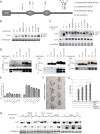
Co-expression of CK1ϵ with FLAG-Dvl3 retards electrophoretic migration and induces phosphorylation-dependent shift of Dvl (PS-Dvl3) (A) and induces TCF/LEF-dependent transcription as shown by TopFlash assay. Samples were analyzed by one-way analysis of variance followed by Tukey post tests. ***, p < 0.001, n = 3 (B). C, representative example of Coomassie Brilliant Blue R-stained gel used for MS/MS analysis. See the details “Experimental Procedures.”
FIGURE 2.
FIGURE 2.
A, Dvl3 phosphorylation (summary of MS/MS data). Position and dynamics of detected phosphorylations is indicated by color coded lines: green, less frequent in CK1ϵ induced samples; blue, constitutive; orange, more often in CK1ϵ-induced samples; red, only in CK1ϵ-induced samples. The yellow/violet lines indicate hDvl3 protein coverage in the MS/MS analysis (yellow, significant identification; violet, insignificant identification). C1, C2, C3, C4, Ser-280, and Ser-311 labels indicate the position of residues mutated in the study. B, secondary structure of Dvl3 as predicted by PONDR-FIT software (upper panel). Structured regions are indicated by values <0.5 and unstructured by values >0.5. Accuracy of the prediction is confirmed by the identification of DIX, PDZ, and DEP domains as regions with secondary structure. In silico mutation of all identified phosphorylated Ser/Thr sites in the basic region and C terminus for Asp did not lead to the massive changes in the secondary structure (lower panel).
FIGURE 3.
FIGURE 3.
Phospho-preventing mutations of Ser-280/Ser-311 in the PDZ domain blocks activation of the Wnt/β-catenin pathway. A–C, HEK 293t cells were transfected with the indicated Dvl3 constructs, TopFlash reporter plasmid, plasmid encoding Renilla luciferase as an internal control, and CK1ϵ. A, mutations of Ser-280 and Ser-311 prevent efficient activation of Wnt/β-catenin by Dvl3. B, the function of Ser-280 is conserved in Dvl1 and Dvl2. Dvl1-S282A and Dvl2-S298A, corresponding to Dvl3 S280A, activated the Wnt/β-catenin pathway less efficiently than WT Dvl1 and Dvl2, respectively. C, in CK1ϵ-treated samples no significant differences between the WT Dvl3 and the mutant Dvl3 were detected. Samples were analyzed by one-way analysis of variance followed by Tukey post tests. *, p < 0.05; ***, p < 0.001; number of experiments ≥4. D, ventral overexpression of XDvl3 induced partial secondary body axes in Xenopus embryos. This phenotype reflects activation of the Wnt/β-catenin pathway in vivo. Images of one normal embryo and one embryo with a secondary axis are shown in larger magnification in addition to representative sets of embryos injected as indicated. The secondary body axes are indicated by arrows. E, the graph shows the average percentage of embryos that developed a secondary body axis from at least three independent experiments; error bars indicate the standard error, and asterisks indicate statistically significant deviation compared with overexpression of Dvl3-WT (t test, p > 0.99). F, interaction sites of Idax (in yellow), Fzd7 (in blue), and Tmem88 (in green) proteins determined for mouse Dvl1 PDZ domain (PDB ID 1MC7). Phylogenetically conserved Ser-280 and Ser-311 phosphorylation sites in human Dvl3 PDZ are shown in red. G, left panel, schematic representation of secondary structure of mouse Dvl1 PDZ domain. Central panel, modeled electrostatic surface potential of mDvl1 PDZ. Right panel, electrostatic surface potential of mDvl1 PDZ bearing phosphomimicking mutations at positions corresponding to Ser-280 and Ser-311 of human Dvl3 PDZ. Phosphorylation sites are highlighted in magenta (magenta arrow). Calculations of electrostatic surface potential were performed in UCSF Chimera software.
FIGURE 4.
FIGURE 4.
Functional analysis of phosphorylation sites in clusters C1–C4. A, representation of Dvl structure with the positions of mutated residues. B and C, HEK293t cells were transfected with the indicated plasmids, and the electrophoretic migration of Dvl3 in the absence (B) and presence (C) of CK1ϵ was analyzed by Western blotting with anti-FLAG antibody. Mutations in C2, C2+3, and C2+3+4 blocked PS-Dvl3 induction by CK1ϵ (C). The protein expression levels of individual mutants were comparable. D, the ability of individual Dvl3 mutants to interact with CK1ϵ was tested by immunoprecipitation (IP) of CK1ϵ and FLAG-Dvl3 and subsequent analysis by Western blotting (total cell lysate (TCL)). E, HEK293t cells were transfected with the indicated plasmids, and the ability to activate TCF/LEF-dependent transcription was assessed by TopFlash system. None of the C1, C2, C2+3, C2+3+4 mutants showed any statistically significant difference. Statistical differences were analyzed by one-way analysis of variance followed by Tukey post tests. Number of experiments n ≥ 3. F and G, XDvl3 S625A/S628A/S631A (XDvl3 C2 S-A) induced secondary body axes in Xenopus embryos at the same rate as XDvl3 WT. F, the image shows a representative set of embryos injected as indicated. G, the graph summarizes the average percentage of embryos that developed a secondary body axis from at least three independent experiments; error bars indicate the standard error. n.s., not significant. H, HEK293t cells were transfected with the indicated plasmids, and the electrophoretic mobility of Dvl3 after V5-Fzd5 coexpression was analyzed by Western blotting. With the exception of Dvl3-K435M, electrophoretic mobility of all Dvl3 mutants was indistinguishable from wild type.
FIGURE 5.
FIGURE 5.
Analysis of subcellular localization of individual Dvl pools. A, representation of typical distribution patterns of Dvl3. (i), punctate; (ii), even, (iii), membrane. B, HEK293t cells were transfected with corresponding plasmids and fixed, and Dvl3 was stained with anti-FLAG antibody. Distribution pattern of Dvl3 was analyzed in at least 100 cells. CK1ϵ is unable to promote even localization of Dvl3 in C2, C3, and C4 S-A mutants, whereas V5-Fzd5 changes the intracellular distribution of Dvl3 to membranous in all tested constructs with the exception of Dvl3K435M, which served as a positive control. C, HEK293t cells were transfected with corresponding plasmids and treated as indicated with 10 μm CK1δ/ϵ inhibitor PF-670462. Inhibition of CK1δ/ϵ activity resulted in faster migration of FLAG-hDvl3 in control and CK1ϵ-induced conditions. In conditions with FLAG-hDvl3 coexpressed with V5-Fzd5 and dominant negative (DN) CK1ϵ-P3, acceleration of FLAG-hDvl3 electrophoretic mobility was observed only in mutant K435M. This finding confirms that CK1ϵ is and K435 is not dispensable for V5-Fzd5-induced phosphorylation of FLAG-hDvl3. D, phospho-Ser-643-Dvl3 was detected using anti-Ser(P)-643-Dvl3-specific antibody by Western blotting. Total Dvl3 was detected by anti-FLAG antibody. Anti-Ser(P)-643-Dvl3-specific antibody does not detect Dvl3 C2+C3 S-A mutant, but it strongly recognizes Dvl3 co-expressed with CK1ϵ. E, inhibition of endogenous CK1ϵ blocks signal detected by anti-Ser(P)-643-Dvl3 specific antibody. Decline in signal intensity was confirmed by Western blot quantification. CK1ϵ overexpression serves as a positive control. F, phospho-Ser-643-Dvl3 was detected by immunocytochemistry in HEK293t cells transfected with the indicated combination of plasmids. The signal of anti-Ser(P)-643-Dvl3 antibody (red) is negligible for Dvl3 expressed either alone or in combination with Fzd5 but strong for evenly distributed Dvl3 after CK1ϵ co-expression. Total Dvl3 detected by anti-FLAG antibody is shown in green. All confocal images were acquired using the same laser/detector settings and subsequently quantified using ImageJ software. Graphs show the overlap of fluorescence intensity peaks of individual channels along profiles indicated in the merged micrographs by a white line. A.U., arbitrary units.

References

    1. Clevers H. (2006) Wnt/β-catenin signaling in development and disease. Cell 127, 469–480 - PubMed
    1. MacDonald B. T., Tamai K., He X. (2009) Wnt/β-catenin signaling: components, mechanisms, and diseases. Dev. Cell 17, 9–26 - PMC - PubMed
    1. Schwarz-Romond T., Fiedler M., Shibata N., Butler P. J., Kikuchi A., Higuchi Y., Bienz M. (2007) The DIX domain of Dishevelled confers Wnt signaling by dynamic polymerization. Nat. Struct. Mol. Biol. 14, 484–492 - PubMed
    1. Gao C., Chen Y. G. (2010) Dishevelled: The hub of Wnt signaling. Cell. Signal. 22, 717–727 - PubMed
    1. Axelrod J. D., Miller J. R., Shulman J. M., Moon R. T., Perrimon N. (1998) Differential recruitment of Dishevelled provides signaling specificity in the planar cell polarity and Wingless signaling pathways. Genes Dev. 12, 2610–2622 - PMC - PubMed

Publication types

MeSH terms

Associated data

LinkOut - more resources