

Defining the structure and receptor binding domain of the leaderless bacteriocin LsbB
- PMID: 24993828
- PMCID: PMC4156037
- DOI: 10.1074/jbc.M114.579698
Defining the structure and receptor binding domain of the leaderless bacteriocin LsbB
Abstract
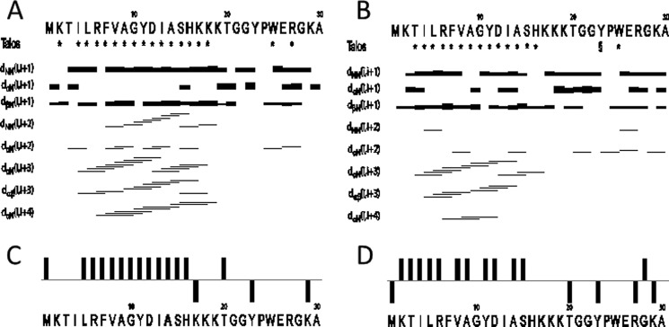
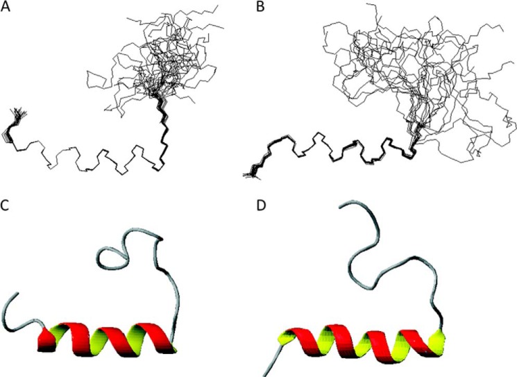
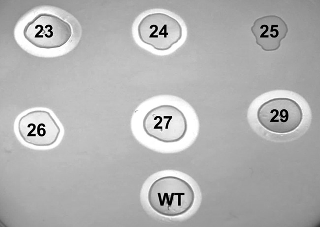
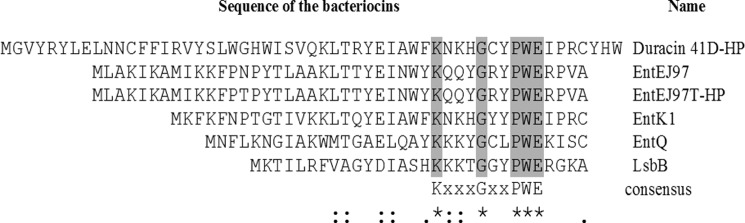
LsbB is a class II leaderless lactococcal bacteriocin of 30 amino acids. In the present work, the structure and function relationship of LsbB was assessed. Structure determination by NMR spectroscopy showed that LsbB has an N-terminal α-helix, whereas the C-terminal of the molecule remains unstructured. To define the receptor binding domain of LsbB, a competition assay was performed in which a systematic collection of truncated peptides of various lengths covering different parts of LsbB was used to inhibit the antimicrobial activity of LsbB. The results indicate that the outmost eight-amino acid sequence at the C-terminal end is likely to contain the receptor binding domain because only truncated fragments from this region could antagonize the antimicrobial activity of LsbB. Furthermore, alanine substitution revealed that the tryptophan in position 25 (Trp(25)) is crucial for the blocking activity of the truncated peptides, as well as for the antimicrobial activity of the full-length bacteriocin. LsbB shares significant sequence homology with five other leaderless bacteriocins, especially at their C-terminal halves where all contain a conserved KXXXGXXPWE motif, suggesting that they might recognize the same receptor as LsbB. This notion was supported by the fact that truncated peptides with sequences derived from the C-terminal regions of two LsbB-related bacteriocins inhibited the activity of LsbB, in the same manner as found with the truncated version of LsbB. Taken together, these structure-function studies provide strong evidence that the receptor-binding parts of LsbB and sequence-related bacteriocins are located in their C-terminal halves.
Keywords: Antimicrobial Peptide (AMP); Bacteriocin Receptor; Leaderless Bacteriocin; Nuclear Magnetic Resonance (NMR); Peptide Interaction; Receptor; Structural Biology; Zn-dependent Protease.
© 2014 by The American Society for Biochemistry and Molecular Biology, Inc.
Figures
Similar articles
-
LsbB Bacteriocin Interacts with the Third Transmembrane Domain of the YvjB Receptor.Appl Environ Microbiol. 2016 Aug 15;82(17):5364-74. doi: 10.1128/AEM.01293-16. Print 2016 Sep 1. Appl Environ Microbiol. 2016. PMID: 27342562 Free PMC article.
-
A Zn-dependent metallopeptidase is responsible for sensitivity to LsbB, a class II leaderless bacteriocin of Lactococcus lactis subsp. lactis BGMN1-5.J Bacteriol. 2013 Dec;195(24):5614-21. doi: 10.1128/JB.00859-13. Epub 2013 Oct 11. J Bacteriol. 2013. PMID: 24123824 Free PMC article.
-
Expression of bacteriocin LsbB is dependent on a transcription terminator.Microbiol Res. 2015 Oct;179:45-53. doi: 10.1016/j.micres.2015.06.011. Epub 2015 Jul 16. Microbiol Res. 2015. PMID: 26411894
-
New type non-lantibiotic bacteriocins: circular and leaderless bacteriocins.Benef Microbes. 2012 Mar 1;3(1):3-12. doi: 10.3920/BM2011.0047. Benef Microbes. 2012. PMID: 22348904 Review.
-
Pediocin-like antimicrobial peptides (class IIa bacteriocins) and their immunity proteins: biosynthesis, structure, and mode of action.J Pept Sci. 2005 Nov;11(11):688-96. doi: 10.1002/psc.699. J Pept Sci. 2005. PMID: 16059970 Review.
Cited by
-
Bacteriocins Revitalize Non-Effective Penicillin G to Overcome Methicillin-Resistant Staphylococcus pseudintermedius.Antibiotics (Basel). 2022 Nov 24;11(12):1691. doi: 10.3390/antibiotics11121691. Antibiotics (Basel). 2022. PMID: 36551348 Free PMC article.
-
The role of site-2-proteases in bacteria: a review on physiology, virulence, and therapeutic potential.Microlife. 2023 May 3;4:uqad025. doi: 10.1093/femsml/uqad025. eCollection 2023. Microlife. 2023. PMID: 37223736 Free PMC article. Review.
-
Flow cytometric detection of vancomycin-resistant Enterococcus faecium in urine using fluorescently labelled enterocin K1.Sci Rep. 2023 Jul 6;13(1):10930. doi: 10.1038/s41598-023-38114-9. Sci Rep. 2023. PMID: 37414859 Free PMC article.
-
Use of Fecal Slurry Cultures to Study In Vitro Effects of Bacteriocins on the Gut Bacterial Populations of Infants.Probiotics Antimicrob Proteins. 2020 Sep;12(3):1218-1225. doi: 10.1007/s12602-019-09614-w. Probiotics Antimicrob Proteins. 2020. PMID: 31788767
-
Structural features of many circular and leaderless bacteriocins are similar to those in saposins and saposin-like peptides.Medchemcomm. 2017 Jan 11;8(2):276-285. doi: 10.1039/c6md00607h. eCollection 2017 Feb 1. Medchemcomm. 2017. PMID: 30108744 Free PMC article. Review.
References
-
- Nes I. F., Diep D. B., Håvarstein L. S., Brurberg M. B., Eijsink V., Holo H. (1996) Biosynthesis of bacteriocins in lactic acid bacteria. Antonie van Leeuwenhoek 70, 113–128 - PubMed
-
- Riley M. A. (1998) Molecular mechanisms of bacteriocin evolution. Annu. Rev. Genet. 32, 255–278 - PubMed
-
- Benmechernene Z., Fernandez-No I., Kihal M., Böhme K., Calo-Mata P., Barros-Velazquez J. (2013) Recent patents on bacteriocins: food and biomedical applications. Recent Pat. DNA Gene Seq. 7, 66–73 - PubMed
-
- Arthur T. D., Cavera V. L., Chikindas M. L. (2014) On bacteriocin delivery systems and potential applications. Future Microbiol. 9, 235–248 - PubMed
Publication types
MeSH terms
Substances
Associated data
- Actions
- Actions
LinkOut - more resources
Full Text Sources
Other Literature Sources
