

A modified prenatal growth assessment score for the evaluation of fetal growth in the third trimester using single and composite biometric parameters
- PMID: 24993892
- PMCID: PMC5951292
- DOI: 10.3109/14767058.2014.934218
A modified prenatal growth assessment score for the evaluation of fetal growth in the third trimester using single and composite biometric parameters
Abstract
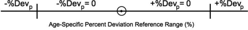
Objective: To define modified Prenatal Growth Assessment Scores (mPGAS) for single and composite biometric parameters and determine their reference ranges in normal fetuses.
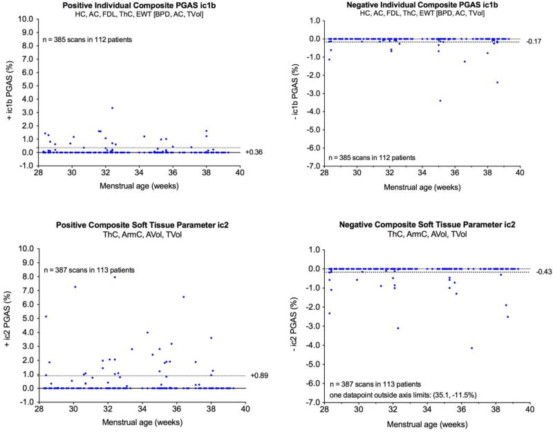
Methods: Nine anatomical parameters (ap) were measured and the weight estimated (EWTa, EWTb) in a longitudinal study of 119 fetuses with normal neonatal growth outcomes. Expected third trimester size trajectories, obtained from second trimester Rossavik size models, were used in calculating Percent Deviations (% Dev's) and their age-specific reference ranges in each fetus. The components of individual % Dev's values outside their reference ranges, designated +iapPGAS, -iapPGAS, were averaged to give +apPGAS and -apPGAS values for the 3rd trimester. The +iapPGAS and -iapPGAS values for different combinations of ap (c1a (HC, AC, FDL, ThC, EWTa), c1b (HC, AC, FDL, ThC, EWTb), c2 (ThC, ArmC, AVol, TVol), c3 (HC, AC, FDL, EWTa)) were then averaged to give +icPGAS and -icPGAS values at different time points or at the end of the third trimester (+cPGAS, -cPGAS). Values for iapPGAS, ic1bPGAS, and ic2PGAS were compared to their respective apPGAS or cPGAS reference ranges.
Results: All mPGAS values had one 95% range boundary at 0.0%. Upper boundaries of 1D +apPGAS values ranged from 0.0% (HC) to +0.49% (ThC) and were +0.06%, +2.3% and +1.8% for EWT, AVol and TVol, respectively. Comparable values for -apPGAS were 0.0% (BPD, FDL, HDL), to -0.58% (ArmC), -0.13% (EWT), -0.8% (AVol), and 0.0% (TVol). The +cPGAS, 95% reference range upper boundaries varied from +0.36% (c1b) to +0.89% (c2). Comparable values for -cPGAS lower boundaries were -0.17% (c1b) to -0.43% (c2).
Conclusions: The original PGAS concept has now been extended to individual biometric parameters and their combinations. With the standards provided, mPGAS values can now be tested to see if detection of different types of third trimester growth problems is improved.
Keywords: Individualized growth assessment; Rossavik models; pregnancy; size standards.
Figures
Similar articles
-
Individualized fetal growth assessment: critical evaluation of key concepts in the specification of third trimester size trajectories.J Matern Fetal Neonatal Med. 2014 Apr;27(6):543-51. doi: 10.3109/14767058.2013.833904. Epub 2013 Sep 12. J Matern Fetal Neonatal Med. 2014. PMID: 23962305 Free PMC article.
-
Comparison of fetal size standards obtained with conventional methods and individualized assessment: the effect of adjusting for differences in growth potential.J Matern Fetal Neonatal Med. 2020 Sep;33(18):3170-3176. doi: 10.1080/14767058.2019.1601695. Epub 2019 Apr 30. J Matern Fetal Neonatal Med. 2020. PMID: 30922145
-
Individualized growth assessment of fetal thigh circumference using three-dimensional ultrasonography.Ultrasound Obstet Gynecol. 2008 May;31(5):520-8. doi: 10.1002/uog.5302. Ultrasound Obstet Gynecol. 2008. PMID: 18389488
-
The World Health Organization fetal growth charts: concept, findings, interpretation, and application.Am J Obstet Gynecol. 2018 Feb;218(2S):S619-S629. doi: 10.1016/j.ajog.2017.12.010. Am J Obstet Gynecol. 2018. PMID: 29422204 Review.
-
Individualized growth assessment: conceptual framework and practical implementation for the evaluation of fetal growth and neonatal growth outcome.Am J Obstet Gynecol. 2018 Feb;218(2S):S656-S678. doi: 10.1016/j.ajog.2017.12.210. Am J Obstet Gynecol. 2018. PMID: 29422206 Free PMC article. Review.
Cited by
-
Fetal growth pathology score: a novel ultrasound parameter for individualized assessment of third trimester growth abnormalities.J Matern Fetal Neonatal Med. 2018 Apr;31(7):866-876. doi: 10.1080/14767058.2017.1300646. Epub 2017 Mar 20. J Matern Fetal Neonatal Med. 2018. PMID: 28277911 Free PMC article.
-
Ultrasound parameters of arteries and heart in normal fetuses.Cardiovasc Ultrasound. 2024 Jul 29;22(1):9. doi: 10.1186/s12947-024-00328-w. Cardiovasc Ultrasound. 2024. PMID: 39075466 Free PMC article.
-
Second trimester growth velocities: assessment of fetal growth potential in SGA singletons.J Matern Fetal Neonatal Med. 2019 Mar;32(6):939-946. doi: 10.1080/14767058.2017.1395849. Epub 2017 Nov 7. J Matern Fetal Neonatal Med. 2019. PMID: 29057683 Free PMC article.
-
Third trimester growth restriction patterns: individualized assessment using a fetal growth pathology score.J Matern Fetal Neonatal Med. 2018 Aug;31(16):2155-2163. doi: 10.1080/14767058.2017.1337741. Epub 2017 Jul 6. J Matern Fetal Neonatal Med. 2018. PMID: 28573931 Free PMC article.
-
The use of angiogenic biomarkers in maternal blood to identify which SGA fetuses will require a preterm delivery and mothers who will develop pre-eclampsia.J Matern Fetal Neonatal Med. 2016;29(8):1214-28. doi: 10.3109/14767058.2015.1048431. J Matern Fetal Neonatal Med. 2016. PMID: 26303962 Free PMC article.
References
-
- Nyberg DA, Abuhamad A, Ville Y. Ultrasound assessment of abnormal fetal growth. Semin Perinatol. 2004;28:3–22. - PubMed
-
- Dudley NJ. A systematic review of the ultrasound estimation of fetal weight. Ultrasound Obstet Gynecol. 2005;25:80–9. - PubMed
-
- Melamed N, Yogev Y, Meizner I, Mashiach R, Bardin R, Ben-Haroush A. Sonographic fetal weight estimation: which model should be used? J Ultrasound Med. 2009;28:617–29. - PubMed
-
- Mikolajczyk RT, Zhang J, Betran AP, Souza JP, Mori R, Gulmezoglu AM, Merialdi M. A global reference for fetal-weight and birthweight percentiles. Lancet. 2011;377:1855–61. - PubMed
-
- Mayer C, Joseph KS. Fetal growth: a review of terms, concepts and issues relevant to obstetrics. Ultrasound Obstet Gynecol. 2013;41:136–45. - PubMed
Publication types
MeSH terms
Grants and funding
LinkOut - more resources
Full Text Sources
Other Literature Sources
Medical
Miscellaneous