

The destiny of Ca(2+) released by mitochondria
- PMID: 24994533
- PMCID: PMC4276810
- DOI: 10.1007/s12576-014-0326-7
The destiny of Ca(2+) released by mitochondria
Abstract
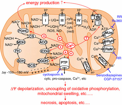
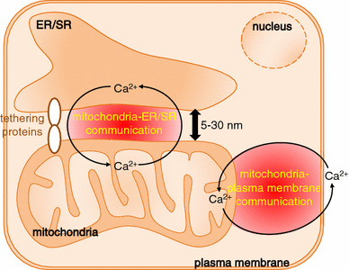
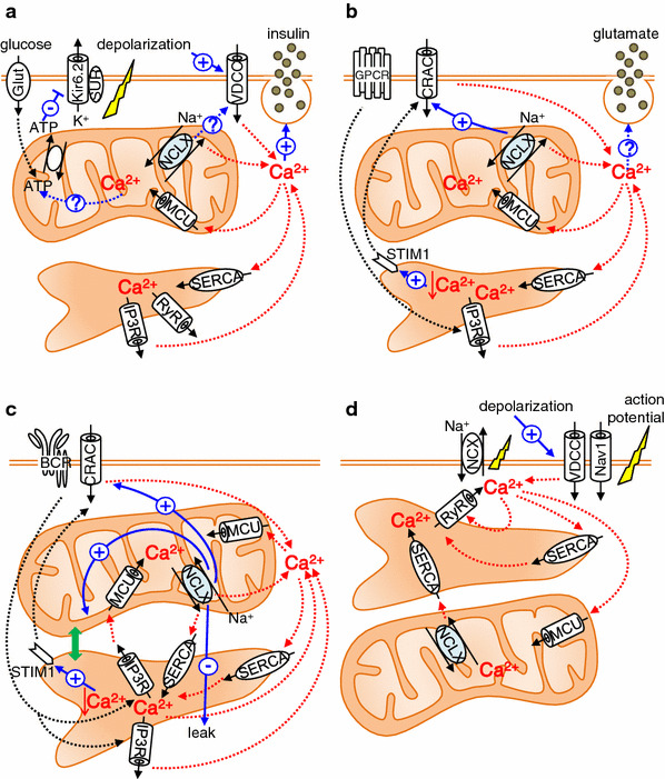
Mitochondrial Ca(2+) is known to regulate diverse cellular functions, for example energy production and cell death, by modulating mitochondrial dehydrogenases, inducing production of reactive oxygen species, and opening mitochondrial permeability transition pores. In addition to the action of Ca(2+) within mitochondria, Ca(2+) released from mitochondria is also important in a variety of cellular functions. In the last 5 years, the molecules responsible for mitochondrial Ca(2+) dynamics have been identified: a mitochondrial Ca(2+) uniporter (MCU), a mitochondrial Na(+)-Ca(2+) exchanger (NCLX), and a candidate for a mitochondrial H(+)-Ca(2+) exchanger (Letm1). In this review, we focus on the mitochondrial Ca(2+) release system, and discuss its physiological and pathophysiological significance. Accumulating evidence suggests that the mitochondrial Ca(2+) release system is not only crucial in maintaining mitochondrial Ca(2+) homeostasis but also participates in the Ca(2+) crosstalk between mitochondria and the plasma membrane and between mitochondria and the endoplasmic/sarcoplasmic reticulum.
Conflict of interest statement
The authors declare that they have no conflict of interest.
Figures
Similar articles
-
NCLX protein, but not LETM1, mediates mitochondrial Ca2+ extrusion, thereby limiting Ca2+-induced NAD(P)H production and modulating matrix redox state.J Biol Chem. 2014 Jul 18;289(29):20377-85. doi: 10.1074/jbc.M113.540898. Epub 2014 Jun 4. J Biol Chem. 2014. PMID: 24898248 Free PMC article.
-
Mitochondrial VDAC, the Na+/Ca2+ Exchanger, and the Ca2+ Uniporter in Ca2+ Dynamics and Signaling.Adv Exp Med Biol. 2017;981:323-347. doi: 10.1007/978-3-319-55858-5_13. Adv Exp Med Biol. 2017. PMID: 29594867 Review.
-
Calcium signalling: fishing out molecules of mitochondrial calcium transport.Curr Biol. 2010 Oct 26;20(20):R888-91. doi: 10.1016/j.cub.2010.09.035. Curr Biol. 2010. PMID: 20971432 Free PMC article.
-
Functional properties and mode of regulation of the mitochondrial Na+/Ca2+ exchanger, NCLX.Semin Cell Dev Biol. 2019 Oct;94:59-65. doi: 10.1016/j.semcdb.2019.01.009. Epub 2019 Jan 30. Semin Cell Dev Biol. 2019. PMID: 30658153 Review.
-
Enjoy the Trip: Calcium in Mitochondria Back and Forth.Annu Rev Biochem. 2016 Jun 2;85:161-92. doi: 10.1146/annurev-biochem-060614-034216. Epub 2016 May 4. Annu Rev Biochem. 2016. PMID: 27145841 Review.
Cited by
-
The lack of slow force response in failing rat myocardium: role of stretch-induced modulation of Ca-TnC kinetics.J Physiol Sci. 2019 Mar;69(2):345-357. doi: 10.1007/s12576-018-0651-3. Epub 2018 Dec 18. J Physiol Sci. 2019. PMID: 30560346 Free PMC article.
-
Total Matrix Ca2+ Modulates Ca2+ Efflux via the Ca2+/H+ Exchanger in Cardiac Mitochondria.Front Physiol. 2020 Sep 16;11:510600. doi: 10.3389/fphys.2020.510600. eCollection 2020. Front Physiol. 2020. PMID: 33041851 Free PMC article.
-
MicroRNA-138 and MicroRNA-25 Down-regulate Mitochondrial Calcium Uniporter, Causing the Pulmonary Arterial Hypertension Cancer Phenotype.Am J Respir Crit Care Med. 2017 Feb 15;195(4):515-529. doi: 10.1164/rccm.201604-0814OC. Am J Respir Crit Care Med. 2017. PMID: 27648837 Free PMC article.
-
Reconsidering the Role of Mitochondria in Aging.J Gerontol A Biol Sci Med Sci. 2015 Nov;70(11):1334-42. doi: 10.1093/gerona/glv070. Epub 2015 May 20. J Gerontol A Biol Sci Med Sci. 2015. PMID: 25995290 Free PMC article. Review.
-
Sepsis‑induced cardiac dysfunction and pathogenetic mechanisms (Review).Mol Med Rep. 2023 Dec;28(6):227. doi: 10.3892/mmr.2023.13114. Epub 2023 Oct 20. Mol Med Rep. 2023. PMID: 37859613 Free PMC article. Review.
References
-
- Bernardi P. Mitochondrial transport of cations: channels, exchangers, and permeability transition. Physiol Rev. 1999;79:1127–1155. - PubMed
Publication types
MeSH terms
Substances
LinkOut - more resources
Full Text Sources
Other Literature Sources
Miscellaneous
