. 2014 Jul 4;345(6192):90-4.
doi: 10.1126/science.1251487.

Vessel formation. De novo formation of a distinct coronary vascular population in neonatal heart

Affiliations

Vessel formation. De novo formation of a distinct coronary vascular population in neonatal heart

Xueying Tian et al. Science. .

Abstract

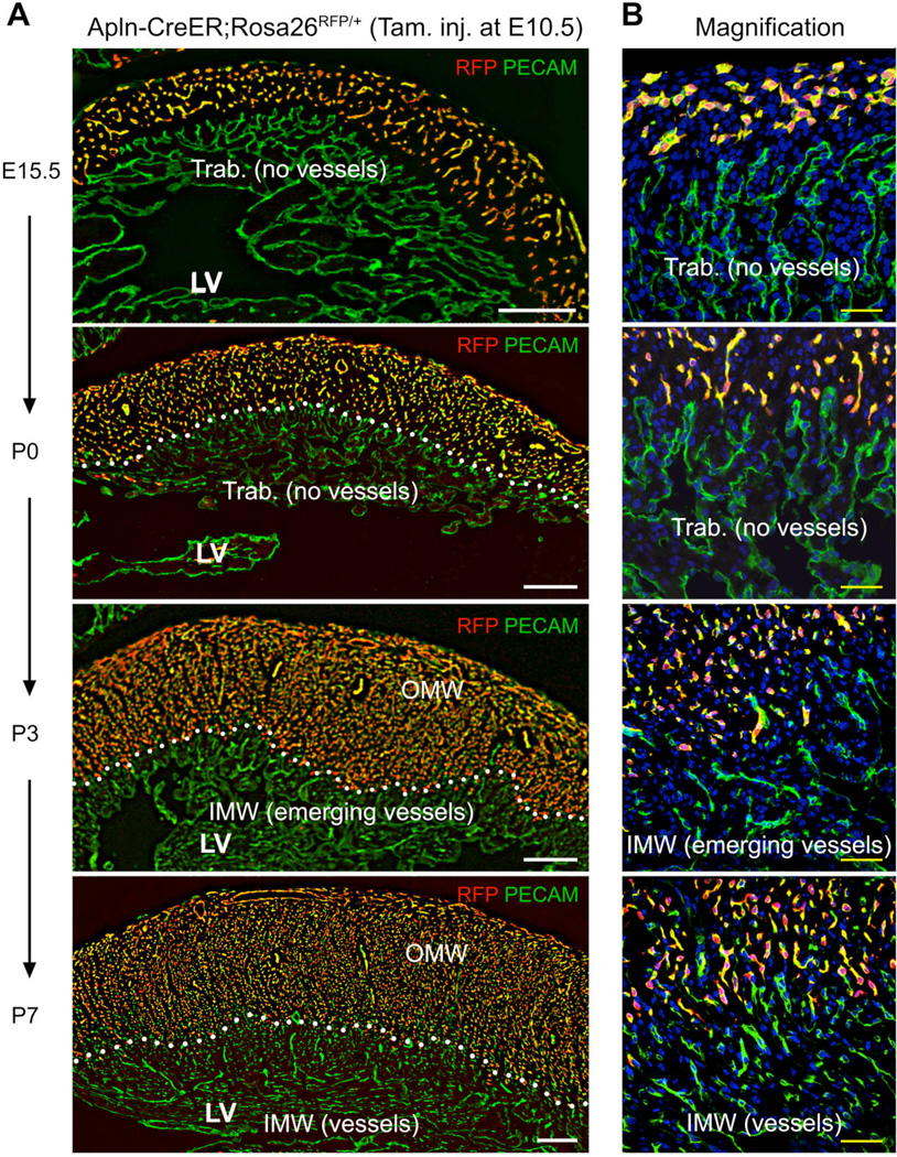
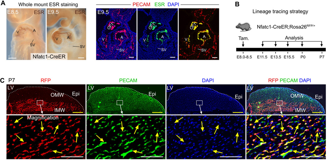
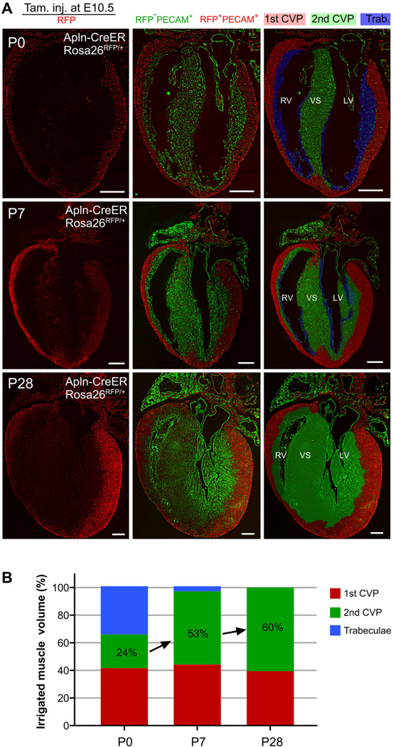
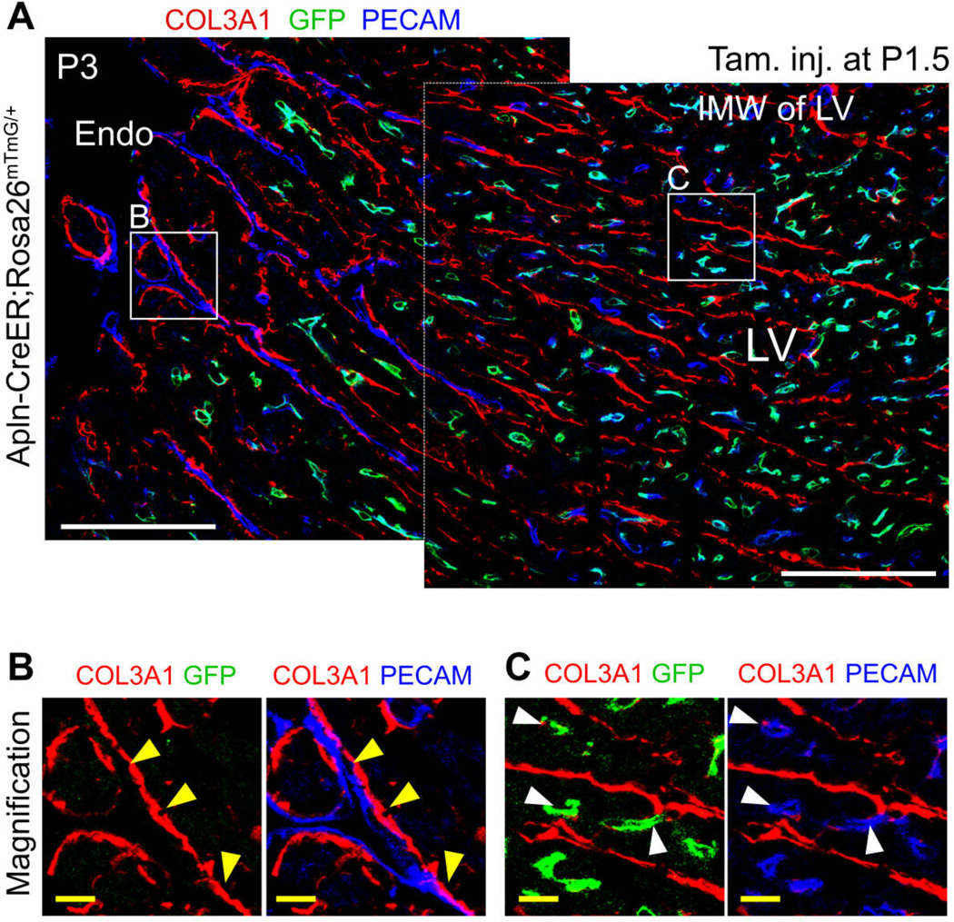
The postnatal coronary vessels have been viewed as developing through expansion of vessels formed during the fetal period. Using genetic lineage tracing, we found that a substantial portion of postnatal coronary vessels arise de novo in the neonatal mouse heart, rather than expanding from preexisting embryonic vasculature. Our data show that lineage conversion of neonatal endocardial cells during trabecular compaction generates a distinct compartment of the coronary circulation located within the inner half of the ventricular wall. This lineage conversion occurs within a brief period after birth and provides an efficient means of rapidly augmenting the coronary vasculature. This mechanism of postnatal coronary vascular growth provides avenues for understanding and stimulating cardiovascular regeneration following injury and disease.


Figures

Fig. 1
Fig. 1. Derivatives of embryonic coronary vessels do not expand into the inner myocardial wall of the neonatal heart
(A, B) Embryonic coronary vessels were RFP-labeled by tamoxifen treatment of Apln-CreER;Rosa26RFP/+ embryos at E10.5. In the neonatal heart, these VECs, derived from embryonic coronary vessels, occupied the outer portion of the compact myocardium, defined as the Outer Myocardial Wall (OMW). The remaining, RFP negative portion of the compact myocardium was defined as the Inner Myocardial Wall (IMW). White dotted lines highlight the inner border of left ventricular myocardium occupied by embryonically labeled vessels. From P0 to P7, Trabecular myocardium (Trab.) transformed into compact myocardium where new coronary vessels arise. White bar = 200 µm; yellow bar = 50 µm. LV, left ventricle.
Fig. 2
Fig. 2. Neonatal endocardial cells contribute to the de novo-formed coronary vessels in the inner myocardial wall
(A) CreER was expressed in atrial and ventricular endocardium but not sinus venosus at E8.5 and E9.5. CreER was visualized by staining for the estrogen receptor (ESR) portion of the CreER fusion protein. V, ventricle; A, atrium; OFT, outflow tract; SV, sinus venosus. (B) Lineage tracing strategy using Nfatc1-CreER;Rosa26RFP/+ line. Tamoxifen was injected at E8.0 to E8.5, and hearts were collected at later stages. (C) Immunostaining of genetic marker RFP and endothelial cell marker PECAM on P7 heart sections. Nfatc1-CreER labeled endocardial cells contribute to the majority of VECs (76.7 ± 5.4%, yellow arrows) in the inner myocardial wall (IMW), but not VECs in the outer myocardial wall (OMW). White bar = 100 µm, yellow bar = 500 µm. 3 – 5 embryos were examined at each time point.
Fig. 3
Fig. 3. 1st and 2nd CVP irrigate mutually exclusive regions of the myocardium of the adult heart
(A) Long axis sections of mouse heart delineating the temporal evolution of 1st CVP (red), 2nd CVP (green), and trabecular regions (blue) during the neonatal to adult heart transition. The 1st CVP was defined as the vessels genetically labeled (PECAM+;RFP+, pseudo color red) by tamoxifen treatment of Apln-CreER;Rosa26RFP/+ embryos at E10.5. The 2nd CVP was defined as the remaining unlabeled intramyocardial vessels (PECAM+;RFP, pseudo coloar green). Trabecular myocardium was identified as the myocardium lacking intramyocardial vessels (PECAM;RFP). Shading in the right column indicates regions annotated as occupied by 1st CVP, 2nd CVP or trabecular myocardium based on staining patterns. Images are representative of 3 - 6 hearts for each time point. White bar = 0.5 mm. (B) Relative ventricular myocardial volume occupied by 1st CVP, 2nd CVP or trabecular myocardium during postnatal heart growth. Note the rapid expansion of the myocardium irrigated by 2nd CVP and diminution of the trabecular myocardium between P0 – P7. n = 3 – 6 for each time point.
Fig. 4
Fig. 4. Neonatal endocardial cells were trapped in the inner myocardial wall
(A) Coronary vessels forming in the myocardial wall were labeled by tamoxifen treatment of Apln-CreER;Rosa26mTmG/+ at P1.5 and analyzed at P3. Region (B) represents the luminal surface and contains un-remodeled trabecular myocardium that persists at this stage. Yellow arrowheads indicate PECAM+ endothelial cells that are not labeled by Apln-CreER and that reside on the basement membrane, indicative of endocardial cells. Region (C) represents the inner myocardial wall, containing PECAM+ endothelial cells that are labeled by Apln-CreER and that reside within myocardium rather than on COL3A1 basement membrane, identifying them as tube-like coronary VECs (white arrowheads). White bar = 100 µm, yellow bar = 10 µm.

Comment in

References

    1. Riley P, Smart N. Cardiovasc Res. 2011;91:260. - PubMed
    1. Yi B, Wernet O, Chien K. J Clin Invest. 2010;120:20. - PMC - PubMed
    1. Smart N, et al. Nature. 2007;445:177. - PubMed
    1. Porrello E, et al. Science. 2011;331:1078. - PMC - PubMed
    1. Bennett HS. Am. J. Anat. 1936;60:27–53.

Publication types