. 2014 Jul 4;345(6192):98-101.
doi: 10.1126/science.1254312.

Opposing unfolded-protein-response signals converge on death receptor 5 to control apoptosis

Affiliations

Opposing unfolded-protein-response signals converge on death receptor 5 to control apoptosis

Min Lu et al. Science. .

Abstract

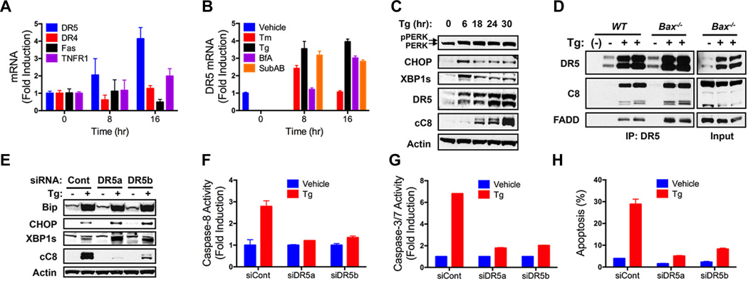
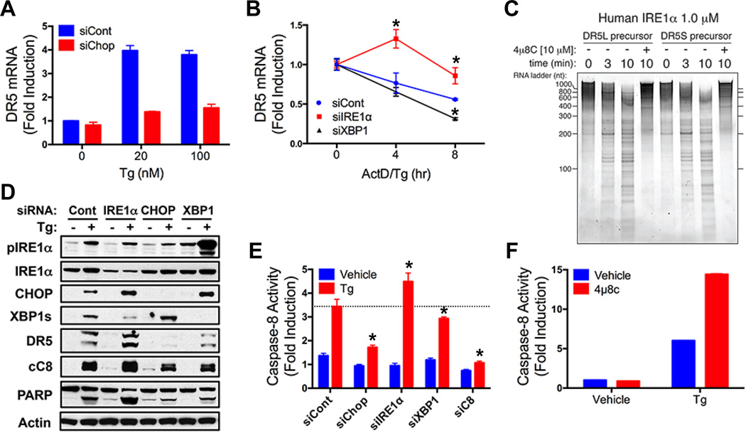
Protein folding by the endoplasmic reticulum (ER) is physiologically critical; its disruption causes ER stress and augments disease. ER stress activates the unfolded protein response (UPR) to restore homeostasis. If stress persists, the UPR induces apoptotic cell death, but the mechanisms remain elusive. Here, we report that unmitigated ER stress promoted apoptosis through cell-autonomous, UPR-controlled activation of death receptor 5 (DR5). ER stressors induced DR5 transcription via the UPR mediator CHOP; however, the UPR sensor IRE1α transiently catalyzed DR5 mRNA decay, which allowed time for adaptation. Persistent ER stress built up intracellular DR5 protein, driving ligand-independent DR5 activation and apoptosis engagement via caspase-8. Thus, DR5 integrates opposing UPR signals to couple ER stress and apoptotic cell fate.


Figures

Fig. 1
Fig. 1. Unmitigated ER stress triggers apoptosis via caspase-8
(A) KMS11 cells were treated with SubAB and analyzed by immunoblot. cC8: cleaved caspase-8; cC3: cleaved caspase-3. (B) SK-MES-1 cells were treated with BfA (24 hr) and analyzed by immunoblot. (C and D) Wildtype (WT) or Bax−/− HCT116 cells were treated with Tg (100 nM) and analyzed by immunoblot (C), or caspase-8 activity assay (24 hr) (D). (E and F) HCT116 cells were transfected (48 hr) with control siRNA, or a single (C8a), or an independent pool (C8b) of caspase-8 siRNAs, or caspase-2 siRNA. Cells were treated with Tg (100 nM) and analyzed by immunoblot (E) or FACS to measure apoptosis by subG1 DNA content (F). Graphs depict means ± SD of triplicates (D) or duplicates (F).
Fig. 2
Fig. 2. Unmitigated ER stress activates caspase-8 via DR5
(A) HCT116 cells were treated with Tg (100 nM) and mRNA levels were measured by QPCR (normalized to GAPDH). (B) HCT116 cells were treated with Tm (1 µg/ml), Tg (100 nM), BfA (1 µg/ml), or SubAB (1 µg/ml) and analyzed by QPCR (normalized to GAPDH). (C) HCT116 cells were treated with Tg (100 nM) and analyzed by immunoblot. (D) WT or Bax−/− HCT116 cells were treated as in C (24 hr), subjected to DR5 IP, and analyzed by immunoblot. (E–H) HCT116 cells were treated as in D and analyzed for caspase-8 activity (F), caspase-3/7 activity (G) and apoptosis (H). Graphs depict means ± SD of triplicates.
Fig. 3
Fig. 3. Unmitigated ER stress engages caspase-8 by inducing ligand-independent intracellular DR5 activation
(A) HCT116 cells were transfected (48 hr) with control siRNA or siRNA targeting Apo2L/TRAIL or caspase-8. Cells were treated with Tg (100 nM, 24 hr) and analyzed for apoptosis. (B) HCT116 cells were treated (24 hr) with Apo2L/TRAIL (1 µg/ml) or Tg (100 nM) in presence of vehicle or DR4-Fc plus DR5-Fc (10 µg/ml each) and analyzed for apoptosis. (C) SK-MES-1 cells were treated (24 hr) with indicated ER stressors and analyzed by immunofluorescence with DR5-specific antibody. (D) SK-MES-1 cells were treated with Tg (20 nM, 24 hr) and analyzed by immunofluorescence for DR5 or RACS1. (E and F) HCT116 cells were treated with Tg (100 nM, 24 hr) extracted with 1% Triton X-100, and subjected to size exclusion chromatography; fractions were analyzed by DR5 IP and immunoblot (E) or caspase-8 activity assay (F): Control: direct DR5 IP from Tg-treated cells. Graphs depict means ± SD of duplicates (B) or triplicates (F).
Fig. 4
Fig. 4. DR5 integrates opposing UPR signals to control apoptosis
(A) HCT116 cells were transfected (48 hr) with control or CHOP siRNA, treated with Tg (8 hr), and analyzed by QPCR (normalized to GAPDH). (B) HCT116 cells were transfected (48 hr) with control, IRE1α or XBP1s siRNA, treated with actinomycin D (2 µg/ml) plus Tg (20 nM), and DR5 mRNA was measured as in A. (C) Purified recombinant human IRE1α comprising the kinase and RNase domains (KR43) was incubated with in vitro-transcribed DR5S or DR5L mRNA in the presence of vehicle or 4µ8c (10 µM). Reactions were resolved on 6% TBE-Urea PAGE gels and stained with SYBR Gold. (D and E) HCT116 cells were transfected (48 hr) with control, IRE1α, CHOP, or XBP1s siRNA, treated with Tg (100 nM, 24 hr) and analyzed by immunoblot (D) or caspase-8 activity assay (E). (F) HCT116 cells were treated with Tg (100 nM, 24 hr) in presence of vehicle or 4µ8c (30 µM) and analyzed for caspase-8 activity. Graphs depict means ± SD of triplicates.

Comment in

  • Apoptosis. DR5 unfolds ER stress.
    Zlotorynski E. Zlotorynski E. Nat Rev Mol Cell Biol. 2014 Aug;15(8):498-9. doi: 10.1038/nrm3843. Epub 2014 Jul 16. Nat Rev Mol Cell Biol. 2014. PMID: 25027654

References

    1. Walter P, Ron D. Science. 2011;334:1081–1086. - PubMed
    1. Hetz C. Nat. Rev. Mol. Cell Biol. 2012;13:89–102. - PubMed
    1. Korennykh A, Walter P. Annu Rev Cell Dev Biol. 2012;28:251–277. - PubMed
    1. Hollien J, Weissman JS. Science. 2006;313:104–107. - PubMed
    1. Wang S, Kaufman RJ. J. Cell Biol. 2012;197:857–867. - PMC - PubMed

Publication types

MeSH terms