

Comparison of multiple parameters obtained on 3T pulsed arterial spin-labeling, diffusion tensor imaging, and MRS and the Ki-67 labeling index in evaluating glioma grading
- PMID: 24994829
- PMCID: PMC7965191
- DOI: 10.3174/ajnr.A4018
Comparison of multiple parameters obtained on 3T pulsed arterial spin-labeling, diffusion tensor imaging, and MRS and the Ki-67 labeling index in evaluating glioma grading
Abstract
Background and purpose: Pulsed arterial spin-labeling, DTI, and MR spectroscopy provide useful data for tumor evaluation. We evaluated multiple parameters by using these pulse sequences and the Ki-67 labeling index in newly diagnosed supratentorial gliomas.
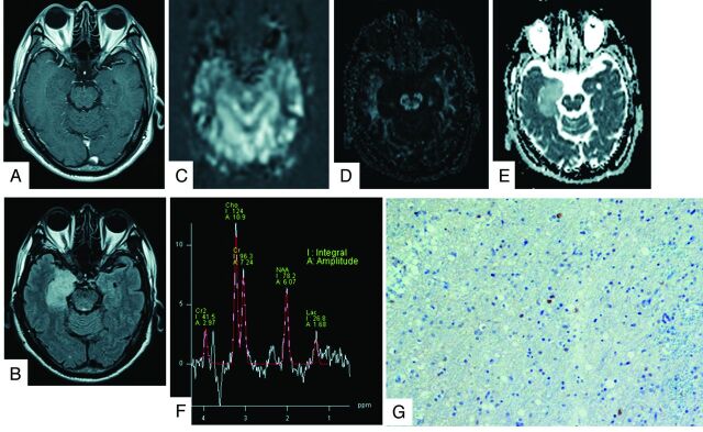
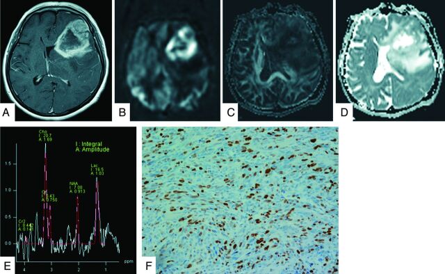
Materials and methods: All 32 patients, with grade II (3 each of diffuse astrocytoma, oligodendroglioma, and oligoastrocytoma), grade III (3 anaplastic astrocytomas, 4 anaplastic oligodendrogliomas, and 1 anaplastic oligoastrocytoma), and grade IV (14 glioblastomas and 1 glioblastoma with an oligodendroglioma component) cases underwent pulsed arterial spin-labeling, DTI, and MR spectroscopy studies by using 3T MR imaging. The following variables were used to compare the tumors: relative cerebral blood flow, fractional anisotropy; ADC tumor/normal ratios; and the Cho/Cr, NAA/Cho, NAA/Cr, and lactate/Cr ratios. A logistic regression and receiver operating characteristic analysis were used to assess parameters with a high sensitivity and specificity to identify the threshold values for separate grading. We compared the Ki-67 index with various MR imaging parameters in tumor specimens.
Results: Significant correlations were observed between the Ki-67 index and the mean, maximum, and minimum ADC, Cho/Cr, and lactate/Cr ratios. The receiver operating characteristic analysis showed that the combination of the minimum ADC and Cho/Cr ratios could differentiate low-grade and high-grade gliomas, with a sensitivity and specificity of 87.0% and 88.9%, respectively. The mean and maximum relative cerebral blood flow ratios were used to classify glioblastomas from other-grade astrocytomas, with a sensitivity and specificity of 92.9% and 83.3%, respectively.
Conclusions: Our findings indicate that pulsed arterial spin-labeling, DTI, and MR spectroscopy are useful for predicting glioma grade. Additionally, the parameters obtained on DTI and MR spectroscopy closely correlated with the proliferative potential of gliomas.
© 2014 by American Journal of Neuroradiology.
Figures
Similar articles
-
Measurements of diagnostic examination performance using quantitative apparent diffusion coefficient and proton MR spectroscopic imaging in the preoperative evaluation of tumor grade in cerebral gliomas.Eur J Radiol. 2011 Nov;80(2):462-70. doi: 10.1016/j.ejrad.2010.07.017. Epub 2010 Aug 13. Eur J Radiol. 2011. PMID: 20708868
-
In the assessment of supratentorial glioma grade: the combined role of multivoxel proton MR spectroscopy and diffusion tensor imaging.Clin Radiol. 2011 Oct;66(10):953-60. doi: 10.1016/j.crad.2011.05.001. Epub 2011 Jun 12. Clin Radiol. 2011. PMID: 21663899
-
Perfusion Parameter Obtained on 3-Tesla Magnetic Resonance Imaging and the Ki-67 Labeling Index Predict the Overall Survival of Glioblastoma.World Neurosurg. 2021 May;149:e469-e480. doi: 10.1016/j.wneu.2021.02.002. Epub 2021 Feb 7. World Neurosurg. 2021. PMID: 33567368
-
The role of imaging in the management of adults with diffuse low grade glioma: A systematic review and evidence-based clinical practice guideline.J Neurooncol. 2015 Dec;125(3):457-79. doi: 10.1007/s11060-015-1908-9. Epub 2015 Nov 3. J Neurooncol. 2015. PMID: 26530262
-
The use of advanced neuroimaging modalities in the evaluation of low-grade glioma in adults: a literature review.Neurosurg Focus. 2024 Feb;56(2):E3. doi: 10.3171/2023.11.FOCUS23649. Neurosurg Focus. 2024. PMID: 38301240 Review.
Cited by
-
Current Clinical Brain Tumor Imaging.Neurosurgery. 2017 Sep 1;81(3):397-415. doi: 10.1093/neuros/nyx103. Neurosurgery. 2017. PMID: 28486641 Free PMC article. Review.
-
Quantitative magnetic resonance imaging and radiogenomic biomarkers for glioma characterisation: a systematic review.Br J Radiol. 2018 Dec;91(1092):20170930. doi: 10.1259/bjr.20170930. Epub 2018 Jun 29. Br J Radiol. 2018. PMID: 29902076 Free PMC article.
-
Three-dimensional pulsed continuous arterial spin labeling and intravoxel incoherent motion imaging of nasopharyngeal carcinoma: correlations with Ki-67 proliferation status.Quant Imaging Med Surg. 2021 Apr;11(4):1394-1405. doi: 10.21037/qims-20-349. Quant Imaging Med Surg. 2021. PMID: 33816177 Free PMC article.
-
Correlation Between Apparent Diffusion Coefficient and the Ki-67 Proliferation Index in Grading Pediatric Glioma.J Comput Assist Tomogr. 2023 Mar-Apr 01;47(2):322-328. doi: 10.1097/RCT.0000000000001400. J Comput Assist Tomogr. 2023. PMID: 36957971 Free PMC article.
-
The value of arterial spin labelling in adults glioma grading: systematic review and meta-analysis.Oncotarget. 2019 Feb 22;10(16):1589-1601. doi: 10.18632/oncotarget.26674. eCollection 2019 Feb 22. Oncotarget. 2019. PMID: 30899427 Free PMC article.
References
-
- Warmuth C, Gunther M, Zimmer C. Quantification of blood flow in brain tumors: comparison of arterial spin labeling and dynamic susceptibility weighted contrast-enhanced MR imaging. Radiology 2003;228:523–32 - PubMed
Publication types
MeSH terms
Substances
LinkOut - more resources
Full Text Sources
Other Literature Sources
Medical