. 2014 Sep 1;307(5):G499-507.
doi: 10.1152/ajpgi.00141.2014. Epub 2014 Jul 3.

Allergen-induced resistin-like molecule-α promotes esophageal epithelial cell hyperplasia in eosinophilic esophagitis

Affiliations

Allergen-induced resistin-like molecule-α promotes esophageal epithelial cell hyperplasia in eosinophilic esophagitis

Parm Mavi et al. Am J Physiol Gastrointest Liver Physiol. .

Erratum in

Abstract

Resistin-like molecule (Relm)-α is a secreted, cysteine-rich protein belonging to a newly defined family of proteins, including resistin, Relm-β, and Relm-γ. Although resistin was initially defined based on its insulin-resistance activity, the family members are highly induced in various inflammatory states. Earlier studies implicated Relm-α in insulin resistance, asthmatic responses, and intestinal inflammation; however, its function still remains an enigma. We now report that Relm-α is strongly induced in the esophagus in an allergen-challenged murine model of eosinophilic esophagitis (EoE). Furthermore, to understand the in vivo role of Relm-α, we generated Relm-α gene-inducible bitransgenic mice by using lung-specific CC-10 promoter (CC10-rtTA-Relm-α). We found Relm-α protein is significantly induced in the esophagus of CC10-rtTA-Relm-α bitransgenic mice exposed to doxycycline food. The most prominent effect observed by the induction of Relm-α is epithelial cell hyperplasia, basal layer thickness, accumulation of activated CD4(+) and CD4(-) T cell subsets, and eosinophilic inflammation in the esophagus. The in vitro experiments further confirm that Relm-α promotes primary epithelial cell proliferation but has no chemotactic activity for eosinophils. Taken together, our studies report for the first time that Relm-α induction in the esophagus has a major role in promoting epithelial cell hyperplasia and basal layer thickness, and the accumulation of activated CD4(+) and CD4(-) T cell subsets may be responsible for partial esophageal eosinophilia in the mouse models of EoE. Notably, the epithelial cell hyperplasia and basal layer thickness are the characteristic features commonly observed in human EoE.

Keywords: eosinophils; epithelial cells; esophagus.


Figures

Fig. 1.
Fig. 1.
Resistin-like molecule (Relm)-α mRNA and protein expression following aeroallergen or food allergen-induced experimental eosinophilic esophagitis (EoE). The experimental EoE was induced following aeroallergen and food [ovalbumin (OVA) or peanut] allergen challenge in mice, and esophageal RNA and homogenate was obtained for Relm-α expression in the esophagus. The Relm-α mRNA expression and protein levels in the esophagus were examined by performing qPCR and ELISA analysis. Relm-α mRNA and protein in the esophagus from Aspergillus-challenged mice (A and D), OVA-challenged mice (B and E), and peanut-challenged mice (C and F) are shown. The data are expressed as means ± SD; n = 10 mice/group.
Fig. 2.
Fig. 2.
Generation of doxycyline (DOX)-inducible CC-10-Relm-α bitransgenic mice and the analysis of Relm-α expression in the esophagus. The transgenic constructs present in the tetracycline reverse-transactivator transgenic mouse line CC10-rtTA (expressing rtTA cDNA under the regulation of the lung-specific, rat, 2.3-kb CC-10 promoter) and the tetO-Relm-α transgenic mice [containing germline Relm-α cDNA under the regulation of the tetracycline operator (tetO)] are shown (A). The Relm-α bitransgenic mice were fed DOX food or normal food (no DOX) for 6 wk, and esophageal RNA from homogenate was isolated and analyzed for Relm-α mRNA and protein expression via quantitative RT-PCR and ELISA, and data of Relm-α mRNA (B) and Relm-α protein (C) levels are shown. The data are expressed as means ± SD; n = 8 mice/group.
Fig. 3.
Fig. 3.
The detection of activated CD4+ T cells and eosinophils in the esophagus of Relm-α bitransgenic mice. Relm-α bitransgenic mice were exposed to DOX or no DOX food for 6 wk, and the induction of activated CD4+ T cells and eosinophils in the esophagus was analyzed using flow cytometric and immunohistochemical analysis. A representative dot-blot for activated CD4+ T cells using anti-CD69 and anti-CD4 antibodies and analyzed by flow cytometer is shown in A and B, and the absolute number of esophageal activated CD4+ and CD4 T cells is shown in C (data are means ± SD; n = 3 experiments, 4 mice in each group/experiment). AC show all live esophageal CD45+ and CD45 cells of the esophagus. The CD45+ cells were gated and analyzed for activated CD4+/CD69+ cells in the esophagus of no DOX and DOX food-exposed mice (A and B). A representative photomicrograph of DOX and no DOX-exposed mouse esophageal section following antiserum against mouse eosinophil major basic protein [anti-major basic protein (MBP)] immunostaining is shown in D and E, and the number of eosinophils is quantitated in the esophageal section are shown in F. The original magnification of the photomicrograph is ×125, and the eosinophils are marked by arrows. The number of eosinophils in the esophagus is expressed as eosinophils/mm2. Data are expressed as means ± SD (n = 12, P < 0.001).
Fig. 4.
Fig. 4.
Eosinophil active cytokines and chemokines are not induced in the esophagus of 6 wk exposed Relm-α bitransgenic mice. The quantitative real-time PCR analyses of IL-5, IL-13, eotaxin-1, and eotaxin-2 mRNA levels in the esophagus of Relm-α bitransgenic mice following 6 wk of no DOX and DOX-exposed mice were analyzed and are shown (AD). The data are expressed as means ± SD; n = 10 mice/group.
Fig. 5.
Fig. 5.
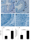
Immunohistochemical detection of 5′-bromodeoxyuridine-positive (BrdU+) epithelial cells in the esophagus of Relm-α bitransgenic mice. Relm-α bitransgenic mice were exposed to no DOX and DOX food for 6 wk, and the BrdU+ cells were analyzed in the esophagus by performing anti-BrdU immunostaining. A representative photomicrograph of mouse esophageal sections following anti-BrdU immunostaining is shown following no DOX and DOX food exposure (original magnification ×400; A and B) and thickened basal layer following no DOX and DOX food exposure (original magnification ×1,000; C and D). The %BrdU+ cell numbers in no DOX and DOX food-exposed mice in esophageal tissue sections were counted in the esophageal epithelial layer of the esophagus and shown in E. The epithelial cell thickening was measured and shown in F. Data are expressed as means ± SD; n = 12 mice.
Fig. 6.
Fig. 6.
Epithelial cell hyperplasia in Aspergillus-challenged wild-type and Relm-α gene-deficient mice. The epithelial cell hyperplasia was measured in the esophagus of wild-type mice and Relm-α gene-deficient mice following saline and Aspergillus challenge by counting the percent anti- proliferating cell nuclear antigen (PCNA)-positive immunostained cells and shown in A. The anti-MBP+ eosinophils were also counted in the saline and Aspergillus-challenged wild-type (+/+) mice and Relm-α gene-deficient mice (−/−) and shown in B. Data is expressed as means ± SD; n = 12 mice.
Fig. 7.
Fig. 7.
Mouse primary esophageal epithelial cell (mPECC) proliferation following different concentrations of recombinant Relm-α treatment. The characteristics of isolated and cultured mouse primary epithelial cells (mPEEC) were verified by immunostaining with mouse anti-cytokeratin antibody and tested by performing FACS analysis (A). The mouse primary esophageal epithelial cell proliferation following 48 h of Relm-α (0, 1, 50, 100, 250, 500, and 1,000 ng/ml) exposure was examined, and the optical density data and cell counts of each concentration are shown (B and C). The three independent experiments were performed in triplicate. Data are expressed as means ± SD; n = 3 experiments.

References

    1. Akei HS, Mishra A, Blanchard C, Rothenberg ME. Epicutaneous antigen exposure primes for experimental eosinophilic esophagitis in mice. Gastroenterology 129: 985–994, 2005 - PubMed
    1. Arora AS, Yamazaki K. Eosinophilic esophagitis: asthma of the esophagus? Clin Gastroenterol Hepatol 2: 523–530, 2004 - PubMed
    1. Banerjee RR, Lazar MA. Dimerization of resistin and resistin-like molecules is determined by a single cysteine. J Biol Chem 276: 25970–25973, 2001 - PubMed
    1. Beltowski J. Adiponectin and resistin–new hormones of white adipose tissue. Int Med J Exp Clin Res 9: RA55–RA61, 2003 - PubMed
    1. Blagoev B, Kratchmarova I, Nielsen MM, Fernandez MM, Voldby J, Andersen JS, Kristiansen K, Pandey A, Mann M. Inhibition of adipocyte differentiation by Resistin like molecule alpha (RELM alpha): biochemical characterization of its oligomeric nature (Abstract). J Biol Chem 19: 19, 2002 - PubMed

Publication types

MeSH terms