

Intrinsic stiffness of extracellular matrix increases with age in skeletal muscles of mice
- PMID: 24994884
- PMCID: PMC4137235
- DOI: 10.1152/japplphysiol.00256.2014
Intrinsic stiffness of extracellular matrix increases with age in skeletal muscles of mice
Abstract
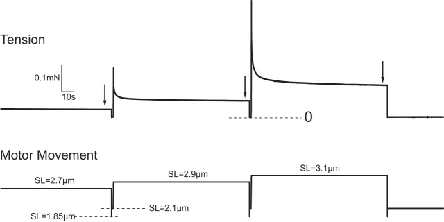
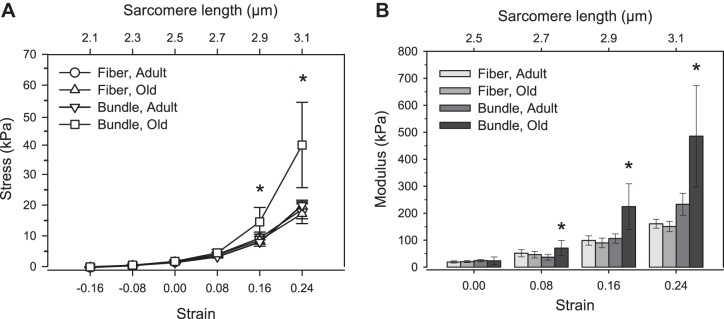
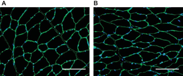
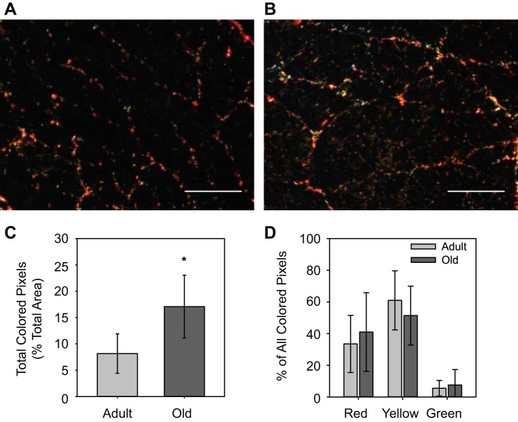
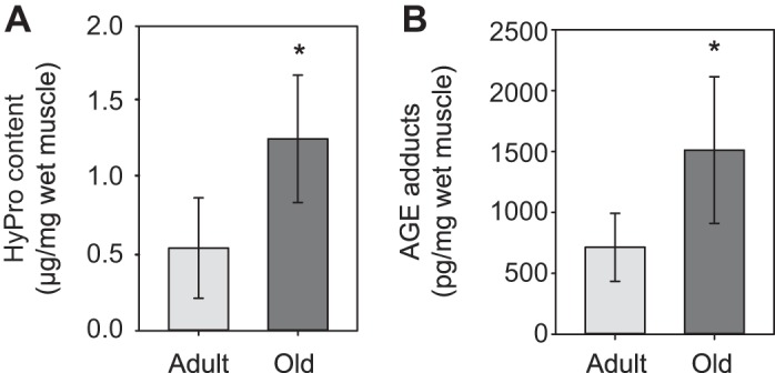
Advanced age is associated with increases in muscle passive stiffness, but the contributors to the changes remain unclear. Our purpose was to determine the relative contributions of muscle fibers and extracellular matrix (ECM) to muscle passive stiffness in both adult and old animals. Passive mechanical properties were determined for isolated individual muscle fibers and bundles of muscle fibers that included their associated ECM, obtained from tibialis anterior muscles of adult (8-12 mo old) and old (28-30 mo old) mice. Maximum tangent moduli of individual muscle fibers from adult and old muscles were not different at any sarcomere length tested. In contrast, the moduli of bundles of fibers from old mice was more than twofold greater than that of fiber bundles from adult muscles at sarcomere lengths >2.5 μm. Because ECM mechanical behavior is determined by the composition and arrangement of its molecular constituents, we also examined the effect of aging on ECM collagen characteristics. With aging, muscle ECM hydroxyproline content increased twofold and advanced glycation end-product protein adducts increased threefold, whereas collagen fibril orientation and total ECM area were not different between muscles from adult and old mice. Taken together, these findings indicate that the ECM of tibialis anterior muscles from old mice has a higher modulus than the ECM of adult muscles, likely driven by an accumulation of densely packed extensively crosslinked collagen.
Keywords: age crosslinking; collagen; muscle mechanics; passive tension; tangent modulus.
Copyright © 2014 the American Physiological Society.
Figures
References
-
- Arruda EM, Mundy K, Calve S, Baar K. Denervation does not change the ratio of collagen I and collagen III mRNA in the extracellular matrix of muscle. Am J Physiol Regul Integr Comp Physiol 292: R983–R987, 2007 - PubMed
-
- Bailey AJ, Paul RG, Knott L. Mechanisms of maturation and ageing of collagen. Mech Ageing Dev 106: 1–56, 1998 - PubMed
-
- Baudry S, Lecoeuvre G, Duchateau J. Age-related changes in the behavior of the muscle-tendon unit of the gastrocenemius medialis during upright stance. J Appl Physiol 112: 296–304, 2012 - PubMed
-
- Bojsen-Moller J, Magnusson SP, Rasmussen LR, Kjaer M, Aagaard P. Muscle performance during maximal isometric and dynamic contractions is influenced by the stiffness of the tendinous structures. J Appl Physiol 99: 986–994, 2005 - PubMed
Publication types
MeSH terms
Substances
Grants and funding
LinkOut - more resources
Full Text Sources
Other Literature Sources