. 2014 Jul 22;111(29):10666-71.
doi: 10.1073/pnas.1404271111. Epub 2014 Jul 3.

25-Hydroxycholesterol acts as an amplifier of inflammatory signaling

Affiliations

25-Hydroxycholesterol acts as an amplifier of inflammatory signaling

Elizabeth S Gold et al. Proc Natl Acad Sci U S A. .

Abstract

Cross-talk between sterol regulatory pathways and inflammatory pathways has been demonstrated to significantly impact the development of both atherosclerosis and infectious disease. The oxysterol 25-hydroxycholesterol (25HC) plays multiple roles in lipid biosynthesis and immunity. We recently used a systems biology approach to identify 25HC as an innate immune mediator that had a predicted role in atherosclerosis and we demonstrated a role for 25HC in foam cell formation. Here, we show that this mediator also has several complex roles in the antiviral response. The host response to viruses involves gene regulatory circuits with multiple feedback loops and we show here that 25HC acts as an amplifier of inflammatory signaling in macrophages. We determined that 25HC amplifies inflammatory signaling, at least in part, by mediating the recruitment of the AP-1 components FBJ osteosarcoma oncogene (FOS) and jun proto-oncogene (JUN) to the promoters of a subset of Toll-like receptor-responsive genes. Consistent with previous reports, we found that 25HC inhibits in vitro infection of airway epithelial cells by influenza. Surprisingly, we found that deletion of Ch25h, the gene encoding the enzyme responsible for 25HC production, is protective in a mouse model of influenza infection as a result of decreased inflammatory-induced pathology. Thus, our study demonstrates, for the first time to our knowledge, that in addition to its direct antiviral role, 25HC also regulates transcriptional responses and acts as an amplifier of inflammation via AP-1 and that the resulting alteration in inflammatory response leads to increased tissue damage in mice following infection with influenza.


Conflict of interest statement

The authors declare no conflict of interest.

Figures

Fig. 1.
Fig. 1.
The oxysterol 25HC amplifies the macrophage transcriptional response following stimulation with PIC. (A) The expression level of Ch25h in WT and Ifnar−/− macrophages after 12 h of stimulation with the indicated TLR agonists was determined by microarray analysis. Dashed line indicates basal expression level. The data are the average of three independent biological replicates. (B) BMDMs from three WT and three Ch25h−/− mice were stimulated with PIC for 18 h and RNA was analyzed by microarray. The expression of all genes up-regulated by more than twofold (P < 0.01) following PIC stimulation of WT macrophages is shown. Genes that are differentially expressed in macrophages derived from WT and Ch25h−/− mice by more than twofold with P < 0.01 are shown with red dots. (C) The expression of the subset of genes that are differentially expressed between the two genotypes (red dots in B) is plotted. (D) The expression of the indicated genes from the microarray data are plotted. (E and F) BMDMs from three WT and three Ch25h−/− mice were treated with 5 μM 25HC for 1 h and then stimulated with 6 μg/mL PIC for 18 h. The level of Il6 mRNA was determined by real-time PCR and the level of IL6 protein was determined by ELISA.
Fig. 2.
Fig. 2.
The oxysterol 25HC affects inflammatory signaling mediated by several PRRs via a non–LXR-dependent pathway. (A) Macrophages derived from three WT and three Ch25h−/− mice were stimulated with the indicated agonists at the indicated time points and RNA was analyzed by real-time PCR. Data shown are the average from the three biological replicates. (B) Expression of Il6 in Lxrαβ−/− BMDMs treated with 5 μM 25HC for 1 h and stimulated with 6 μg/mL PIC for 6 or 18 h were determined by real-time PCR. Data shown are the average of three replicates.
Fig. 3.
Fig. 3.
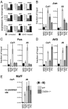
The effect of 25HC on PIC-induced inflammatory responses is mediated by AP-1. (A) BMDMs from three WT mice were stimulated with 25HC for 18 h and RNA expression was analyzed by microarray. Average expression levels of the indicated genes are shown. (B–E) Chromatin from unstimulated BMDMs or BMDMs stimulated with 6 μg/mL PIC for 18 h was immunoprecipitated with the indicated antibodies. Binding of the indicated transcription factors to their predicted sites was determined by real-time PCR. Data are expressed as the percentage of DNA in the input chromatin detected in the IP. NRS, normal rabbit serum control. Light gray, WT; dark gray, Ch25h−/−.
Fig. 4.
Fig. 4.
Ch25h is deleterious in an in vivo model of influenza infection due to increased inflammation. (A) Expression of Ch25h in the lungs of mice infected with PR8, X31, or mock infected measured by real-time PCR. Mice were infected with 105 pfu of virus and the data shown are the average from four mice. (B) Survival curve of wild-type and Ch25h−/− mice (11 mice per group) infected with 200 pfu of PR8. The percentage of mice not reaching criteria for being killed (defined as >25% total body weight loss) is shown. The P value indicates the significance of the difference in the Kaplan–Meier survival curves as assessed by the log-rank test. Data shown are representative of two separate experiments. (C) RNA was extracted from lungs isolated from infected mice at the indicated time points and viral load was assessed by real-time PCR for influenza M gene. (Light bars, WT; dark bars, Ch25h−/−). (D) LET1a cells derived from WT and Ch25h−/− mice were infected in vitro with PR8. The percentage of cells positive for influenza NP protein was determined by flow cytometry. The data are the average of three separate samples (light bars, WT; dark bars, Ch25h−/−. *P < 0.05). (E) Following 18 h of pretreatment of WT LET1a cells with the indicated concentrations of 25HC, cells were infected with PR8 for 6 h and the percentage of nucleoprotein (NP) positive cells was determined by flow cytometry. The data are the average of three independent samples. (F) At the indicated times following infection with 200 pfu PR8, lungs were extracted and histologic changes were analyzed in a blinded manner. The average total scores based on a sum of 18 histologic criteria are shown (light bars, WT; dark bars, Ch25h−/−. ****P < 0.0001, **P < 0.01). There were 21 WT and 21 Ch25h−/− mice used in this experiment and the data are representative of two separate experiments. (G) Representative histologic images from mice at day 3 following infection. (Top) Representative hematoxylin and eosin-stained lung sections from WT (C57BL6) (Left) and Ch25h−/− (Right) mice. (Scale bar, 8 mm.) Dark pink regions are affected by influenza-induced changes. (Middle) Representative influenza NP-1 antigen (brown) staining in the distal lung (boxed regions, Top). Note the WT bronchiole (b) has intense signal within epithelial cells and exudate (e) with positive alveolar lining cells (arrowheads, presumptive macrophages) and pneumocytes. In contrast, the Ch25h−/− lung has a single + cell with alveolar macrophage morphology (arrowhead). (Scale bar, 400 μm.) (Bottom) Higher power of proximal circled region demonstrating typical influenza-induced changes including necrotizing bronchiolitis (between arrowheads) and alveolitis. Similar, yet less severe, changes are present in the KO lung. Bronchiole (b), exudate (e), and alveoli (a) are as indicated. (Scale bar, 400 μm.) (H) The level of the indicated cytokine mRNA in whole lungs isolated from WT and Ch25h−/− mice at the indicated time points was determined using real-time PCR (light bars, WT; dark bars, Ch25h−/−; differences shown as *P < 0.05). (I) The level of IL6 and macrophage colony stimulating factor protein in bronchiolar lavage (BAL) fluid was determined by multiplexed immunoassay (light bars, WT, dark bars, Ch25h−/−; *P < 0.05). Data shown are averaged from the 29 WT and 29 Ch25h−/− mice used in this experiment.

References

    1. Chawla A, et al. PPAR-gamma dependent and independent effects on macrophage-gene expression in lipid metabolism and inflammation. Nat Med. 2001;7(1):48–52. - PubMed
    1. Ogawa S, et al. Molecular determinants of crosstalk between nuclear receptors and toll-like receptors. Cell. 2005;122(5):707–721. - PMC - PubMed
    1. Castrillo A, et al. Crosstalk between LXR and toll-like receptor signaling mediates bacterial and viral antagonism of cholesterol metabolism. Mol Cell. 2003;12(4):805–816. - PubMed
    1. Joseph SB, Castrillo A, Laffitte BA, Mangelsdorf DJ, Tontonoz P. Reciprocal regulation of inflammation and lipid metabolism by liver X receptors. Nat Med. 2003;9(2):213–219. - PubMed
    1. Jiang C, Ting AT, Seed B. PPAR-gamma agonists inhibit production of monocyte inflammatory cytokines. Nature. 1998;391(6662):82–86. - PubMed

Publication types

MeSH terms

Associated data