Review
. 2014 Aug 1;127(Pt 15):3205-15.
doi: 10.1242/jcs.153197. Epub 2014 Jul 2.

Establishment of epithelial polarity--GEF who's minding the GAP?

Affiliations
Review

Establishment of epithelial polarity--GEF who's minding the GAP?

Siu P Ngok et al. J Cell Sci. .

Abstract

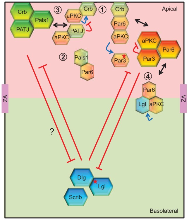
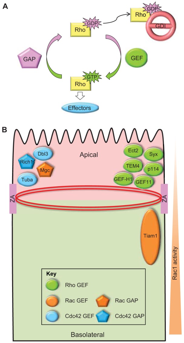
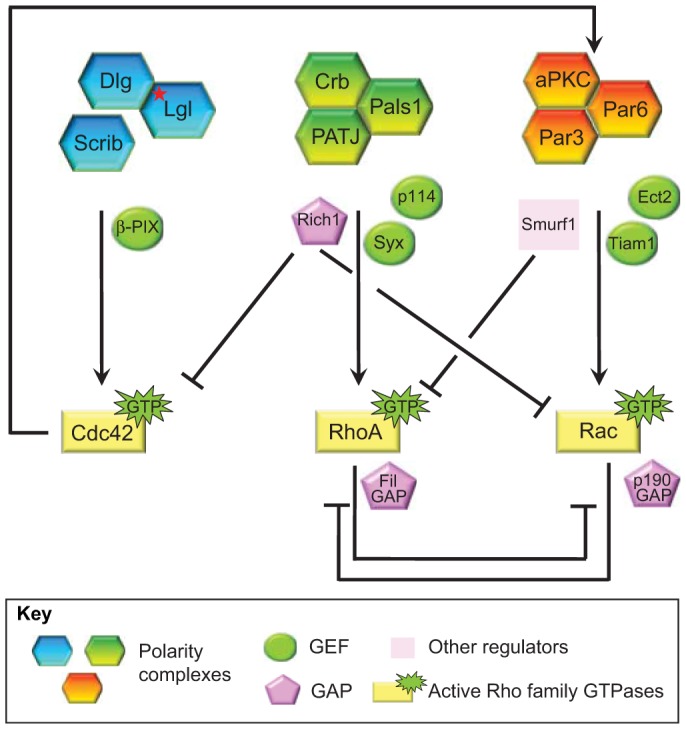
Cell polarization is a fundamental process that underlies epithelial morphogenesis, cell motility, cell division and organogenesis. Loss of polarity predisposes tissues to developmental disorders and contributes to cancer progression. The formation and establishment of epithelial cell polarity is mediated by the cooperation of polarity protein complexes, namely the Crumbs, partitioning defective (Par) and Scribble complexes, with Rho family GTPases, including RhoA, Rac1 and Cdc42. The activation of different GTPases triggers distinct downstream signaling pathways to modulate protein-protein interactions and cytoskeletal remodeling. The spatio-temporal activation and inactivation of these small GTPases is tightly controlled by a complex interconnected network of different regulatory proteins, including guanine-nucleotide-exchange factors (GEFs), GTPase-activating proteins (GAPs), and guanine-nucleotide-dissociation inhibitors (GDIs). In this Commentary, we focus on current understanding on how polarity complexes interact with GEFs and GAPs to control the precise location and activation of Rho GTPases (Crumbs for RhoA, Par for Rac1, and Scribble for Cdc42) to promote apical-basal polarization in mammalian epithelial cells. The mutual exclusion of GTPase activities, especially that of RhoA and Rac1, which is well established, provides a mechanism through which polarity complexes that act through distinct Rho GTPases function as cellular rheostats to fine-tune specific downstream pathways to differentiate and preserve the apical and basolateral domains. This article is part of a Minifocus on Establishing polarity.

Keywords: Cell adhesion; Cell polarity; Crumbs; Par3; Rho GTPase; Scribble.


Figures

Fig. 1.
Fig. 1.
The polarity complex ‘triangle’ – cooperative and antagonistic crosstalk to regulate epithelial apical–basal polarity. The three main protein complexes that regulate epithelial polarization are the apical Crumbs (Crb, Pals1 and PATJ) and Par (Par3, Par6 and aPKC) complexes, and the basolateral Scribble complex (Scrib, Dlg and Lgl). The interaction of Dlg with Scribble is probably indirect (Mathew et al., 2002), and with Lgl is phosphorylation-dependent (red star) (Zhu et al., 2014). Apical and basolateral domains are demarcated by the zonula adherens (ZA), a specialized cell–cell adhesion zone. Members of these three complexes interconnect with each other, allowing mutual exclusion and positive feedback (red, blue and black lines) between different complexes at distinct locations. For example, Crb interacts with the Par6–aPKC complex (Hurd et al., 2003; Kempkens et al., 2006), which is required for the regulation of Par3 localization (Morais-de-Sá et al., 2010; Walther and Pichaud, 2010), modulating junction formation (Lemmers et al., 2004), as well as epithelial morphogenesis and polarity (1) (Nam and Choi, 2003; Walther and Pichaud, 2010). Par6, in turn, interferes with the binding of PATJ to Pals1 (2) (Wang et al., 2004). Although the enzymatic activity of aPKC is inhibited by Par3 in the Par complex (Lin et al., 2000), aPKC can phosphorylate Par3, Crumbs and Lgl (blue arrow), thus bridging and regulating all three polarity complexes (Morais-de-Sá et al., 2010; Plant et al., 2003; Sotillos et al., 2004). Par3 phosphorylation results in its more basal localization relative to the Par6–aPKC complex (Morais-de-Sá et al., 2010). Phosphorylation of Crumbs is crucial for its apical localization and might be inhibited by the presence of PATJ (3) (Sotillos et al., 2004). Lgl competes with Par3 for binding to the Par6–aPKC complex (4); however, once phosphorylated by aPKC, Lgl is released and localizes to the basolateral area (Yamanaka et al., 2003). Despite the importance of the Scribble complex in the basolateral exclusion of Crumbs (Bilder et al., 2000; Bilder and Perrimon, 2000), the physical interactions between members of these two complexes that could underlie this effect are currently unclear.
Fig. 2.
Fig. 2.
The activation of Rho family GTPases is spatio-temporally controlled by regulatory molecules. (A) Rho family GTPases, including RhoA, Rac1 and Cdc42 cycle between the active GTP-bound and the inactive GDP-bound states. Once activated, Rho GTPases interact with downstream effectors, which lead to diverse signaling pathways and biological processes, such as cytoskeletal organization, cell junction formation, cell polarization and cell motility. The on–off cycle of Rho family GTPases is tightly controlled by three classes of regulatory proteins: GEFs catalyze the exchange of GDP for GTP; GAPs enhance GTPase activity to hydrolyze GTP back to GDP; and GDIs associate with GDP-bound Rho GTPases and prevent nucleotide dissociation. (B) In polarized mammalian epithelial cells, several GEFs and GAPs have been shown to localize either apically at mature junctions (zonula adherens, ZA) that encompass the circumferential actomyosin ring or to sub-apical areas of cell–cell contact. Localization of the Cdc42 GEF Tuba at apical junctions activates Cdc42 to promote junctional actin organization (Otani et al., 2006) and lumenogenesis (Bryant et al., 2010; Qin et al., 2010). Interestingly, the Cdc42 GAP Rich1, which also localizes to apical junctions, is required for junction integrity (Wells et al., 2006). A number of RhoA GEFs have been identified at apical junctions. Specifically, Ect2, Syx, p114RhoGEF (p114), TEM4 and ARHGEF11 (GEF11) promote the localization and activity of RhoA at the ZA to support junction integrity (Ngok et al., 2012; Ratheesh et al., 2012; Terry et al., 2011; Itoh et al., 2012; Ngok et al., 2013). In contrast, GEF-H1-regulated RhoA activity is negatively controlled to prevent junction disassembly (Aijaz et al., 2005; Guillemot et al., 2008; Samarin et al., 2007). Although the precise localization of activated RhoA and Cdc42 in polarized epithelial cells is still unclear, an apical–basal gradient of Rac1 activity has been observed with higher Rac1 activity at more basal regions due to the localized activation of the RacGEF TIAM1, which is required for proper junction formation (Mack et al., 2012). Consistent with this, MgcRacGAP localizes and acts apically (Guillemot et al., 2014). Collectively, the studies support a key role for the precise spatio-temporal activation of Rho family GTPases in junction formation and epithelial organization.
Fig. 3.
Fig. 3.
Crosstalk between polarity complexes and Rho family GTPases. The Scribble polarity complex is coupled to Cdc42 activation through the Rac and Cdc42 GEF β-PIX (Osmani et al., 2006). Upon Cdc42 activation, the Par complex mediates the activation of Rac1 through the Rac GEF Tiam1 (Mack et al., 2012; Mertens et al., 2005). Rac1 activation can also be induced by the Par complex by the Rho GEF Ect2 (Liu et al., 2004). In response to TGF-β stimulation, the Par complex interacts with the E3 ubiquitin ligase Smurf1, resulting in RhoA degradation (Ozdamar et al., 2005). By contrast, the Crumbs complex recruits Rho GEFs to the apical domain to signal RhoA activation. For instance, p114RhoGEF (p114) is recruited by PATJ to cell–cell junctions (Nakajima and Tanoue, 2011; Terry et al., 2011), whereas the Rho GEF Syx is recruited to cell junctions by Mupp1 (Ngok et al., 2012), a paralog of PATJ, resulting in the local activation of RhoA and downstream signaling. The Crumbs complex also interacts with the Rac and Cdc42 GAP Rich1 through the Pals1–Amot complex and downregulates the activities of Rac1 and Cdc42 (Wells et al., 2006; Yi et al., 2011). Although they might not directly associate with polarity complexes, p190RhoGAP and FilGAP can mediate mutual antagonism of RhoA and Rac1 signaling downstream of polarity complexes.

Comment in

  • ERM proteins at a glance.
    McClatchey AI. McClatchey AI. J Cell Sci. 2014 Aug 1;127(Pt 15):3199-204. doi: 10.1242/jcs.098343. Epub 2014 Jun 20. J Cell Sci. 2014. PMID: 24951115 Free PMC article.

Similar articles

Cited by

References

    1. Adachi M., Hamazaki Y., Kobayashi Y., Itoh M., Tsukita S., Furuse M., Tsukita S. (2009). Similar and distinct properties of MUPP1 and Patj, two homologous PDZ domain-containing tight-junction proteins. Mol. Cell. Biol. 29, 2372–2389 10.1128/MCB.01505-08 - DOI - PMC - PubMed
    1. Adams C. L., Nelson W. J. (1998). Cytomechanics of cadherin-mediated cell-cell adhesion. Curr. Opin. Cell Biol. 10, 572–577 10.1016/S0955-0674(98)80031-8 - DOI - PubMed
    1. Adams C. L., Chen Y. T., Smith S. J., Nelson W. J. (1998). Mechanisms of epithelial cell-cell adhesion and cell compaction revealed by high-resolution tracking of E-cadherin-green fluorescent protein. J. Cell Biol. 142, 1105–1119 10.1083/jcb.142.4.1105 - DOI - PMC - PubMed
    1. Aijaz S., D'Atri F., Citi S., Balda M. S., Matter K. (2005). Binding of GEF-H1 to the tight junction-associated adaptor cingulin results in inhibition of Rho signaling and G1/S phase transition. Dev. Cell 8, 777–786 10.1016/j.devcel.2005.03.003 - DOI - PubMed
    1. Al-Dosari M. S., Al-Owain M., Tulbah M., Kurdi W., Adly N., Al-Hemidan A., Masoodi T. A., Albash B., Alkuraya F. S. (2013). Mutation in MPDZ causes severe congenital hydrocephalus. J. Med. Genet. 50, 54–58 10.1136/jmedgenet-2012-101294 - DOI - PubMed

Publication types