Review
. 2014 May 16:5:110.
doi: 10.3389/fphar.2014.00110. eCollection 2014.

Toward a hierarchy of mechanisms in CaMKII-mediated arrhythmia

Affiliations
Review

Toward a hierarchy of mechanisms in CaMKII-mediated arrhythmia

Kevin P Vincent et al. Front Pharmacol. .

Abstract

Calcium/calmodulin-dependent protein kinase II (CaMKII) activity has been shown to contribute to arrhythmogenesis in a remarkably broad range of cardiac pathologies. Several of these involve significant structural and electrophysiologic remodeling, whereas others are due to specific channelopathies, and are not typically associated with arrhythmogenic changes to protein expression or cellular and tissue structure. The ability of CaMKII to contribute to arrhythmia across such a broad range of phenotypes suggests one of two interpretations regarding the role of CaMKII in cardiac arrhythmia: (1) some CaMKII-dependent mechanism is a common driver of arrhythmia irrespective of the specific etiology of the disease, or (2) these different etiologies expose different mechanisms by which CaMKII is capable of promoting arrhythmia. In this review, we dissect the available mechanistic evidence to explore these two possibilities and discuss how the various molecular actions of CaMKII promote arrhythmia in different pathophysiologic contexts.

Keywords: CaMKII; afterdepolarizations; arrhythmias; cardiovascular diseases; ryanodine receptor.


Figures

FIGURE 1
FIGURE 1
CaMKII regulation of cardiomyocyte electrophysiology and Ca2+ handling. CaMKII exerts acute (black) and transcriptional (blue) regulation ([10]-Wu et al., 2006; [11]-Backs et al., 2006) of many key proteins in cardiac electrophysiology and calcium handling. Acute phosphorylation of LCCs by CaMKII potentiates ICaL and slows inactivation ([1]-Yuan and Bers, 1994). Transcriptional effects are less established but likely downregulate channel expression ([2]-Ronkainen et al., 2011). Phosphorylation of RyR2 by CaMKII promotes Ca2+ release from the SR and is implicated in many proarrhythmic contexts ([3]-Witcher et al., 1991; [4]-Wehrens et al., 2004). Available data suggest CaMKII transcriptional regulation promotes Ca2+ extrusion from the cell by increasing NCX expression and decreasing SERCA ([6]-Mani et al., 2010; [5]-Lu et al., 2011). Ito and IK1 are enhanced by acute CaMKII phosphorylation ([7]-Li et al., 2006), and transcriptional downregulation of these currents is a well-established effect of long term CaMKII activity. This reduces repolarization reserve and may destabilize resting membrane potential ([8]-Wagner et al., 2009). Acute regulatory effects of CaMKII on NaV1.5 enhance inactivation, decrease availability and potentiate INaL, but again any transcriptional regulation is unclear ([9]-Wagner et al., 2006). Acute phosphorylation of PLN disinhibits SERCA and enhances SR Ca2+ reuptake ([12]-Karczewski et al., 1997).
FIGURE 2
FIGURE 2
CaMKII positive feedback mechanisms involved in cardiac disease. As a Ca2+ regulated kinase, CaMKII is sensitive to any physiological mechanism that alters intracellular Ca2+ cycling. Many of its own catalytic actions result in such alterations, and two gain-of-function effects that create positive feedback by enhancing Ca2+ cycling occur at ICaL (left) and INa (right). CaMKII regulation of INa is thought to increase intracellular Na+ via enhanced late INa, which in turn reduces the thermodynamic potential for Ca2+ extrusion via NCX. CaMKII regulation of ICaL more directly enhances Ca2+ cycling by increasing Ca2+ influx through slightly elevated peak current and slowed inactivation.
FIGURE 3
FIGURE 3
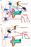
Mechanisms of CaMKII-mediated afterdepolarizations. (A) Pathological CaMKII regulation can trigger diastolic SR Ca2+ release resulting in electrogenic Ca2+ extrusion by NCX and DADs (red arrows). This results from pro-arrhythmic CaMKII regulation at multiple target proteins, which together drive 3 mechanisms through which CaMKII promotes SCR. First, CaMKII elicits gain-of-function effects at ICaL and SERCA (via PLN-mediated disinhibition), thereby increasing Ca2+ influx and resulting in Ca2+ overload (mechanism 1, blue arrows). Second, CaMKII hyper-phosphorylation of NaV1.5 can elevate intracellular Na+ resulting in Na+-induced Ca2+-overload by decreasing Ca2+ efflux through NCX (mechanism 2, black arrows). Third, CaMKII directly phosphorylates RyR2, which has been shown to promote SCR and has been strongly implicated in a number of different models of arrhythmogenic disease (mechanism 3, red arrow). (B) CaMKII phosphorylation of depolarizing currents reduces repolarization reserve and promotes EADs (red arrows) in certain disease contexts. Altered LCC gating due to CaMKII hyper-activity can elicit EADs by increasing and prolonging ICaL (mechanism 4, blue arrows). NaV1.5 phosphorylation increases INaL and may lead to non-equilibrium reactivation of INa (mechanism 5, black arrows), both of which can trigger EADs.

References

    1. Ai X., Curran J. W., Shannon T. R., Bers D. M., Pogwizd S. M. (2005). Ca2+/calmodulin-dependent protein kinase modulates cardiac ryanodine receptor phosphorylation and sarcoplasmic reticulum Ca2+ leak in heart failure. Circ. Res. 97 1314–1322 10.1161/01.RES.0000194329.41863.89 - DOI - PubMed
    1. Aiba T., Barth A. S., Hesketh G. G., Hashambhoy Y. L., Chakir K., Tunin R. S., et al. (2013). Cardiac resynchronization therapy improves altered Na channel gating in canine model of dyssynchronous heart failure. Circ. Arrhythm. Electrophysiol. 6 546–554 10.1161/CIRCEP.113.000400 - DOI - PMC - PubMed
    1. Aiba T., Hesketh G. G., Barth A. S., Liu T., Daya S., Chakir K., et al. (2009). Electrophysiological consequences of dyssynchronous heart failure and its restoration by resynchronization therapy. Circulation 119 1220–1230 10.1161/CIRCULATIONAHA.108.794834 - DOI - PMC - PubMed
    1. Anderson M. E., Braun A. P., Schulman H., Premack B. A. (1994). Multifunctional Ca2+/calmodulin-dependent protein kinase mediates Ca(2+)-induced enhancement of the L-type Ca2+ current in rabbit ventricular myocytes. Circ. Res. 75 854–861 10.1161/01.RES.75.5.854 - DOI - PubMed
    1. Ashpole N. M., Herren A. W., Ginsburg K. S., Brogan J. D., Johnson D. E., Cummins T. R., et al. (2012). Ca2+/calmodulin-dependent protein kinase II (CaMKII) regulates cardiac sodium channel NaV1.5 gating by multiple phosphorylation sites. J. Biol. Chem. 287 19856–19869. 10.1074/jbc.M111.322537 - DOI - PMC - PubMed

LinkOut - more resources